
Multiplexed quantitative detection of pathogens

Inactive Publication Date: 2007-06-14
PRIMERADX
View PDF7 Cites 123 Cited by
  • Summary
  • Abstract
  • Description
  • Claims
  • Application Information

AI Technical Summary

Benefits of technology

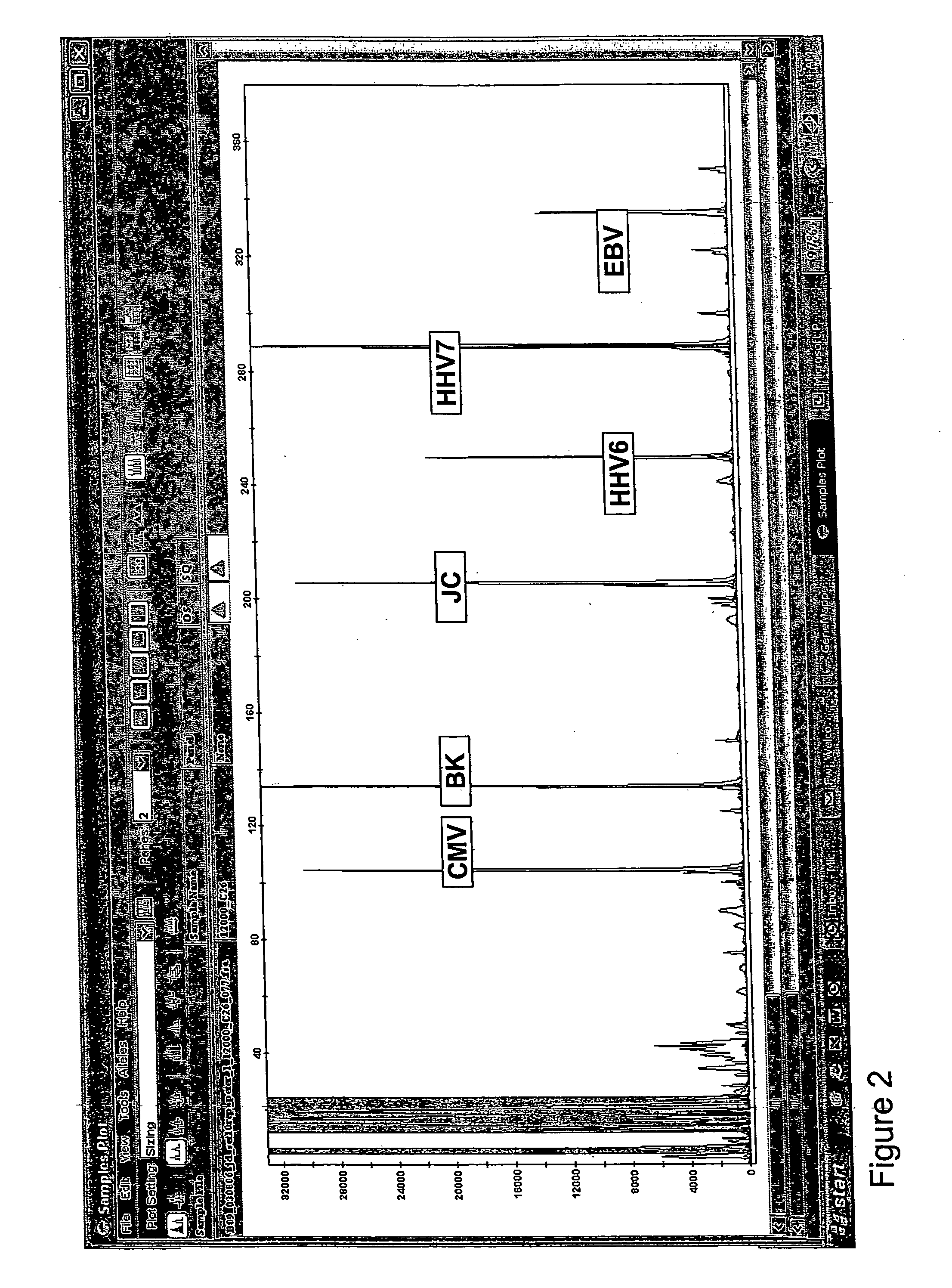
[0008] In one aspect, the methods described herein use internal standards generated through the use of various known concentrations of exogenously added competitor nucleic acids that generate amplification products of known sizes that differ from each other and from the size of the target nucleic acid(s). Size separation by, for example, capillary electrophoresis, coupled with detection by, for example, fluorescence detection, generates a standard curve from the abundance of the amplification products corresponding to the competitor nucleic acids. The standard curve permits the determination of the target nucleic acid concentration(s) in the original sample.
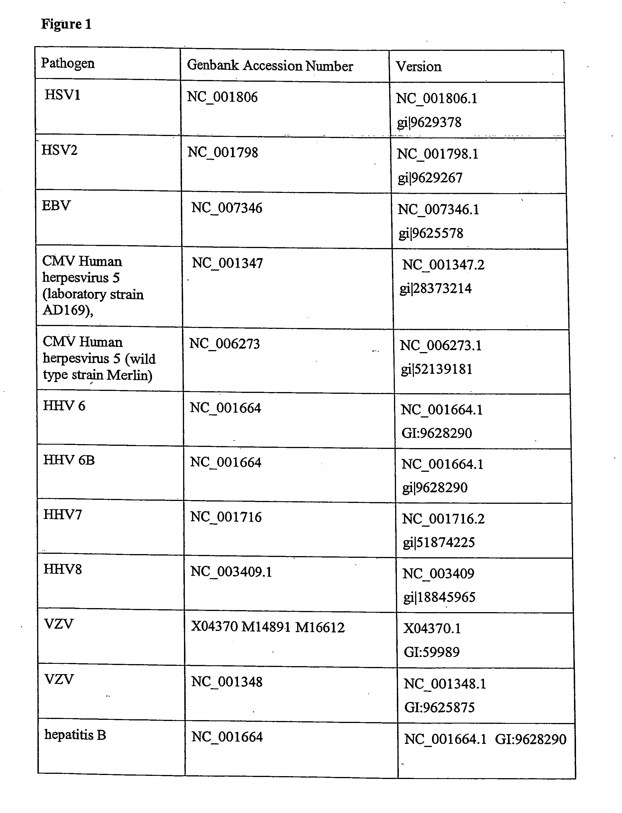
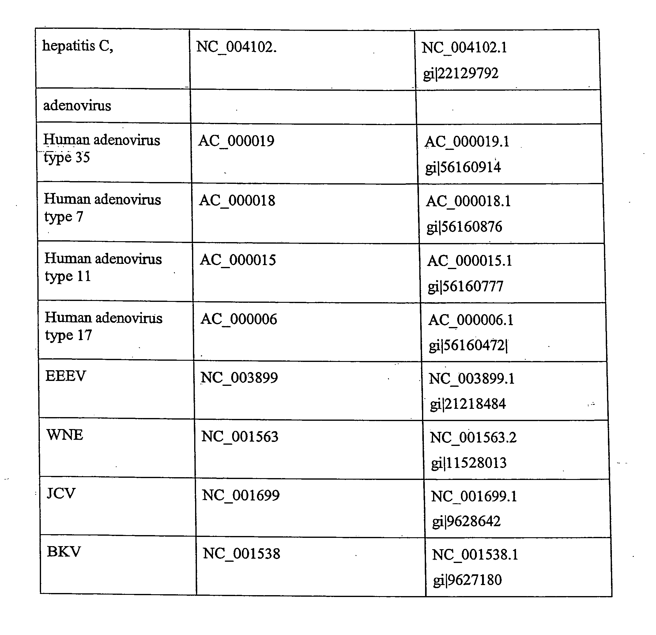
[0009] In one aspect, the methods described herein relate to methods of estimating or determining the level of a pathogen specific target nucleic acid, e.g., a DNA or RNA in a nucleic acid sample, the method comprising: for a given pathogen specific target nucleic acid, selecting a pair of amplification primers that will generate a target amplicon of known length upon amplification of the target, e.g., by PCR or RT-PCR. A set of at least two competitor nucleic acids (e.g., DNA or RNA molecules) is generated, where the competitors yield products of differing lengths but similar amplification efficiencies relative to the target nucleic acid when amplified using the same pair of amplification primers. An amplification reaction is performed in which a sample to be analyzed for target nucleic acid level is mixed with known and differing concentrations of the at least two competitor nucleic acids, followed by separation and detection of the amplified products. The set of competitor nucleic acids provides an internal reference for the determination of target nucleic acid amount in the original sample. This approach is readily adapted to measure multiple pathogen specific target nucleic acids in a single sample in a single run, which permits the generation of an amplification profile for the selected pathogen target gene sequences in a given sample. The profile permits accurate quantitation of the level of pathogen-specific nucleic acid in a given sample.

Problems solved by technology

However, the immunosuppressive therapies need to be carefully monitored because they can cause the recipient to be particularly susceptible to infection by bacteria and viruses that otherwise would be controlled by a normal immune system.

Method used


Image

  • Multiplexed quantitative detection of pathogens
  • Multiplexed quantitative detection of pathogens
  • Multiplexed quantitative detection of pathogens

Examples


example 1

Oligonucleotide Design and Synthesis

[0289] Primers are selected using PrimerSelect software (DNASTAR Inc, Madison, Wis.) based on the following criteria:

[0290] 19-24 nucleotides in length; Melting temperature (Tm) 54.5-58.2° C.; primer stability-45.9 to −39.9 kcal per mole; unique primer 3′ sequence of 7 nucleotides; avoiding self-primer and primer pair formation longer than 2 contiguous bases (ignoring duplexing 8 bases from 3′ end); avoiding internal primer hairpins longer than 2 bases; with minimal 3′ pentamer stability of −8.5 kcal per mole or more.

[0291] In addition, selected primer pairs are assessed for dimer formation in multiplex across different pairs to eliminate any potential dimers with stability less than −6.0 kcal per mole. Furthermore, primers are screened against none-redundant DNA database (Gene Bank, NCBI) using BLAST search program to eliminate any primers with significant (greater than 14 contiguous nucleotides over or 10 contiguous nucleotides from 3′-end) h...

example 2

PCR Amplification and End Detection of Microorganisms

A. One-Step RT-PCR Detection of Microorganism RNA Using Microorganism-Specific Primers

[0292] RNA template is added to the reaction mixture containing 0.25 uM of each RT primer (optional), 0.25 uM of gene-specific PCR primers (one primer of microorganism-specific pair labeled with FAM at 5′ end), a modified 1× Stratagene RT-PCR buffer (Brilliant Single Q-RT-PCR kit cat.#600532), 0.1% Triton X100, 0.2 mM DNTP, 1.5 mM MgCl2, and 1.25U of StrataScript RTase (Stratagene, La Jolla, Calif.) in a total volume of 50 or 100 ul, and overlaid with a mineral oil. Reverse transcription is conducted at 45° C. for 50 min, followed by 2 min incubation at 94 C to inactivate the RTase. Samples are then PCR amplified using a protocol consisting of 44 cycles of 94° C. for 30 seconds, 60° C. for 30 seconds and 72° C. for 1 minute. While ramping up to the first 72 C extension, 1U of thermostable DNA polymerase (Vent exo(−) (New England Biolabs)) is a...

example 3

PCR Amplification and Real-Time Detection of Microorganisms

A. One-Step RT-PCR Detection of Microorganism RNA Using Gene-Specific Primers.

[0296] Briefly, in a total volume of 50 or 100 ul, RNA sample (1-5 ul) is added to the reaction mixture containing 0.25 uM of each RT primer (optional), 0.25 uM of microorganism-specific PCR primers (one primer of microorganism-specific pair labeled with FAM at 5′ end), a modified 1× Stratagene RT-PCR buffer (Brilliant Single Q-RT-PCR kit cat.#600532), 0.1% Triton X100, 0.2 mM dNTP, 1.5 mM MgCl2, and 1.25U of StrataScript RTase (Stratagene, La Jolla, Calif.) and overlaid with a mineral oil. Reverse transcription is conducted at 45 C for 50 min, followed by 2 min incubation at 94° C. to inactivate the RTase. Samples are then PCR amplified using a protocol consisting of 44 cycles of 94° C. for 30 seconds, 60° C. for 30 seconds and 72° C. for 1 minute. While ramping up to the first 72° C. extension, 1U of thermostable DNA polymerase (Vent exo(−) (N...


PUM

PropertyMeasurementUnit
Volumeaaaaaaaaaa
Volumeaaaaaaaaaa
Volumeaaaaaaaaaa

Abstract

The invention allows for the quantitative detection of a plurality of pathogens in a single sample. The method includes the amplification of a sample with a plurality of pathogen-specific primer pairs to generate amplicons of distinct sizes from each of the pathogen specific primer pairs. The method further includes the use of a plurality of competitor polynucleotide targets that correspond to each of the pathogen-specific primer pairs. The competitor polynucleotides are added to the reaction mixture at a known concentration to allow for the quantitation of the amount of pathogen in the sample. The method can be used for monitoring pathogen infection in an individual, preferably an immunocompromised individual.

Description

REFERENCE TO RELATED APPLICATIONS [0001] The present claims priority to and the benefit of U.S. provisional patent application Ser. No. 60 / 735,085, filed Nov. 9, 2005, the entire contents of which are incorporated herein by reference.FIELD OF THE INVENTION [0002] The invention relates to methods and compositions for quantitative testing in a sample for two or more viral, bacterial or protozoan pathogens contemporaneously. More specifically, the invention relates to methods and compositions for quantitative testing in a sample from an individual to detect and / or monitor pathogen infection quantitatively. BACKGROUND OF THE INVENTION [0003] Immune deficiency may result from many different etiologies including hereditary genetic abnormalities (e.g., Chediak-Higashi Syndrome, Severe Combined Immunodeficiency, Chronic Granulomatous Disease, DiGeorge Syndrome) exposure to radiation, chemotherapy, heavy metals or insecticides; or, acquired as a result of bacterial, viral (HIV), parasitic or...

Claims


Application Information

Patent Timeline
IPC IPC(8): C12Q1/70C12Q1/68
CPCC12Q1/6851C12Q1/686C12Q1/6865C12Q1/6888C12Q1/701C12Q1/705C12Q2561/113C12Q2545/10C12Q2537/143C12Q2545/107C12Q2525/204C12Q2600/16
Inventor SLEPNEV, VLADIMIR I.SHIOSAKI, KAZUMIHART, KYLEGARCIA, ELIZABETH
Owner PRIMERADX
Learn More