
Mammalian antigen-presenting T cells and bi-specific T cells

a technology of antigen-presenting t cells and t cells, which is applied in the field of mammalian bispecific t cells, can solve the problems of high risk of disease death, low complete response rate or high incidence of early relapse in patients, and achieve the effect of maintaining in vivo function long-term

Inactive Publication Date: 2007-07-19
CITY OF HOPE
View PDF2 Cites 19 Cited by
  • Summary
  • Abstract
  • Description
  • Claims
  • Application Information

AI Technical Summary

Benefits of technology

[0015] Accordingly, the present invention is directed to bi-specific mammalian T cells and methods for using these bi-specific T cells. More specifically, the invention relates to viral specific T cells that express chimeric anti-tumor receptors. These bi-specific T cells are a source of persistent effector cells that respond to stimulation with viral antigen, allowing the cells to maintain in vivo function long-term.
[0018] In a third aspect, the invention provides a method of improving the in vivo survival of bi-specific T cells through the exogenous administration of interleukin-2 (IL-2).
[0021] In a sixth aspect, the invention provides a method of effectively eliminating bi-specific T cells in vivo by withdrawing administration of the viral antigen recognized by the bi-specific T cell or with-holding viral antigen recognized by the bi-specific T cell.

Problems solved by technology

While these lymphomas are relatively indolent, they are generally considered incurable using conventional treatments.
Patients tend to relapse after therapy, their response to salvage therapy of shorter duration after every relapse, eventually leading to death from disease-related causes.
Patients with low complete response rates or high incidence of early relapse are at especially high risk.
Although capable of initiating T-cell anti-tumor activity upon cross-linking of the extracellular component, some chimeric immunoreceptors currently under consideration for clinical trials only deliver a primary activation signal through a chimeric CD3-ζ domain or FcεRI receptor γ-chain, which may result in an T-cell activation signal that may not be fully competent, based on evidence from well-recognized transgenic mice models.
However, prior methods of identifying and expanding endogenous tumor-specific T cells that can function in vivo to eradicate established disease has been limited by two factors: (i) the difficulty of overcoming or regulating T-cell tolerance to “self” antigens and (ii) down-regulation of major histocompatibility complex MHC molecules on tumor escape-variants by tumor-specific T cells, since recognition of most TAAs is dependent on MHC glycoprotein presentation.
Although adoptive transfer of chimeric receptor-expressing peripheral blood-derived T lymphocytes has resulted in anti-tumor activity in mice, clinical results have so far been disappointing.
The most germane issue appears to be that adoptively transferred chimeric T cells fail to expand and lose their function in vivo in the absence of any immune response directed against the chimeric T cells.
Even under these conditions, however, responsiveness was soon lost.
This problem is exacerbated by the general lack of tumor cell costimulatory molecules essential for the induction and maintenance of a T-cell response.
Although this strategy was effective in maintaining functional activation of the chimeric receptor-modified T cells, it is not conducive to modulating the number of chimeric receptor-modified T cells in vivo for the purposes of coordinating anti-tumor responses in patients, especially those with relapsed malignancies.
The major drawback to using EBV-specific T cells is that neither the patient nor the investigator can control the amount of EBV antigen to which the viral-specific T cells are exposed.
This may result in unpredictable stimulation of the genetically modified T cells leading to possible lack of function or to over-expansion causing potential toxicity or functional inactivation of the over-stimulated T cells.
This is particularly important when the introduced chimeric immunoreceptor also targets normal tissue, because over-stimulated bi-specific T cells may cause unwelcome recognition of normal host tissues.
In addition, there would be no easy way to eliminate the T cells or their activity when it was no longer desired.

Method used


Image

  • Mammalian antigen-presenting T cells and bi-specific T cells
  • Mammalian antigen-presenting T cells and bi-specific T cells
  • Mammalian antigen-presenting T cells and bi-specific T cells

Examples


example 1

Generation of T Cells Expressing MP1 Antigen

[0094] To avoid exposure to infectious virus and circumvent the use of soluble MP1-derived peptide(s), which may not bind to all classical HLA class I antigens, HLA A2+ antigen presenting (AP)-T cells were genetically modified by non-viral gene transfer with the DNA plasmid HyMP1-pMG. Hygromycin phosphotransferase (Hy), which confers resistance to the antibiotic hygromycin B in E. coli and mammalian cells, was expressed from the pMGˆPac vector. This vector is a modification of the pMG vector (InvivoGen, San Diego, Calif.) by site-directed mutagenesis to remove a Pac I RE site at position 307. See FIG. 4.

[0095] The Hy gene plasmid in pMGˆPac was changed to Kanamycin / G418-resistance gene to generate the plasmid intermediate pKEN. Subsequent deletion of the neomycin phosphotransferase gene produced the plasmid pEK. This plasmid was used to express the HyMP1 gene, a fusion of a 972 base pair (bp) fragment of the Hy gene from the DNA plasmid ...

example 2

In Vitro T-Cell Culture System to Expand MP1-Specific CD8+ T Cells using Autologous T Cells Presenting MP1

[0108] A kinetic study determined whether the HyMP1-expressing, genetically modified AP-T cells could directly stimulate expansion of CD8+ MP1-specific T cells in vitro. During three weeks of co-culture with irradiated autologous AP-T cells expressing the HyMP1 gene, flow cytometry was used to demonstrate the expansion of MP1-tetramer+ T cells from a HLA A2+ healthy volunteer donor. HLA A2+ PBMC were co-cultured for 21 days in the presence of low-dose IL-2 (A) without the addition of autologous AP-T cells, or with a 5:1 (Responder:Stimulator) T-cell ratio of γ-irradiated hygromycin-resistant (B) Hy+ AP-T cells (that do not express MP1), or (C) γ-irradiated HyMP1+ AP-T cells. AP-T cells were re-added to the culture system every 7 days. Binding of a control CMV pp65-tetramer on day 21 was negligible. Dead cells were excluded from analysis upon uptake of propidium iodide (PI).

[01...

example 3

MP1-Specific T Cells can be Genetically Modified to Express a CD19-Specific Chimeric Immunoreceptor

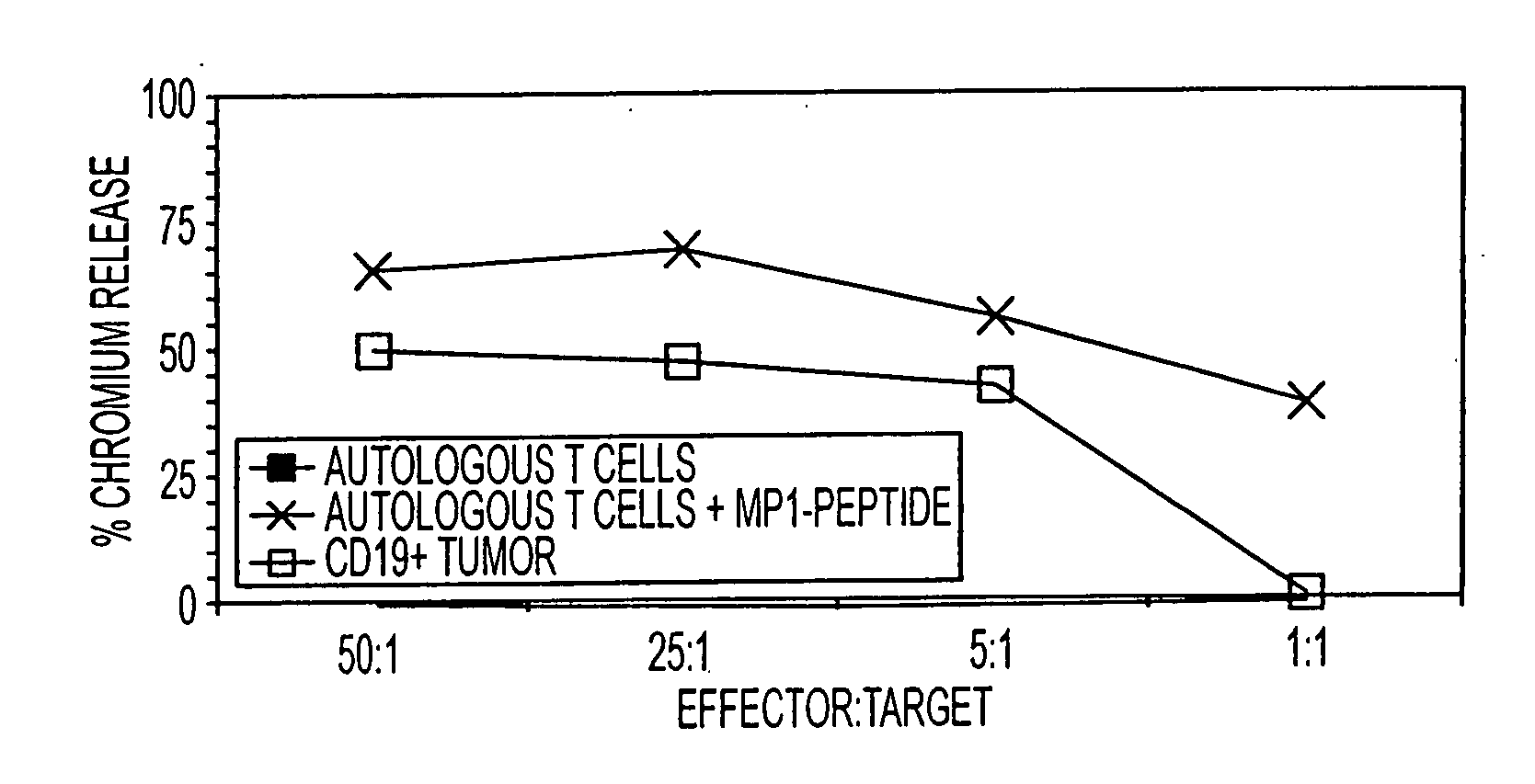
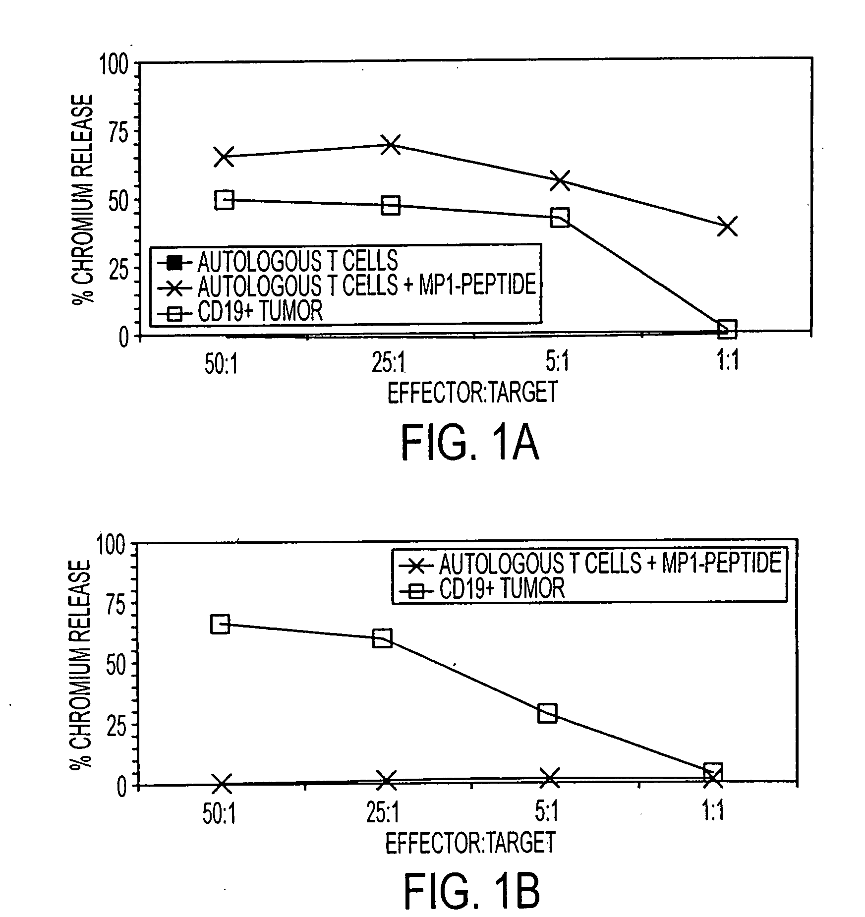
[0114] To determine if MP1-specific T cells could be rendered specific for CD19, the CD19R gene was introduced into MP1-tetramer+ T cells. This genetic modification of T cells was accomplished using non-viral electrotransfer of a DNA expression plasmid designated CD19R / HyTK-pMG which codes for both CD19R and a bifunctional fusion gene thaT combines hygromycin phosphotransferase and herpes virus thymidine kinase (HyTK). The specificity of CD19R is derived from the variable regions of a mouse monoclonal antibody (mAb) specific for CD19, tethered to the T cell via a modified human IgG4 hinge and Fc-fragment attached to the human CD4 transmembrane domain. Upon binding CD19, the genetically modified T cells are activated via the cytoplasmic CD3-ζ chain attached to the chimeric immunoreceptor.

[0115] HLA A2+ T cells were expanded on autologous HyMP1+ AP T cells, FACS sorted for binding to M...


PUM

PropertyMeasurementUnit
concentrationsaaaaaaaaaa
pHaaaaaaaaaa
concentrationsaaaaaaaaaa

Abstract

The present invention is directed to mammalian bi-specific T cells and methods for using these bi-specific T cells. More specifically, the invention relates to viral specific T cells that express chimeric anti-tumor receptors. These bi-specific T cell clones are a source of effector cells that persist in vivo in response to stimulation with viral antigen, leading to long-term function after their transfer to patients with cancer and autoimmune diseases.

Description

[0001] This is a continuation of U.S. application Ser. No. 10 / 797,609, filed Mar. 11, 2004, which claims the benefit of prior co-pending U.S. Provisional Application Ser. No. 60 / 453,197, filed Mar. 11, 2003. The disclosures of both above applications are hereby incorporated by reference in their entirety.STATEMENT OF GOVERNMENT SUPPORT [0002] This application was made in part with Government support under Grant No. PO0 CA30206 and CA33572, funded by the National Cancer Institute, National Institutes of Health, Bethesda, Md. The federal government may have certain rights in this invention.BACKGROUND [0003] The present invention generally relates to mammalian bi-specific T cells and methods for using these bi-specific T cells. More specifically, the invention relates to viral specific T cells, which express chimeric anti-tumor receptors. These bi-specific T cells form a source of effector cells that persist in vivo in response to stimulation with viral antigen, leading to long-term fu...

Claims


Application Information

Patent Timeline
Patent Type & Authority Applications(United States)
IPC IPC(8): A61K39/395A61K39/12A61K39/00A61K48/00C12N5/0783
CPCA61K39/0011A61K2039/5158A61K39/245A61K35/17A61K39/145C12N5/0636A61P35/00A61K39/001112A61K39/001124A61K35/12
Inventor COOPER, LAURENCEJENSEN, MICHAEL
Owner CITY OF HOPE
Learn More