
Self-contained electromagnetic apparatus for treatment of molecules, cells, tissues, and organs within a cerebrofacial area and method for using same

a technology of electromagnetic apparatus and cerebrofacial area, which is applied in the direction of electrotherapy, magnetotherapy using coils/electromagnets, electrotherapy, etc., can solve the problems of inefficient prior art waveforms, insufficient configuration of waveforms, and inability to take into account the dielectric properties of tissue structure as opposed to the properties of isolated cells, etc., to achieve short pulse duration, modulation of angiogenesis and neovascularization, and low peak ampli

Inactive Publication Date: 2007-07-26
RIO GRANDE NEUROSCI
View PDF0 Cites 30 Cited by
  • Summary
  • Abstract
  • Description
  • Claims
  • Application Information

AI Technical Summary

Benefits of technology

[0016] By increasing a range of frequency components transmitted to relevant cellular pathways, hair and other cerebrofacial tissue restoration is advantageously achieved.
[0043] It is another object of the present invention to significantly lower peak amplitudes and shorter pulse duration. This can be accomplished by matching via Power SNR, a frequency range in a signal to frequency response and sensitivity of a target pathway structure such as a molecule, cell, tissue, and organ, in a cerebrofacial area to enable modulation of angiogenesis and neovascularization.

Problems solved by technology

Prior art considerations of EMF dosimetry have not taken into account dielectric properties of tissue structure as opposed to the properties of isolated cells.
However, prior art in this field does not configure waveforms based upon an ion / ligand binding transduction pathway.
Prior art waveforms are inefficient since prior art waveforms apply unnecessarily high amplitude and power to living tissues and cells, require unnecessarily long treatment time, and cannot be generated by a portable device.
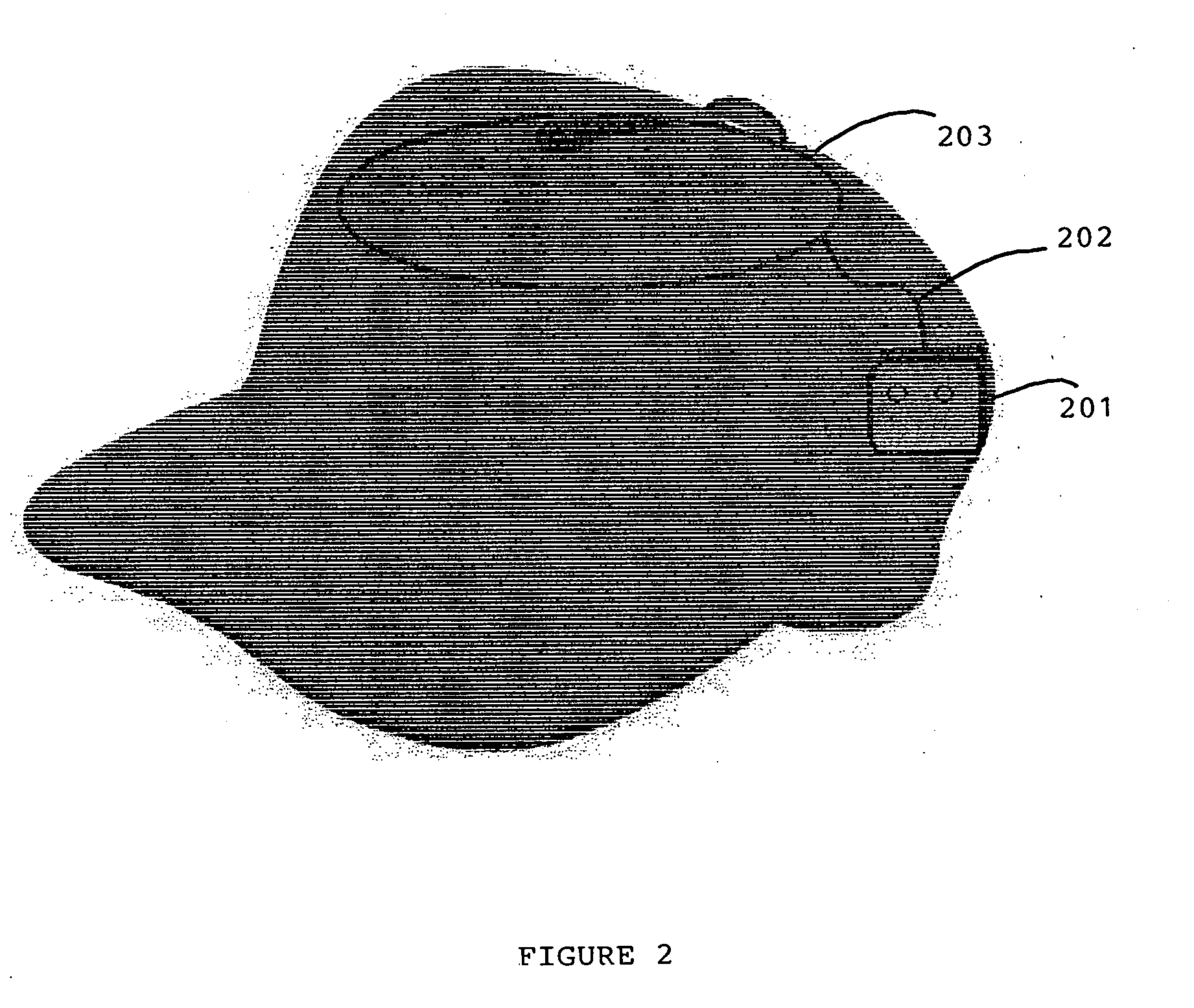
Prior art equipment in this field is bulky, not designed for outdoor use, and not self-contained.

Method used


Image

  • Self-contained electromagnetic apparatus for treatment of molecules, cells, tissues, and organs within a cerebrofacial area and method for using same
  • Self-contained electromagnetic apparatus for treatment of molecules, cells, tissues, and organs within a cerebrofacial area and method for using same
  • Self-contained electromagnetic apparatus for treatment of molecules, cells, tissues, and organs within a cerebrofacial area and method for using same

Examples


example 1

[0071] The Power SNR approach for PMF signal configuration has been tested experimentally on calcium dependent myosin phosphorylation in a standard enzyme assay. The cell-free reaction mixture was chosen for phosphorylation rate to be linear in time for several minutes, and for sub-saturation Ca2+ concentration. This opens the biological window for Ca2+ / CaM to be EMF-sensitive. This system is not responsive to PMF at levels utilized in this study if Ca2+ is at saturation levels with respect to CaM, and reaction is not slowed to a minute time range. Experiments were performed using myosin light chain (“MLC”) and myosin light chain kinase (“MLCK”) isolated from turkey gizzard. A reaction mixture consisted of a basic solution containing 40 mM Hepes buffer, pH 7.0; 0.5 mM magnesium acetate; 1 mg / ml bovine serum albumin, 0.1% (w / v) Tween 80; and 1 mM EGTA12. Free Ca2+ was varied in the 1-7 μM range. Once Ca2+ buffering was established, freshly prepared 70 nM CaM, 160 nM MLC and 2 nM MLCK...

example 2

[0075] According to an embodiment of the present invention use of a Power SNR model was further verified in an in vivo wound repair model. A rat wound model has been well characterized both biomechanically and biochemically, and was used in this study. Healthy, young adult male Sprague Dawley rats weighing more than 300 grams were utilized.

[0076] The animals were anesthetized with an intraperitoneal dose of Ketamine 75 mg / kg and Medetomidine 0.5 mg / kg. After adequate anesthesia had been achieved, the dorsum was shaved, prepped with a dilute betadine / alcohol solution, and draped using sterile technique. Using a #10 scalpel, an 8-cm linear incision was performed through the skin down to the fascia on the dorsum of each rat. The wound edges were bluntly dissected to break any remaining dermal fibers, leaving an open wound approximately 4 cm in diameter. Hemostasis was obtained with applied pressure to avoid any damage to the skin edges. The skin edges were then closed with a 4-0 Ethil...

example 3

[0081] This example illustrates the effects of PRF electromagnetic fields chosen via the Power SNR method on neurons in culture.

[0082] Primary cultures were established from embryonic days 15-16 rodent mesencephalon. This area is dissected, dissociated into single cells by mechanical trituration, and cells are plated in either defined medium or medium with serum. Cells are typically treated after 6 days of culture, when neurons have matured and developed mechanisms that render them vulnerable to biologically relevant toxins. After treatment, conditioned media is collected. Enzyme linked immunosorbent assays (“ELISAs”) for growth factors such as Fibroblast Growth Factor beta (“FGFb”) are used to quantify their release into the medium. Dopaminergic neurons are identified with an antibody to tyrosine hydroxylase (“TH”), an enzyme that converts the amino acid tyrosine to L-dopa, the precursor of dopamine, since dopaminergic neurons are the only cells that produce this enzyme in this sy...


PUM


Abstract

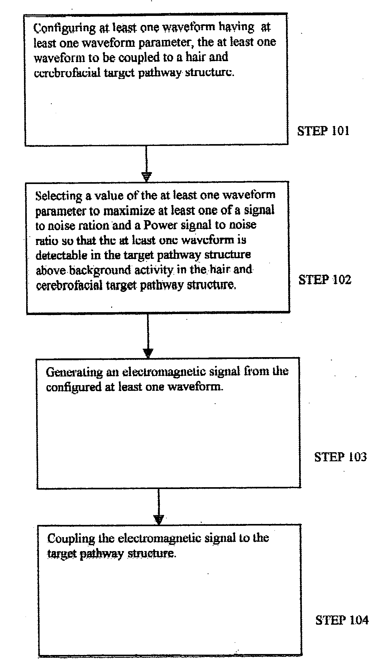
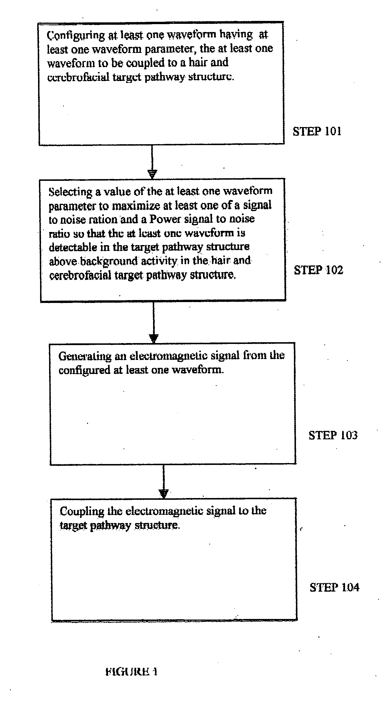
An apparatus and method for electromagnetic treatment for hair restoration and treatment of cerebrofacial molecules, cells, tissues, and organs comprising configuring at least one waveform having at least one waveform parameter, selecting a value of the at least one waveform parameter of the at least one waveform to maximize at least one of a signal to noise ratio and a Power signal to noise ratio in a target pathway structure, using the at least one waveform that maximizes the at least one of a signal to noise ratio and a Power signal to noise ratio in a target pathway structure to generate an electromagnetic signal, and coupling the electromagnetic signal to the hair and cerebrofacial target pathway structure to modulate the hair and cerebrofacial target pathway structure.

Description

BACKGROUND OF THE INVENTION [0001] 1. Field of the Invention [0002] This invention pertains generally to an apparatus and a method for using electromagnetic therapy treatment for hair maintenance and restoration and for treatment of degenerative neurological pathologies and other cerebrofacial conditions, including sleep disorders, by modulation of the interaction of hair, cerebral, neurological, and other tissues with their in situ electromagnetic environment. This invention also relates to a method of modification of cellular and tissue growth, repair, maintenance, and general behavior by application of encoded electromagnetic information to molecules, cells, tissues and organs on humans and animals. More particularly this invention relates to the application of surgically non-invasive coupling of highly specific electromagnetic signal patterns to hair and other cerebrofacial tissue. In particular, an embodiment according to the present invention pertains to using a self-contained...

Claims


Application Information

Patent Timeline
Patent Type & Authority Applications(United States)
IPC IPC(8): A61N1/18
CPCA61N2/02A61N1/328A61N1/00
Inventor PILLA, ARTHUR A.
Owner RIO GRANDE NEUROSCI
Learn More