Oligosaccharide modification and labeling of proteins

a technology of protein and oligosaccharide, applied in the field of protein method, can solve the problem of difficulty in determining how many labels are present on each igg

Active Publication Date: 2007-08-16
LIFE TECH CORP
View PDF99 Cites 78 Cited by
  • Summary
  • Abstract
  • Description
  • Claims
  • Application Information

AI Technical Summary

Benefits of technology

[0010] By labeling antibodies at glycan residues in the Fc portion of the antibody, instead of the traditional labeling of cysteine or lysine residues in the binding region of the antibody, possible disruption of epitope binding is avoided. In addition, the number of glycan residues present on the IgG, is generally known. In contrast, each epitope-specific IgG may have a different number of lysine or cysteine residues in the peptide sequence, and each may have a different ability to be labeled, based on the IgG structure. Therefore, it would generally be difficult to determine how many labels were present on each IgG. Using the methods of the present invention, it is expected that about all of the glycan residues are labeled, which allows for quantitative labeling and detection of the antibodies using, for example, fluorescence-activated cell sorting (FACS).
[0012] Besides the humanization of therapeutic antibodies, there are many possible applications for the methods of the present invention. For example, glycosylation patterns can alter in certain diseases or conditions such as, for example, rheumatoid arthritis and pregnancy. The ability to mix and match oligosaccharides may enable researchers to investigate human diseases involving altered glycosylation in animal models.

Problems solved by technology

Therefore, it would generally be difficult to determine how many labels were present on each IgG.

Method used


Image

  • Oligosaccharide modification and labeling of proteins
  • Oligosaccharide modification and labeling of proteins
  • Oligosaccharide modification and labeling of proteins

Examples


example 1

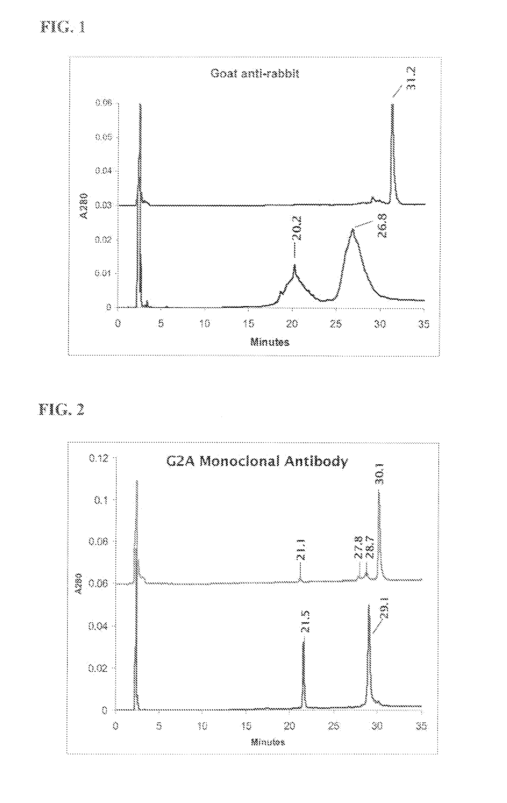
[0233] HPLC verification of monoclonal and polyclonal antibodies with and without oligosaccharide cleavage by Endo H glycosidase.

[0234] Deglycosylation reaction: To 90 μL reduced and alkylated antibody (1 mg / ml) add 40 μL 0.5 M sodium citrate pH 5.5 and 10 μL EndoHf, (1,000,000 U / mL, New England BioLabs). Incubate for 48 hours at 37° C. with rocking. Samples were centrifuged to remove precipitate and then injected directly onto the HPLC. Reversed-phase HPLC was performed on an Agilent Zorbax 300SB-CN column (4.6×150 mm, 3.5 μm) at 75° C. with a flow rate of 0.8 mL / min using a Waters 600E LC system. The mobile phase included water with 0.1% TFA in solvent A and 80% n-propanol, 10% acetonitrile, 10% water with 0.1% TFA in solvent B (conditons of Rehder et al., J. Chrom. A, 1102 (2006) 164-175). Separation was accomplished using a linear gradient from 20 to 40% B over 30 min. Results are depicted in FIG. 1 and FIG. 2.

example 2

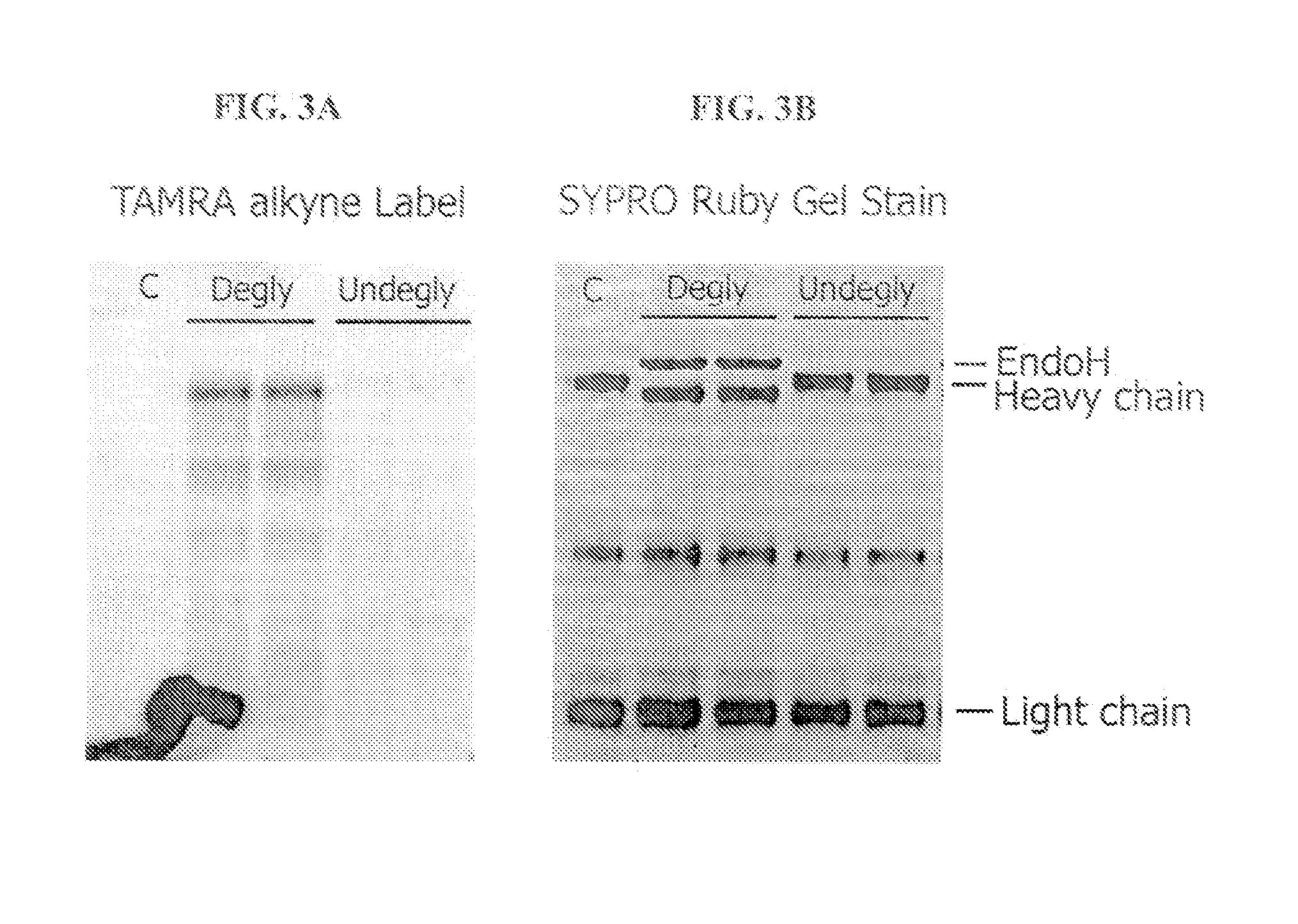
Enzymatic Labeling of Chicken Anti-goat Antibody with and without Oligosaccharide Cleavage by Endo H Glycosidase

[0235] Chicken anti-goat IgG was washed into 50 mM MES pH 6.5 buffer on a VivaSpin 5000 MWCO filter column. For the deglycosylation reaction, an aliquot of washed IgG at 1 mg / mL was incubated 24 hours at 37° C. in 50 mM MES pH 6.5 containing 47.5 U / μL Endo H (New England BioLabs). Washed (undeglycosylated) or EndoHf digested IgG was then azido-labeled with the Click-iT O-GlcNAc enzymatic labeling kit using nondenaturing conditions: 0.5 mg / ml IgG in 50 mM MES pH 6.5, 120 mM NaCl, 11 mM MnCl2, 0.1 mM ZnCl2, 50 μM UDP-GalNAz, 5 U / mL Antarctic phosphatase, and 0.65 mg / mL GalT enzyme, overnight incubation at 4° C. The samples were purified through P10 sizing resin packed into a 0.5 mL spin column into 50 mM Tris pH 8. Collected fractions containing antibody were then labeled with the TAMRA Click-iT™ detection kit (C33370) and purified again through P10 sizing resin packed into...

example 3

Metabolic Labeling of Antibodies with Azido Sugars

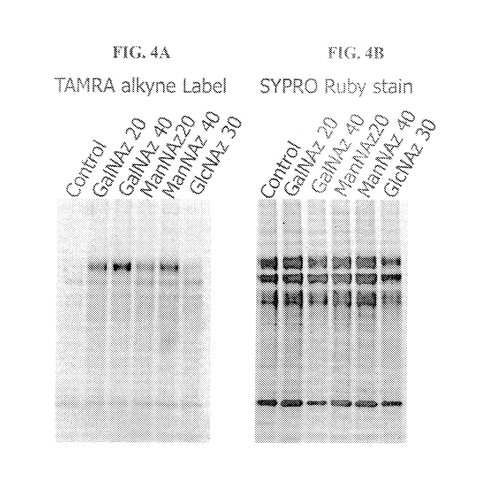
[0236] Mouse M96 hybridoma cells were fed the azido sugar analogues, Ac4GalNAz, Ac4ManNAz or Ac4GlcNAz for five days. The cell supernatant (containing the azido-labeled monoclonal antibody) was collected and proteins were precipitated in chloroform / methanol. Precipitated pellets were resolubilized with 50 μL of 1% SDS, 100 mM TRIS pH 8, labeled with the TAMRA Click-iT™ detection kit (C33370) and 1 μg was analyzed on a 4-12% BIS-TRIS gel using MOPS buffer. The gels were imaged on the BioRad FX imager using the 532 nm laser and 555 nm long pass emission filter (FIG. 4A). The gels were post-stained with SYPRO® Ruby protein gel stain and imaged using the 488 nm laser and 555 nm long pass emission filter (FIG. 4B).


PUM

PropertyMeasurementUnit
fluorescentaaaaaaaaaa
fluorescence-aaaaaaaaaa
HPLCaaaaaaaaaa

Abstract

The present invention generally relates to methods of functionalizing proteins, particularly antibodies, at oligosaccharide linkages, methods of humanizing antibodies by modifying glycosylation, as well as to novel antibodies linked to modified oligosaccharides. The invention further relates to kits that may be used to produce the antibodies of the invention.

Description

[0001] This application claims priority to U.S. Provisional Application No. 60 / 772,221, filed Feb. 10, 2006 and U.S. Provisional Application No. 60 / 804,640, filed Jun. 13, 2006, the contents of which are incorporated by reference as if set forth fully herein.FIELD OF THE INVENTION [0002] The invention generally relates to methods of labeling proteins at novel oligosaccharide linkages, methods of humanizing antibodies by modifying glycosylation, as well as to novel antibodies linked to modified oligosaccharides. BACKGROUND [0003] Isolated or synthesized proteins and antibodies, such as IgGs, are used therapeutically and for diagnostic and research purposes. By labeling antibodies with detectable labels, such as, for example, fluorophores, antibodies can be used to specifically detect target biological molecules or cells. Antibodies may also be tagged with binding reagents, such as, for example, biotin, so that they may be used to specifically bind target biological molecules or cells...

Claims


Application Information

Patent Timeline
Patent Type & Authority Applications(United States)
IPC IPC(8): C12P21/06C07K16/46
CPCA61K47/48092C07K2317/41C12P21/005G01N33/531G01N33/532G01N33/5005Y10T436/17G01N33/533G01N33/534C07K16/00G01N33/583G01N33/581G01N33/582G01N33/5008A61K47/549C07K1/13C12Y302/01096C07K14/47C07K2317/14C07K2317/24
Inventor AGNEW, BRIANCORRY, SCHUYLER B.GEE, KYLE R.
Owner LIFE TECH CORP
Why Eureka
Try Eureka