

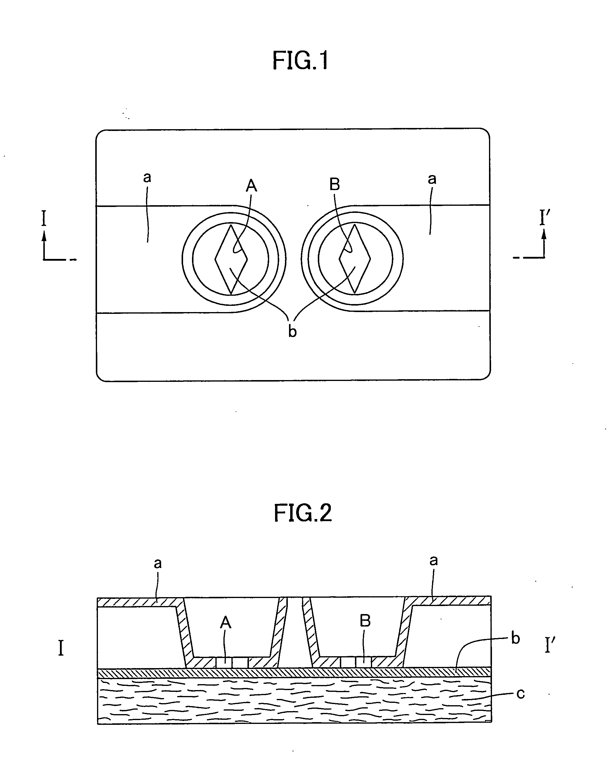
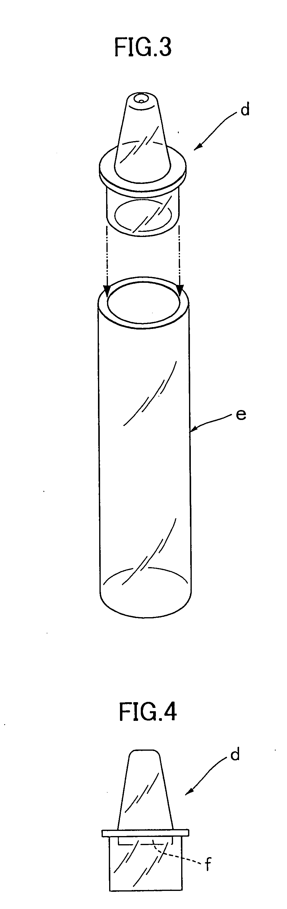
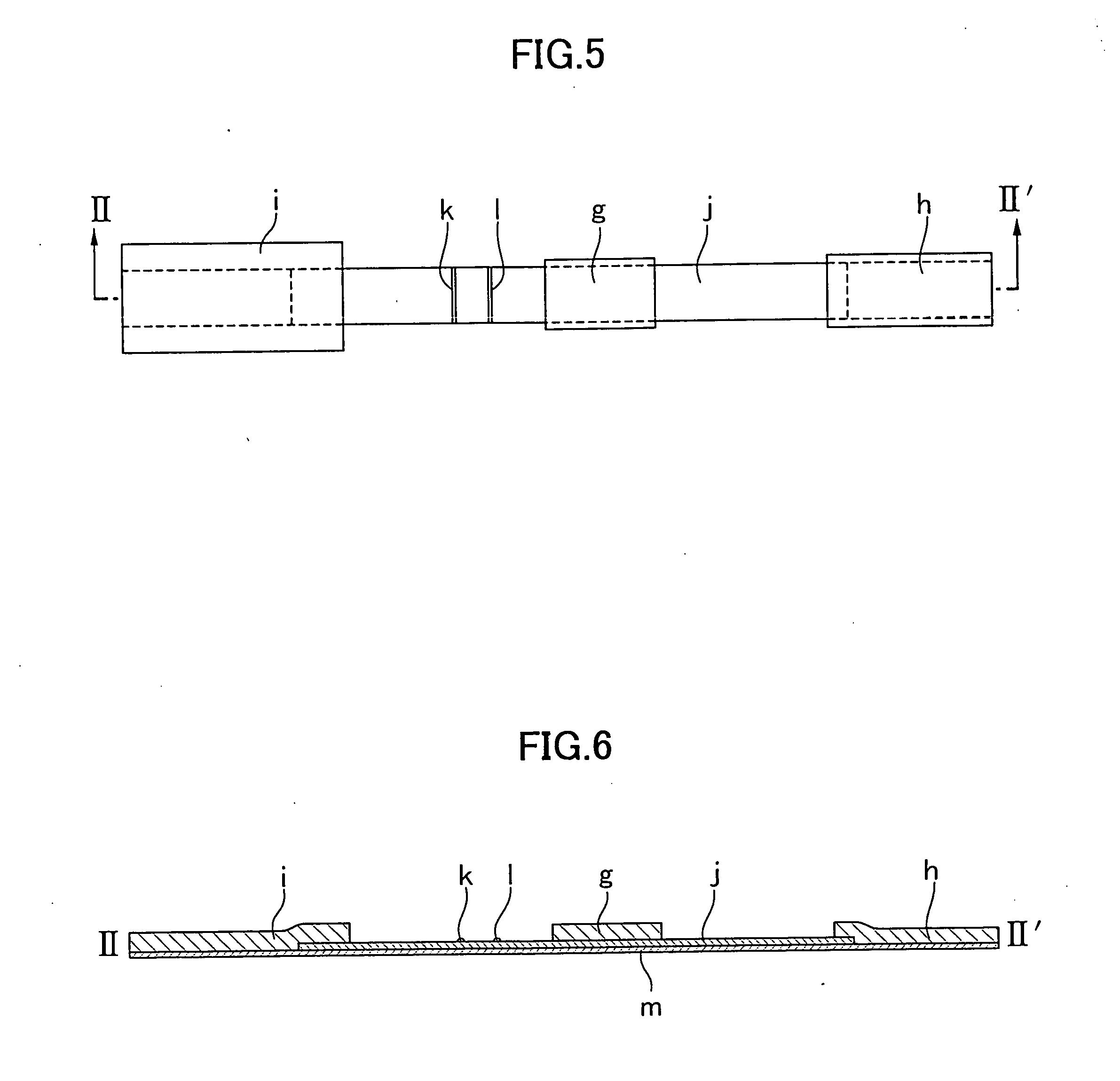
Simple membrane assay method and kit
- Summary
- Abstract
- Description
- Claims
- Application Information
AI Technical Summary
Benefits of technology
Problems solved by technology
Method used
Image

Examples
example
Example 1
Detection of Influenza Virus by Flow-Through Type Immunochromatographic Assay Method
1. Preparation of Monoclonal Antibody
(1) Preparation of Anti-Influenza a Type Virus NP Monoclonal Antibody (Mouse)
[0059] A spleen was ablated from a BALB / c mouse which had been immunized with purified influenza A type virus antigen and kept for a certain period, and it was fused with mouse myeloma cell (P3×63) by the method of Kohler et al. (Kohler et al., Nature, Vol, 256, pp 495-497 (1975)).
[0060] The obtained fused cell (hybridoma) was kept in an incubator at 37° C. and purification (monocloning) of the cell was performed while confirming the antibody activity of the supernatant by ELISA using influenza A type virus NP antigen solid phase plate.
[0061] Two of the obtained cells were each administered intraperitoneally to a pristane-treated BALB / c mouse, after about 2 weeks, each ascites containing an antibody was collected. IgG was purified from each of the obtained ascites by amm...
example 2
Detection of Influenza Virus by Lateral-Flow Type Immunochromatographic Assay Method
1. Preparation of Monoclonal Antibody
(1) Preparation of Anti-Influenza A virus NP Monoclonal Antibody (Mouse)
[0081] This antibody was prepared in the same manner as the method described in [Example 1] 1.(1).
(2) Preparation of Anti-Influenza B Type Virus NP Monoclonal Antibody (Mouse)
[0082] This antibody was prepared in the same manner as the method described in [Example 1] 1.(2).
2. Preparation of Labeled Anti-Influenza Antibody
(1) Preparation of Labeled Anti-Influenza A Type Antibody
[0083] 20 mg of one kind of the purified anti-influenza A type virus NP monoclonal antibodies was dialyzed with 0.1 M acetate buffer (pH 3.8) followed by addition of 10 mg of pepsin and Fab′ digestive treatment was performed for one hour at 37° C. The treated solution was fractionated through ultrogel AcA44 column to yield purified fraction of anti-influenza A type F(ab′)2. The thus obtained fraction was con...
PUM
 Abstract
Description
Claims
Application Information