Cell canaries for biochemical pathogen detection

a biochemical and cell technology, applied in the field of apparatus for pathogen detection and methods of detecting pathogens, can solve the problems of biochemical pathogen detection plagued by false positives, unacceptably high false positive rate, and test just barely more useful than no test at all

Inactive Publication Date: 2007-09-13
SHAPIRO BENJAMIN +3
View PDF5 Cites 216 Cited by
  • Summary
  • Abstract
  • Description
  • Claims
  • Application Information

AI Technical Summary

Benefits of technology

[0009] In a first aspect, the invention is directed to devices for detecting at least one pathogen. The device contains at least one cell that produces a signal upon contact with the pathogen, at least one cell clinic on a surface of a chip for containing the cell; and an on-chip means for detecting the signal. The presence of a pathogen correlates to the production of the signal by the cell. In some cases, more than one signal is detected; in other cases, one cell can detect multiple pathogens, generating distinct signals in response to each pathogen. The cells can be engineered to produce signals that can be detected; modifications can include the introduction of exogenous markers, polynucleotides, viruses, etc. Markers include polypeptides, nanoparticles, polypeptides and dyes. Examples of detectable signals include fluorescence, fluorescence resonance energy transfer, light emission or cessation of light emission; a change, such as a change in cell resistance, cell impedance, cell capacitance, cell ion concentration, cell or medium pH, carbon dioxide concentration, nutrient concentration, cell waste concentration, cellular mechanical properties, cell position, cell number, and temperature. Changes are detected, for example, by sensors. Examples of sensors include those for fluorescence, carbon dioxide, pH, ion concentration, cell resistance, cell impedance, cell capacitance, cell waste, cellular mechanical properties, cell position, cell number nutrient and temperature. Fluorescence sensors can include current mode pixels, APS pixes, and single-photon avalanche detectors. Sensors can also include optical filters, such as notch filters, band-pass filters, and low-pass optical filters. The optical filters can also include absorbing dyes, interference filters, distributed Bragg reflectors, and patterned light-blocking layers. The chip of the can include integrated circuitry, such as that from complementary metal oxide semiconductor technology. The cell clinics can also include actuated lids, such as those containing polypyrrole. The lids can also have semi-permeable membranes. Finally, the device can also contain light sources that direct light to cells in the clinic.

Problems solved by technology

Despite a tremendous amount of research and development efforts, biochemical pathogen detection is plagued with false positive results—that is, the pathogen is detected when not present in a sample.
In some assays, the rate of false positives is unacceptably high, rendering the test just barely more useful than no test at all.
Unacceptable rates of false positives result in part due to the complexity of biological systems, the complex interaction with pathogens, and the inability of current sensor systems to differentiate subtle distinctions between the many possible interactions.
Even those based on molecules such as DNA, RNA, and antibodies are not always able to differentiate between agents that are harmful and similar agents that are benign.
While current systems are valuable tools for detecting some pathogens, the costs, labor, complexity and most importantly, the rate of false results, mitigate their effectiveness.
Furthermore, current systems take approximately 24 hours to determine the pathogen in a sample if there is no other information to narrow down the type.

Method used


Image

  • Cell canaries for biochemical pathogen detection
  • Cell canaries for biochemical pathogen detection
  • Cell canaries for biochemical pathogen detection

Examples


example 1 integrating

MEMS Structures and CMOS Circuits for Bioelectronic Interface with Single Cells (an Example of a Cell Clinic)

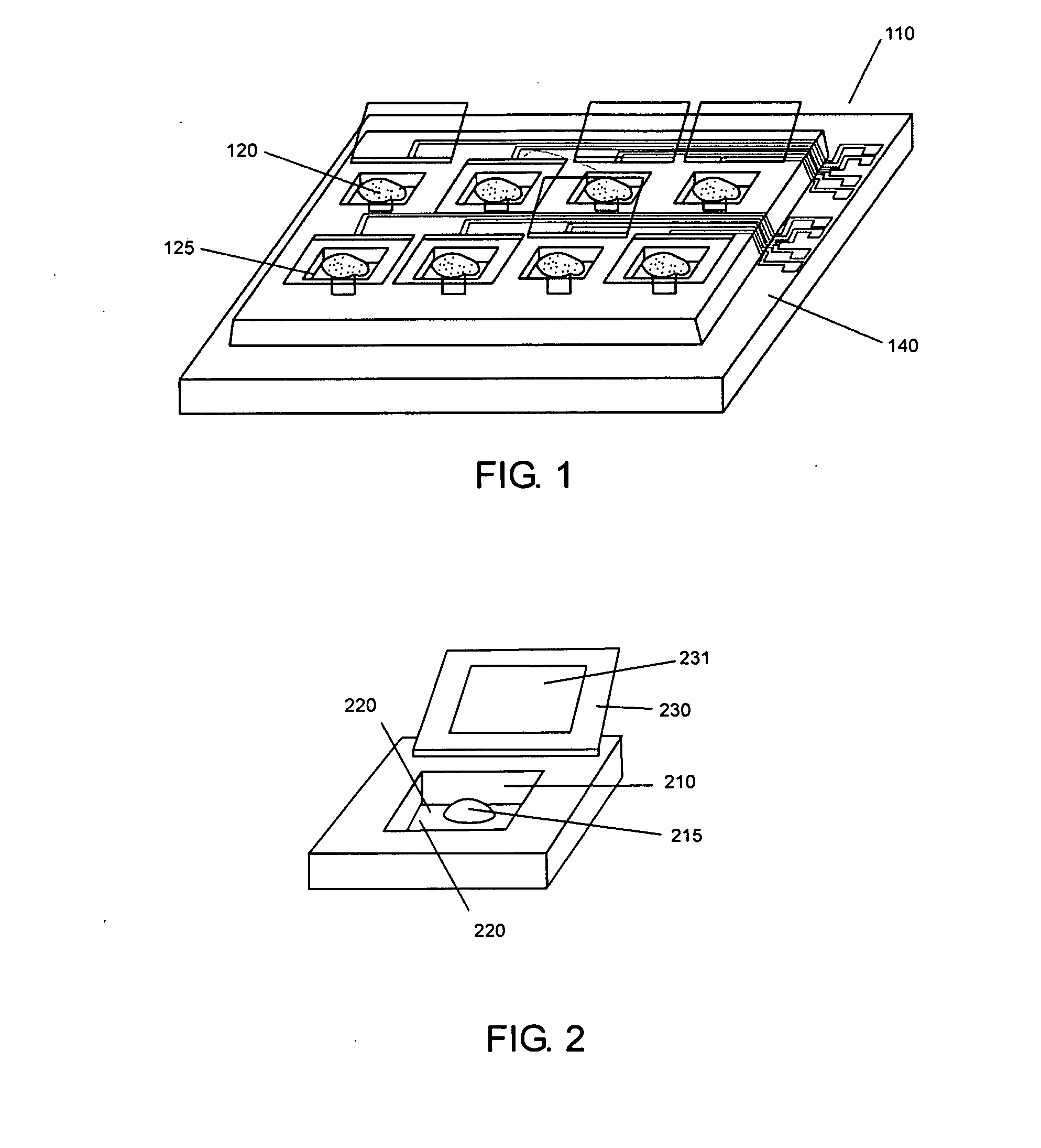
[0153] Microvials 100 μm×100 μm and 10 μm to 20 μm high were made of SU-8 negative photoresist. The microvials were closed by SU-8 / gold lids that were positioned by bilayer polymer actuators of PPy and gold. The clinics were fabricated on silicon wafers with electrodes leading to the hinges and to the interior of the vials. These structures have also been fabricated on top of custom VLSI circuitry designed to record signals from the cells within individual vials. All fabrication steps are performed at low temperature and are compatible with post-processing of the fabricated silicon die.

[0154] Because cells can escape from even deep microvials, a lid is included that can be closed after loading the cells into the vials. PPy doped with dodecylbenezenesulfonate (PPy(DBS)) is deposited over a layer of gold, which acts as the electrode through which potentials are applied as well...

example 2

CMOS Fluorescence Sensor Design and Results

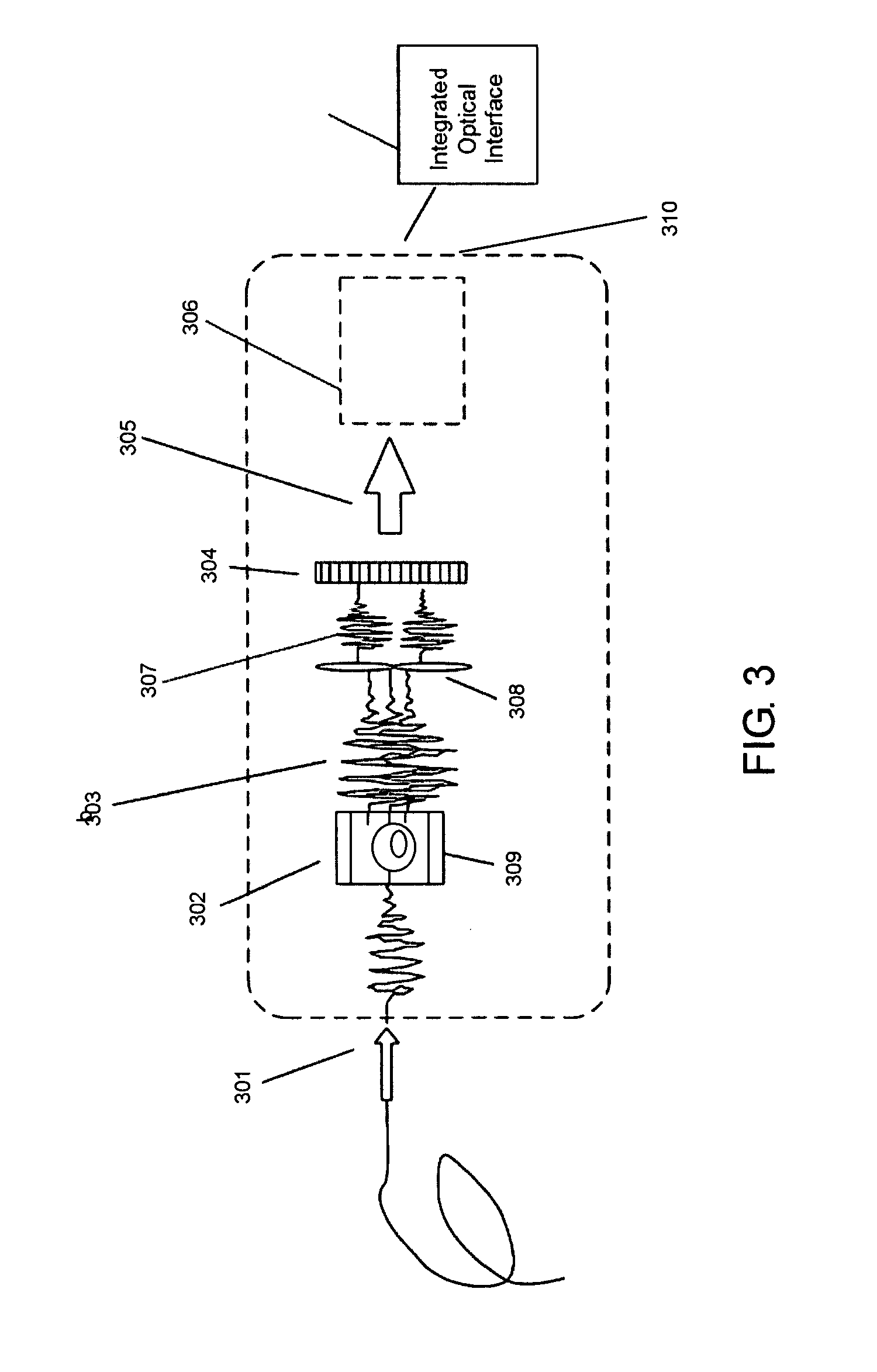
[0163] Fluorescence detection is a mature technology commonly found in microscopy and spectroscopy systems and widely used in biology labs worldwide. Such systems are typically large and require laboratory infrastructure for operation. In this work, the fluorescence sensor has been miniaturized for integration into the cell clinics vials for monitoring cells in real-time.

1. Design

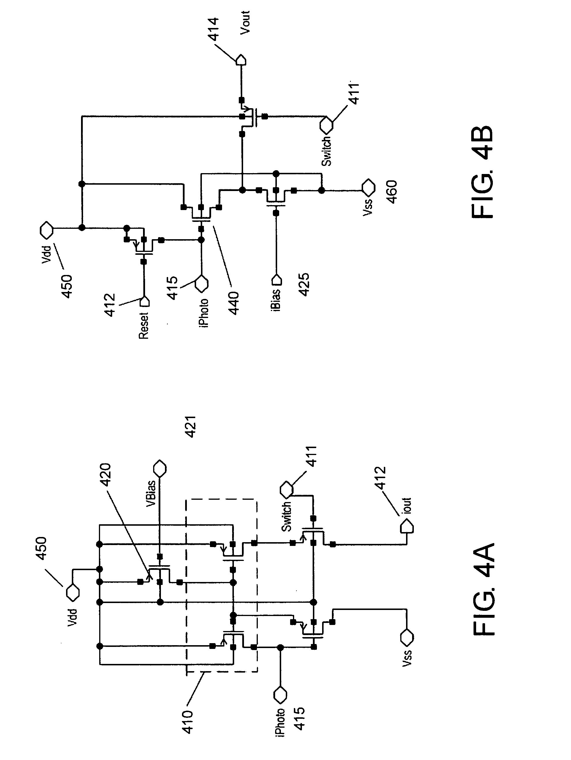
[0164] For the fluorescence sensor, the primary design constraints were sensitivity and spectral selectivity. Fluorescence from the specimen in each cell clinic was expected to be weak (normally 10−4˜10−8 fc (footcandle), and 10−6 fc corresponds to 4.6 photons / 100 μm2 / s) (Herman, 1998). Though collection efficiency for the fluorescence signal was expected to be somewhat more efficient than in normal fluorescence microscopes due to the lack of intermediary optics, the extreme weakness of fluorescence still posed a substantial challenge in the development of an in...

example 3

CMOS Capacitance Sensor for Cell Proximity Detection

[0167] Capacitance measurements are commonly used for applications such as fingerprint sensing, position sensing and interconnect characterization. In this work, the technique was adapted to cell proximity detection for evaluating the surface adhesion properties of living cells in cell clinics.

1. Design

[0168] A custom CMOS capacitance sensor for cell proximity detection has been designed using the topology shown in FIG. 6 (Lee et al., 1999). The physical principle underlying operation of the sensor is charge sharing. The coupling capacitance Ccell is formed by the series combination of the capacitances between the cell and the passivation layer and between the passivation layer and the topmost metal electrode. Ccell varies-with the strength of coupling of the cell to the chip surface.

[0169] The sensor circuit had two nodes, N1 and N2, with parasitic capacitances CN1 and CN2. Charging and discharging of these nodes were controll...


PUM

PropertyMeasurementUnit
fluorescenceaaaaaaaaaa
fluorescence resonance energy transferaaaaaaaaaa
mechanicalaaaaaaaaaa

Abstract

Methods and compositions for the reliable detection of pathogens are presented. The invention uses cells as novel pathogen detection agents, exploiting pathogen-specific pathways and apoptosis.

Description

CROSS-REFERENCE TO RELATED APPLICATION [0001] This application claims priority to 60 / 605,653, filed Aug. 30, 2004, entitled CELL CANARY, the entirety of which is herein incorporated by reference.STATEMENT REGARDING FEDERALLY SPONSORED RESEARCH OR DEVELOPMENT [0002] The subject matter of this application may in part have been funded by the National Science Foundation, ECS0225489, the United States Department of Defense, Md. Procurement H9823004C0470, and the United States Air Force, FA95500410449. The government may have certain rights in this inventionINCORPORATION-BY-REFERENCE OF MATERIAL SUBMITTED ON A COMPACT DISC [0003] Not Applicable. FIELD OF THE INVENTION [0004] The invention relates to apparatus for pathogen detection, methods of detecting pathogens using the apparatus, and methods of making the apparatus. BACKGROUND OF THE INVENTION [0005] Anthrax, plague, smallpox, Clostridium botulinum toxin, salmonella, Ebola virus, and Escherichia coli are just a few of the threats that...

Claims


Application Information

Patent Timeline
Patent Type & Authority Applications(United States)
IPC IPC(8): C12Q1/70C12Q1/68C12M3/00
CPCG01N33/54373C12Q1/04
Inventor SHAPIRO, BENJAMINABSHIRE, PAMELASMELA, ELISABETHWIRTZ, DENIS
Owner SHAPIRO BENJAMIN
Why Eureka
Try Eureka