Transparent Tissue-Visualizng Preparation

Inactive Publication Date: 2007-09-27
SENJU PHARMA CO LTD
View PDF4 Cites 21 Cited by
  • Summary
  • Abstract
  • Description
  • Claims
  • Application Information

AI Technical Summary

Benefits of technology

[0028] In surgical operations on transparent tissues of the eye, i.e., the vitreous body, the lens and the cornea, which otherwise are hardly visible in the operative field, the present invention as defined above is brought into contact with transparent tissues and greatly enhances their visibility, thereby allowing easier manipulation in such surgical operations and, therefore, steady and easier achievement of the purpose of such operations. Since fine particles having no pharmacological activi

Problems solved by technology

As tissues that have proliferated into the vitreous body will form fibers there, which draw the retina and thereby could cause retinal detachment, a condition which, if left untreated, could eventually lead to blindness.
However, the vitreous body is a transparent tissue, whose refractive index differs very little from that of available intraocular irrigating solutions.
Accordingly, when no countermeasure is taken, the transparent tissue lacks visibility in the field seen through an operation microscope, and this makes it hard to locate the tissue, thus making complete removal of it no easy task.
However, in association with application of a steroid preparation to the vitreous body, there have been reported, as their sid

Method used


Image

  • Transparent Tissue-Visualizng Preparation
  • Transparent Tissue-Visualizng Preparation
  • Transparent Tissue-Visualizng Preparation

Examples


production example 1

Prefilled Vial-Type Transparent Tissue-Visualizing Preparation

[0083] An aqueous solution containing 1.11 w / v % of D-mannitol and 1.11 w / v % of Povidone K-30 was filtered through a hydrophilic filter having the pore size of 0.22 μm to make a solution A. An acetone solution of 10 w / v % of PLA0005 (acetone / ethanol=4 / 6) was prepared and filtered through a hydrophobic filter having the pore size of 0.22 μm to make a solution B. Opeguard MA was made a solution C.

[0084] The solutions A and B were mixed at a proportion of 9:1 in the following manner. Briefly, to the solution A, stirred at 700-800 rpm with a stirrer, was added the solution B, at a rate of about 100 μL / sec, to let PLA0005 precipitate as fine particles. The mixture was stirred for about 30 minutes, and aggregation products were removed through a sieve (mesh size 106 μm) to obtain a suspension D. Lyophilization of this in a vial gave a powder form sample E. Prior to use, the solution C was poured into the vial containing the ...

production example 2

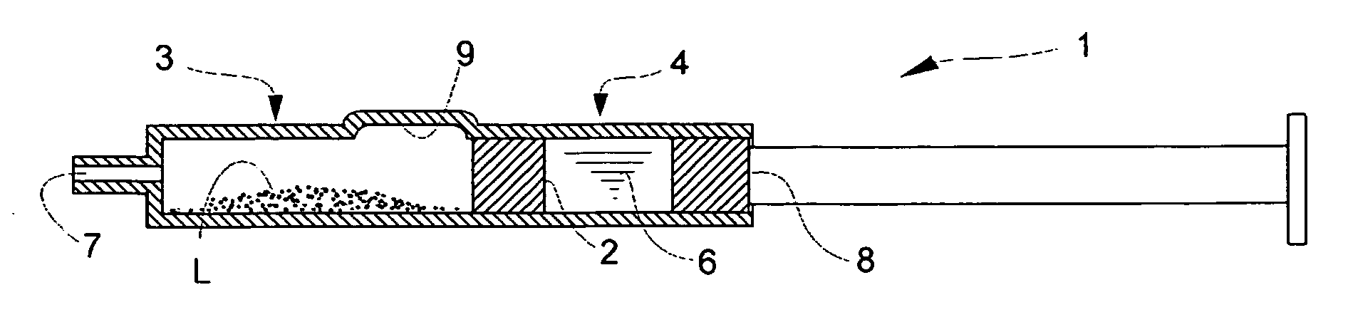
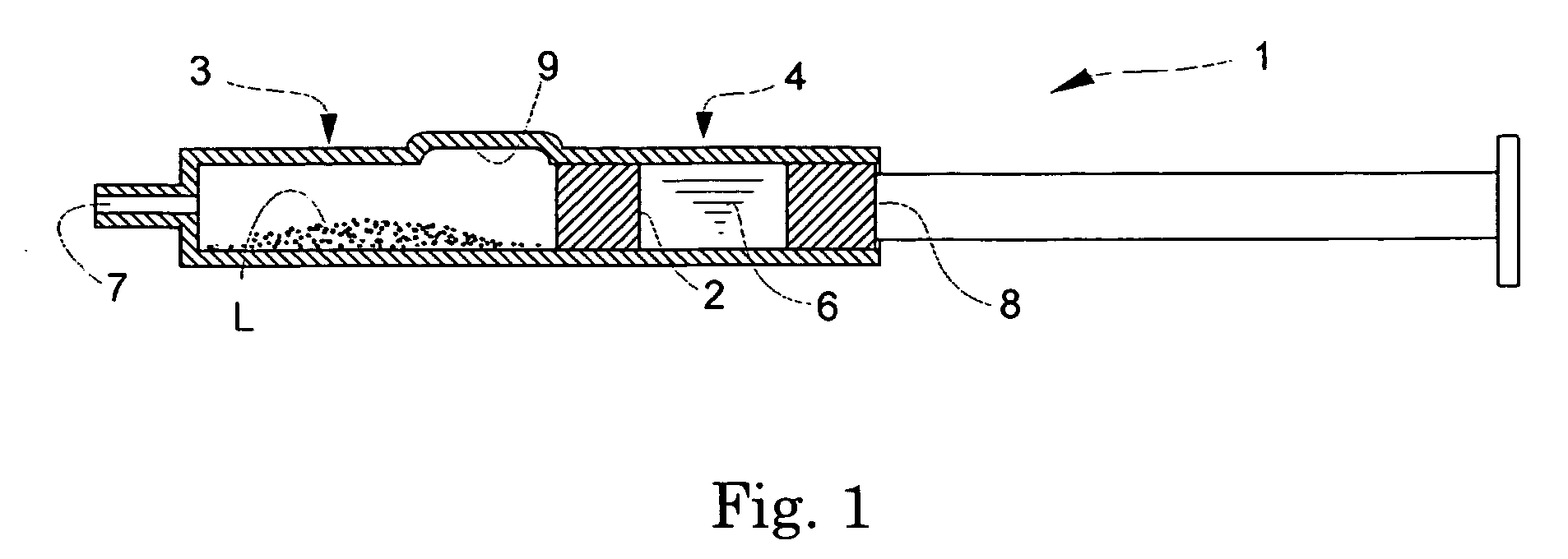
Prefilled Double Chamber Syringe-Type Transparent Tissue-Visualizing Preparation

[0086] An aqueous solution containing 1.11 w / v % of D-mannitol and 1.11 w / v % of Povidone K-30 was filtered through a hydrophilic filter having the pore size of 0.22-μm to make a solution F. On the other hand, a 10 w / v % solution of PLA0005 (acetone / ethanol=4 / 6) was filtered through a hydrophobic filter having the pore size of 0.22 μm to make a solution G. A phosphate buffer (pH 7) containing 0.75% of sodium chloride and 0.16% of potassium chloride was made a liquid H.

[0087] The solutions F and G were mixed at a proportion of 9:1 in the following manner. Briefly, to the solution F, stirred at 700-800 rpm with a stirrer, was added the solution G, at a rate of about 100 μL / sec, to allow PLA0005 to precipitate as fine particles. The mixture was stirred for about 40 minutes (for 10 minutes of which a reduced pressure was applied), and aggregation products were removed through a sieve (mesh size 106 μm) to ...

preparation example 1

Transparent Tissue-Visualizing Preparation Containing Soluble Starch Fine Particles

[0089] A transparent tissue-visualizing preparation of the following formula was prepared in a conventional manner. In the preparation, the average particle size of the soluble starch fine particles was about 50 μm.

Solubile starch1.0 gOpeguard MAto 100 mL


PUM

PropertyMeasurementUnit
Temperatureaaaaaaaaaa
Massaaaaaaaaaa
Sizeaaaaaaaaaa

Abstract

Described is a highly safe means to improve visibility of transparent tissues of the eye, i.e., the vitreous body, the lens or the cornea, in a surgical operation on them. The means comprises fine particles of a macromolecular compound, in particular, those one gram of which cannot be dissolved in less than 30 mL of water within 30 minutes at 20° C., and whose average particle size is 0.01-500 μm, and which, among others, is a biodegradable macromolecular compound and/or a water soluble macromolecular compound.

Description

TECHNICAL FIELD [0001] The present invention relates to a transparent tissue-visualizing preparation which is designed to be infused into or sprinkled over a transparent tissue of the eye in order to enhance visibility of transparent tissues of the eye in surgical operation, as well as to a method to visualize and enhance visibility of a transparent tissue of the eye by application of the preparation. BACKGROUND ART [0002] To receive light from a surrounding environment and project it on the photoreceptor cells, many portions of the eyeball are made of transparent tissues, i.e., the cornea, lens and vitreous body. The vitreous body, which adjoins the retina, provides scaffolds to proliferating tissues formed in the retina in many of retinal diseases including diabetic retinitis. As tissues that have proliferated into the vitreous body will form fibers there, which draw the retina and thereby could cause retinal detachment, a condition which, if left untreated, could eventually lead ...

Claims


Application Information

Patent Timeline
IPC IPC(8): A61F9/00A61K31/718A61K31/722A61K31/765A61K45/00A61P27/02A61P27/12
CPCA61K9/0048A61K31/718A61K31/722A61K31/765A61K49/0091A61K2300/00A61P27/02A61P27/12
Inventor MATSUHISA, KEIICHIINOUE, YUTAKA
Owner SENJU PHARMA CO LTD
Why Eureka
Try Eureka