Instrument and method for the end-to-end reconnection of intestinal tissues

a tissue reconnection and end-to-end technology, applied in the field of end-to-end reconnection of intestinal tissues, can solve the problems of delayed healing and/or inflammation, allergic reactions, limited applicability, etc., and achieve the effects of convenient use, high thermal conductivity, and easy cleaning and sterilization

Inactive Publication Date: 2007-11-29
LIVE TISSUE CONNECT +2
View PDF13 Cites 82 Cited by
  • Summary
  • Abstract
  • Description
  • Claims
  • Application Information

AI Technical Summary

Benefits of technology

[0029] Another embodiment of the intestinal apparatus will allow two ends of intestinal tissue to be reconnected on the serous layers. This tool will be inserted into one end of the intestine through a separate incision and the electrodes of the instrument will clamp the intestinal tissue from the inside of the intestine. The tools encompassed in these embodiments are designed to be used by a surgeon with ordinary level of skill in gastrointestinal surgery. As such, the instruments are designed to not require an exceptional level of dexterity for their proper use. Also, the intestinal tools encompassed in this aspect of the invention may be disassembled as necessary both to make the device easier to use and to allow the device to be easily cleaned, sterilized, and repaired.
[0030] A further aspect of the invention addresses the tendency of tissue to adhere to the electrodes during an electrosurgical procedure. One of these techniques involves fashioning the electrodes out of a material specially selected for this purpose, a copper-molybdenum composite, with high thermal conductivity. The electrodes thereby remove heat from the electrode-tissue connection more rapidly and reduce the potential for the tissue to adhere to the electrode. This technique has been incorporated in the current invention in a manner which advances the technique beyond its current application in surgical equipment.

Problems solved by technology

These techniques have a number of well known disadvantages which include one or more of the following: leaving a foreign body in the tissue, pinching of tissue which can cause delayed healing and / or inflammation, allergic reaction, limited applicability, complexity of use, and the need for expensive equipment.
However, at temperature exceeding 100° C. the tissue becomes dehydrated, its electric resistance increases, which leads to further temperature rise and charring of the tissue.
Still this technique has not been accepted for general clinical use because of the technical complexity of its utilization and because of inadequate surface energy release.
However, the use of the existing bipolar devices for connecting soft tissues other than walls of compressed blood vessels encounters insurmountable difficulties.
Specifically, it has been difficult to correctly set the electrical signal parameters to achieve such aims.
If too little current is applied, then the tissue joint can be spongy, weak and unreliable.
On the other hand, if too much current is applied, then the working surface of the electrode can stick to the tissue so that removal of the electrode causes bleeding and possible injury.
Also, the tissue in the overly-heated zone can become desiccated and charred.
Therefore, such high frequency coagulative devices have seen limited use for only hemostasis of blood vessels of relatively small diameter.
Unless the heating is stopped, severe tissue damage will occur.
Usage of these techniques for other types of tissue creates severe difficulties due to the wide variation in values of impedance which can be encountered due to, for example, tissue structure, thickness, condition of the tissue and condition of the tool surface.
However, this methodology is time-consuming and would be prone to leakage or weak reconnection is if there was either too much or too little overlap between the individual spot welds.
In these cases, using forceps-like electrosurgical devices would not improve significantly over the suturing or stapling method of intestinal reconnection.
The intestinal anastomosis would still be susceptible to leakiness and the post-operative complications that result from that leakiness.

Method used


Image

  • Instrument and method for the end-to-end reconnection of intestinal tissues
  • Instrument and method for the end-to-end reconnection of intestinal tissues
  • Instrument and method for the end-to-end reconnection of intestinal tissues

Examples


fourth embodiment

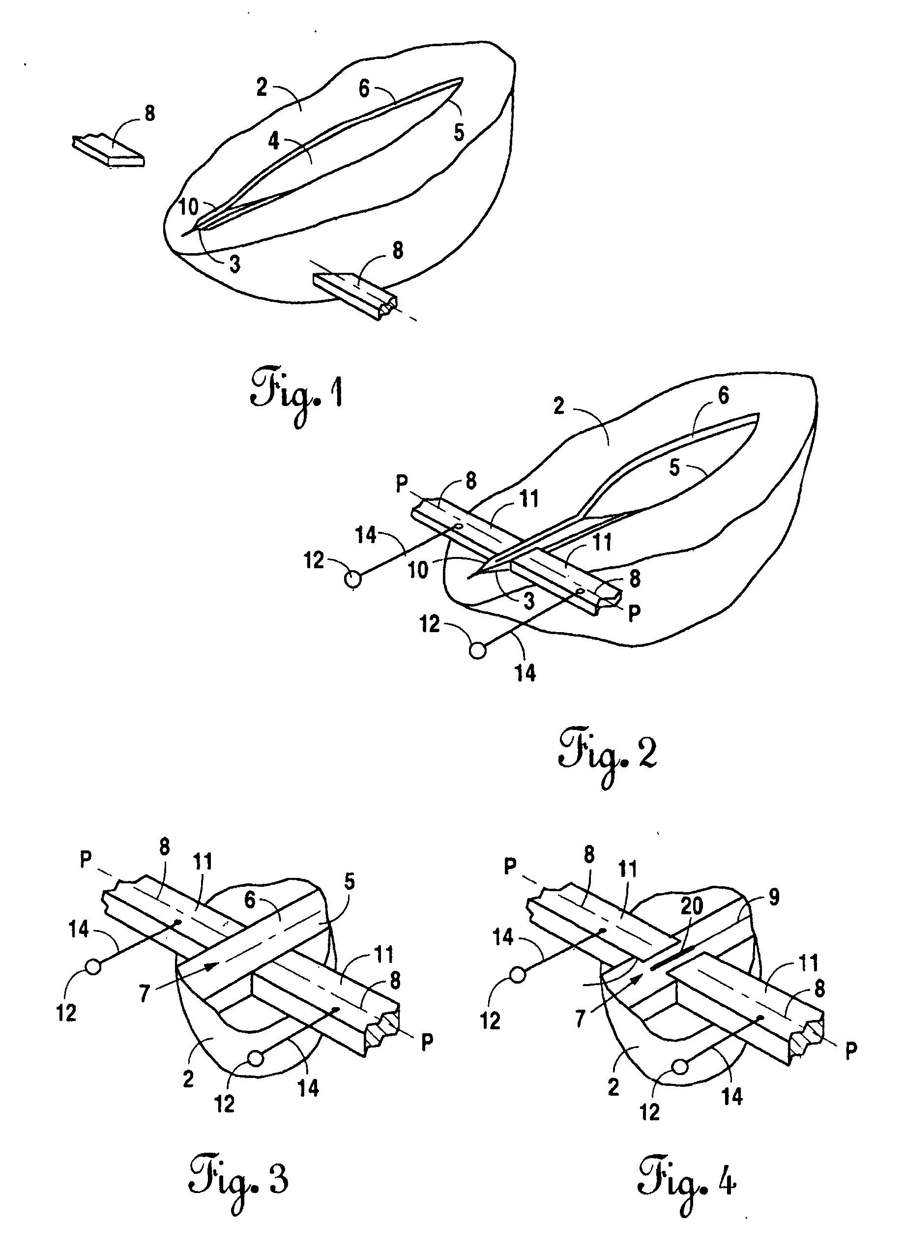
[0090]FIGS. 10-12 show the invention which is designed to bond the entire periphery of the hollow tissue, such as a blood vessel, discussed above in connection as with FIG. 8. The blood vessel is shown in FIG. 10 after it has been cut into parts 30 and 32. Tissue part 30 is inserted into semicircular electrode sleeve 34 attached to the end of arm 36. Similarly, tissue part 32 is inserted into semicircular electrode sleeve 38 attached to the end of arm 40. The axes of sleeves 34 Fund 38 are aligned along line 42, and tissue ends 30a and 32a face each other. As shown in FIG. 11, another semicircular electrode sleeve 35 is placed onto its mate 34 to encircle tissue part 30 therebetween. Electrode 35 is attached to the end of arm 37. Likewise, semicircular electrode sleeve 39 is placed onto its mate 38 to enclose tissue part 32 therebetween. Electrode 39 is attached to the end of arm 41. These various parts can be part of a tool (not shown), the details of which are apparent to one with...

second embodiment

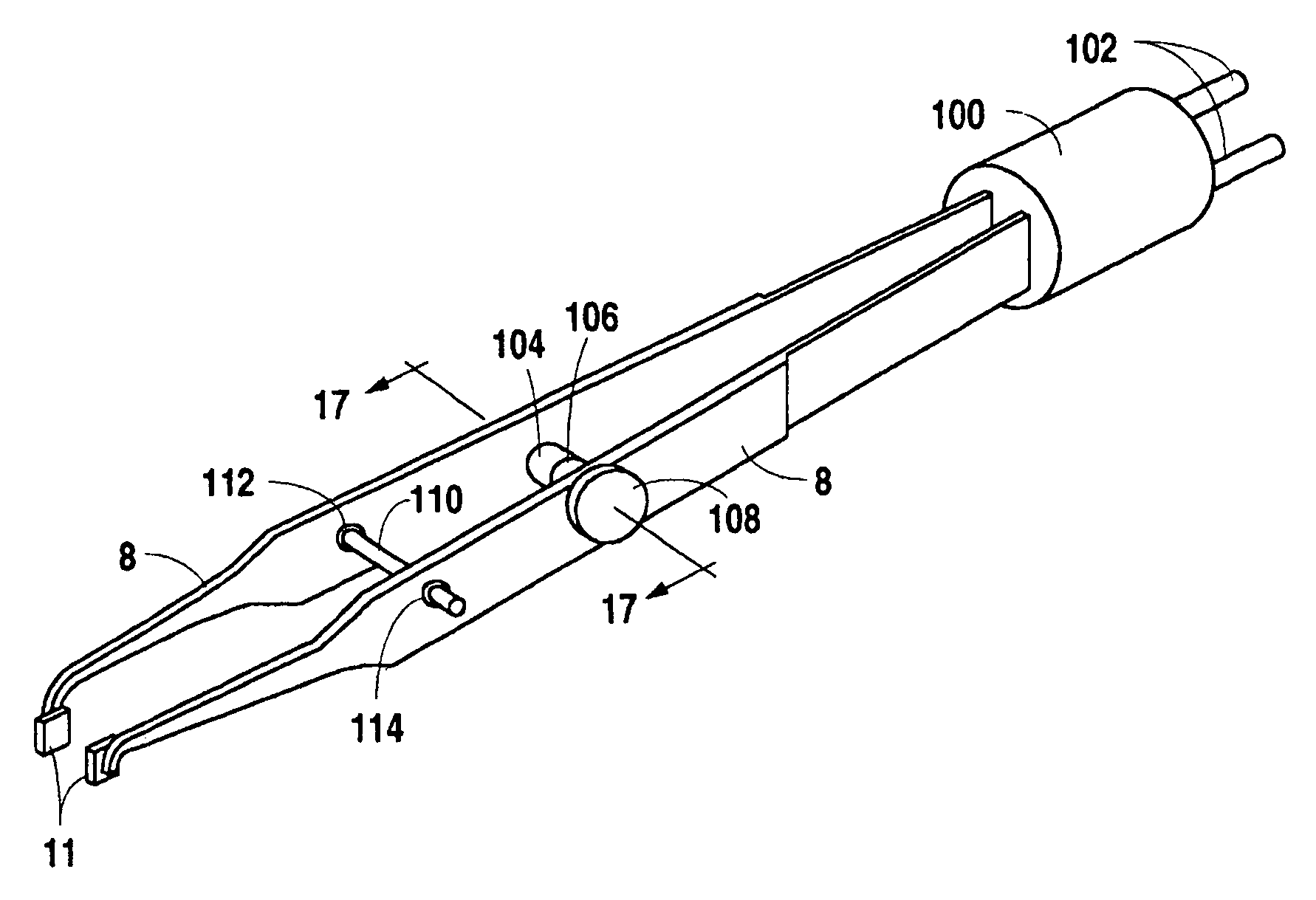
[0202] The tie rod 804 will move laterally through the instrument to move the movable electrode 821 toward the spring-loaded electrode 818 during the reconnection operation. Moving the electrodes together will clamp the tissue and hold it in place during the bonding process. The movement of the tie rod is controlled by an adjusting means. In one embodiment of the instrument, this adjusting means will be comprised of an adjusting nut 812, a flywheel mechanism 811, and an adjusting screw 833 situated inside and coming in contact with the flywheel mechanism 811. In this embodiment as the flywheel 811 is rotated, the adjusting screw 833 will move laterally as its threads and the threads inside the flywheel move in opposition with one another. Since the adjusting screw 833 is connected to the tie rod 804, the tie rod will move laterally within the instrument as the adjusting screw moves laterally. Once the tie rod adjustment has been made, the adjusting nut 812 can be tightened against t...


PUM


Abstract

The current methods for reconnecting intestinal tissues, suturing, or metallic staples, can result in a leaky anastomosis that may result in post-operative infections in the patient's abdominal cavity. By utilizing electrical current to bond or weld the intestinal tissue, this potential for leaky anastomosis can be significantly reduced. This invention provides tools and processes that allow electrical tissue bonding to be used on a hollow tissue, such as an intestine. This invention also discloses a means for further reducing the inherent problem of tissue sticking to the electrodes, by introducing a superior electrode design that uses a composite material, copper-molybdenum (CuMo).

Description

BACKGROUND OF THE INVENTION [0001] 1. Field of the Invention [0002] The present invention is directed to a technique for bonding soft biological tissue to close an incision therein and, in particular, to heating of the tissue with high frequency electric current in combination with compression of the tissue. The present invention provides tools and methods for accomplishing this technique, including tools and methods for accomplishing this technique on a cylindrical hollow tissue, such as an intestine. [0003] 2. Description of Related Art [0004] For purposes of the ensuing discussion, soft biological tissue will be referred to just by the term “tissue” for reasons of simplicity and economy of space, and should be understood to mean any tissue other than bone, such as skin organs, blood vessels and nerves. When tissue is injured, it must be repaired by re-joining the edges of tissue that has been torn or cut. For example, when tissue is cut during a surgical operation, the incision(s...

Claims


Application Information

Patent Timeline
Patent Type & Authority Applications(United States)
IPC IPC(8): A61B18/14
CPCA61B17/11A61B18/1442A61B2019/304A61B2018/00666A61B2018/1462A61B2018/0063A61B2090/034
Inventor PATTON, BORIS E.LEBEDEV, VLADIMIR K.FURMANOV, YURI A.ZAKHARASH, MICHAEL P.PODPRYATOV, SERGEY E.LEBEDEV, ALEXEY V.BUSHTEDT, YURI P.IVANOVA, OLGA N.SHELEST, NIKOLAY A.VASYLCHENKO, VALERIY A.SYDORENKO, DMITRIY F.DUBKO, ANDREY G.TRUNOV, ANATOLII
Owner LIVE TISSUE CONNECT
Why Eureka
Try Eureka