
Breast Cancer Detection and Biopsy

Inactive Publication Date: 2008-01-03
SCI BIOPSY
View PDF10 Cites 28 Cited by
  • Summary
  • Abstract
  • Description
  • Claims
  • Application Information

AI Technical Summary

Benefits of technology

[0019] The soft compression device applies pressure around most of the breast, in contrast to conventional compression devices using flat compression plates, in which the pressure is concentrated in a relatively small part of the breast, so the soft compression device is more comfortable for the patient, and provides better immobilization. Furthermore, ultrasound can be transmitted through the soft compression device, instead of or in addition to x-rays, for providing images for locating the lesion. Optionally, the biopsy needle goes through the soft material of the compression device, so the biopsy needle can come in from any direction, rather than being limited to a small window, and can reach lesions even if they are close to the chest wall. Optionally, if used for a biopsy, the soft compression device is not an integral part of the unit used to perform the biopsy, but is separable from the biopsy unit, and is first placed around the breast and tightened, and then attached to the biopsy unit.
[0094] d) coupling one or both of the flexible band and the base to a biopsy unit, with sufficient spatial stability to enable the biopsy unit to perform an ultrasound guided biopsy;

Problems solved by technology

Open surgery does not require any imaging system, but has all of the inherent disadvantages of an invasive procedure.
X-ray guidance necessarily causes radiation exposure.
However, currently available ultrasonic guidance systems typically rely upon “free hand” positioning.
This is a disadvantage because it requires uninterrupted co-ordination, concentration and significant physical effort throughout a procedure which may last several minutes.
Because a 2D image is used to represent the 3D relationship spatial between the biopsy needle and the target lesion, a significant amount of trial and error is involved, even for a skilled operator.
This can lead to frustration and / or fatigue for the operator.
In addition, because breast is pliant, manipulation in the form of contact from the biopsy apparatus and / or the US transducer may cause the target lesion to move within the breast.
(Ultrasound in Obstetrics and Gynecology (2001) 18(5):520-524) teaches use of a Mammotome® for ultrasound guided biopsy but concludes that failure rate of Mammotome® biopsy was likely due to an error in positioning of the needle.
Since ultrasonic procedures are often conducted on an outpatient basis, a patient may not be able to wash away the applied coupling media in a satisfactory manner.
Some patients view this as a disadvantage of ultrasound procedures.

Method used


Image

  • Breast Cancer Detection and Biopsy
  • Breast Cancer Detection and Biopsy
  • Breast Cancer Detection and Biopsy

Examples


Embodiment Construction

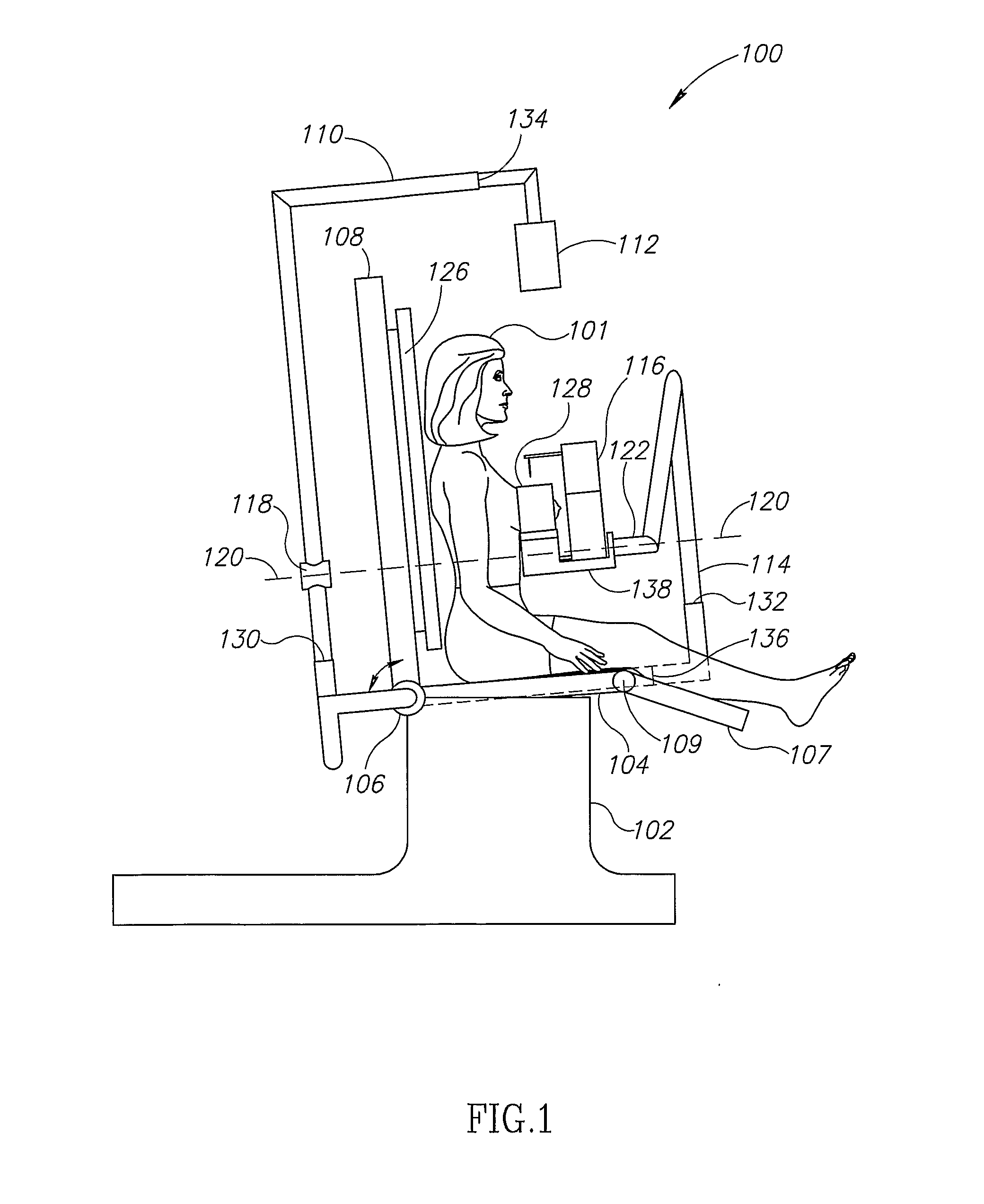
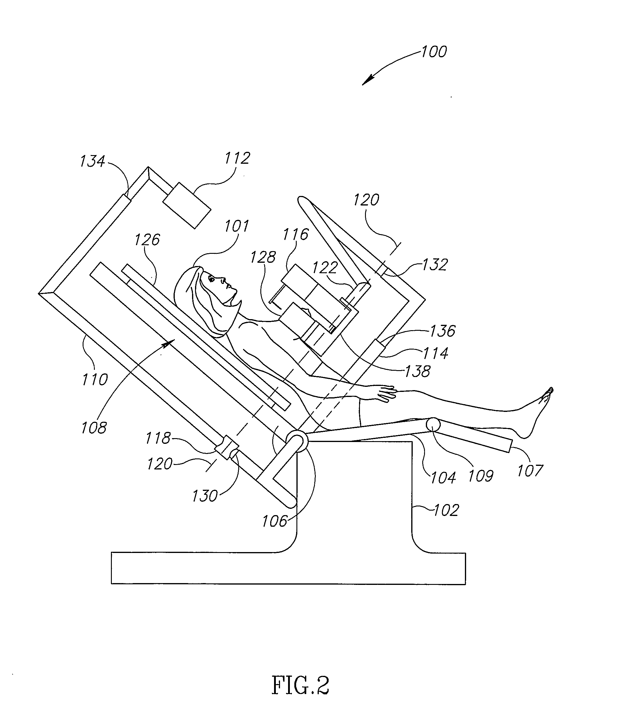
[0120]FIGS. 1 and 2 show a patient 101 in a reclining chair 100, in different positions. In FIG. 1, chair 100 is in an upright position. Chair 100 is similar to a dentist's chair, and in fact a commercially available dentist's chair is optionally used, with modifications as will be described. A rotatable joint 106 is supported by a base 102. Seat 104 and back 108 are attached to joint 106, and independently rotate around joint 106. Joint 106 optionally allows the back of the chair to tilt back by any of a continuous range of angles up to, for example, 90 degrees, which would put the patient in a completely supine position. Optionally, independently of the back, seat 104 optionally can tilt up by any of a continuous range of angles up to, for example, 10 degrees or 15 degrees. Alternatively, seat 104 is not free to tilt at all, but remains horizontal, or at a fixed angle, relative to base 102. Alternatively, the angle between seat 104 and back 108 does not change when the chair tilts...


PUM


Abstract

A system for performing a breast biopsy. The system includes a breast compression device, an ultrasound imaging unit and a biopsy module capable directing a biopsy needle to a lesion in the breast, guided at least in part by ultrasound images. The system may optionally include a piece of furniture, such as table or a reclining chair.

Description

RELATED APPLICATIONS [0001] This application is a continuation in part of PCT Application PCT / IL2004 / 000854 filed on Sep. 15, 2004, which designated the United States and which is entitled “Breast Cancer Detection and Biopsy”, which application is fully incorporated herein by reference.FIELD OF THE INVENTION [0002] The field of the invention is devices and methods for breast biopsy. BACKGROUND OF THE INVENTION [0003] Breast biopsy may be performed either in open surgery or with needle biopsy. [0004] Open surgery does not require any imaging system, but has all of the inherent disadvantages of an invasive procedure. [0005] Needle biopsy, although less invasive, requires an imaging system so that the surgeon can locate the target lesion from which the biopsy is to be taken. [0006] Needle biopsy may be guided using ultrasound (US) or X-ray. [0007] X-ray guidance necessarily causes radiation exposure. [0008] A typical skilled practitioner can easily detect cysts by US and some exception...

Claims


Application Information

Patent Timeline
IPC IPC(8): A61B8/00
CPCA61B8/0825A61B10/0041A61B2019/206A61B2019/205A61B19/201A61B90/11A61B90/17A61B90/18
Inventor GROSS, JACOB
Owner SCI BIOPSY
Learn More