Medical devices having a temporary radiopaque coating

a technology of medical devices and radiopaque coating, which is applied in the direction of prosthesis, catheter, surgery, etc., can solve the problems of limited use of ct angiography in this contex

Inactive Publication Date: 2008-01-10
BOSTON SCI SCIMED INC
View PDF9 Cites 169 Cited by
  • Summary
  • Abstract
  • Description
  • Claims
  • Application Information

AI Technical Summary

Benefits of technology

[0005]There is a need for a medical device such as a stent that is temporarily radiopaque for imaging under fluoroscopy during implantation, but loses its radiopacity after implantation to allow for subsequent imaging under more sensitive radiologic imaging modalities.

Problems solved by technology

But the use of CT angiography in this context has been limited because of distortions (artifacts) in the image caused by the metallic stent, which are sufficiently radiopaque for fluoroscopic visualization, but are often too radiopaque for high sensitivity CT imaging.

Method used


Image

  • Medical devices having a temporary radiopaque coating
  • Medical devices having a temporary radiopaque coating

Examples


Embodiment Construction

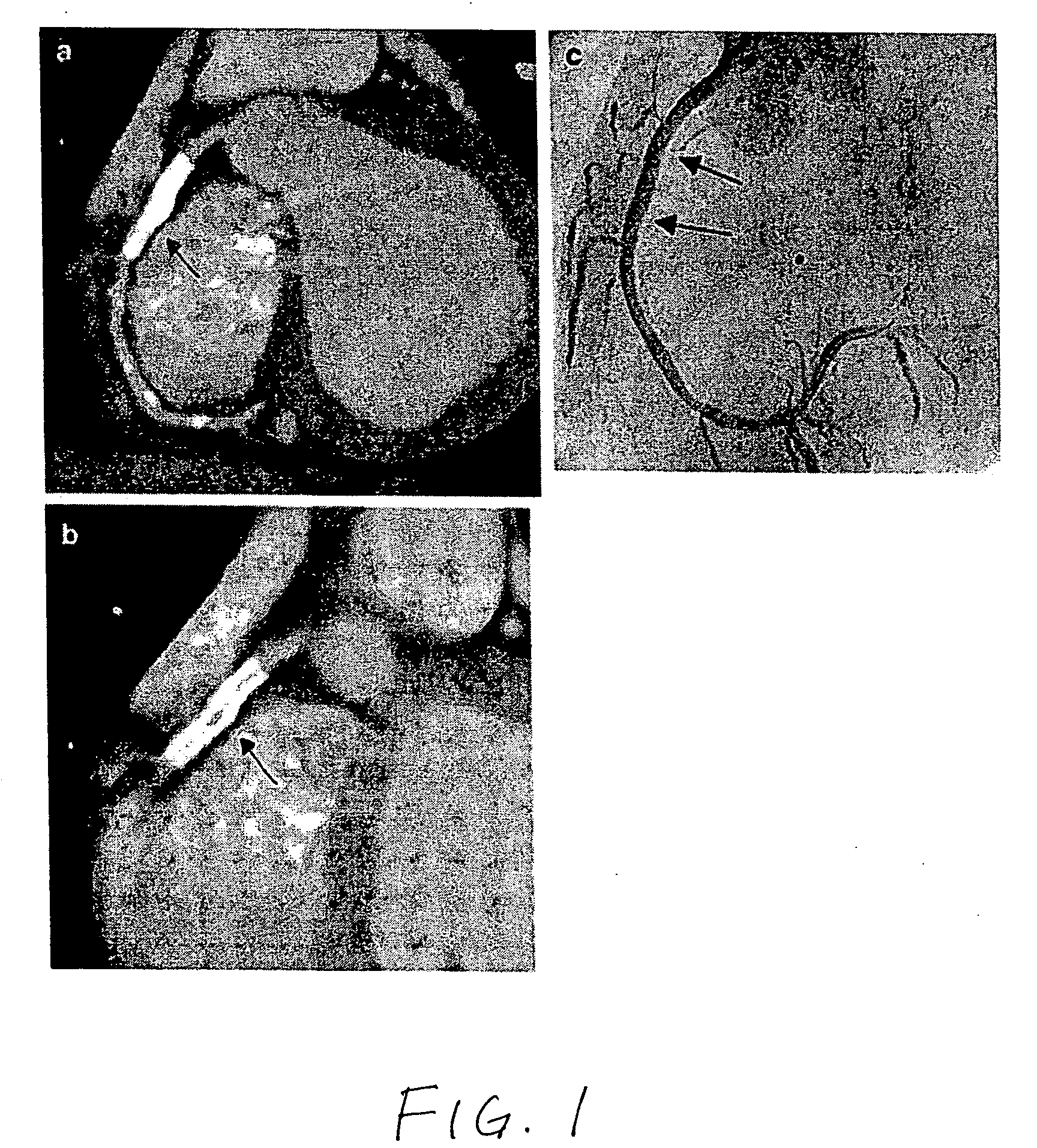
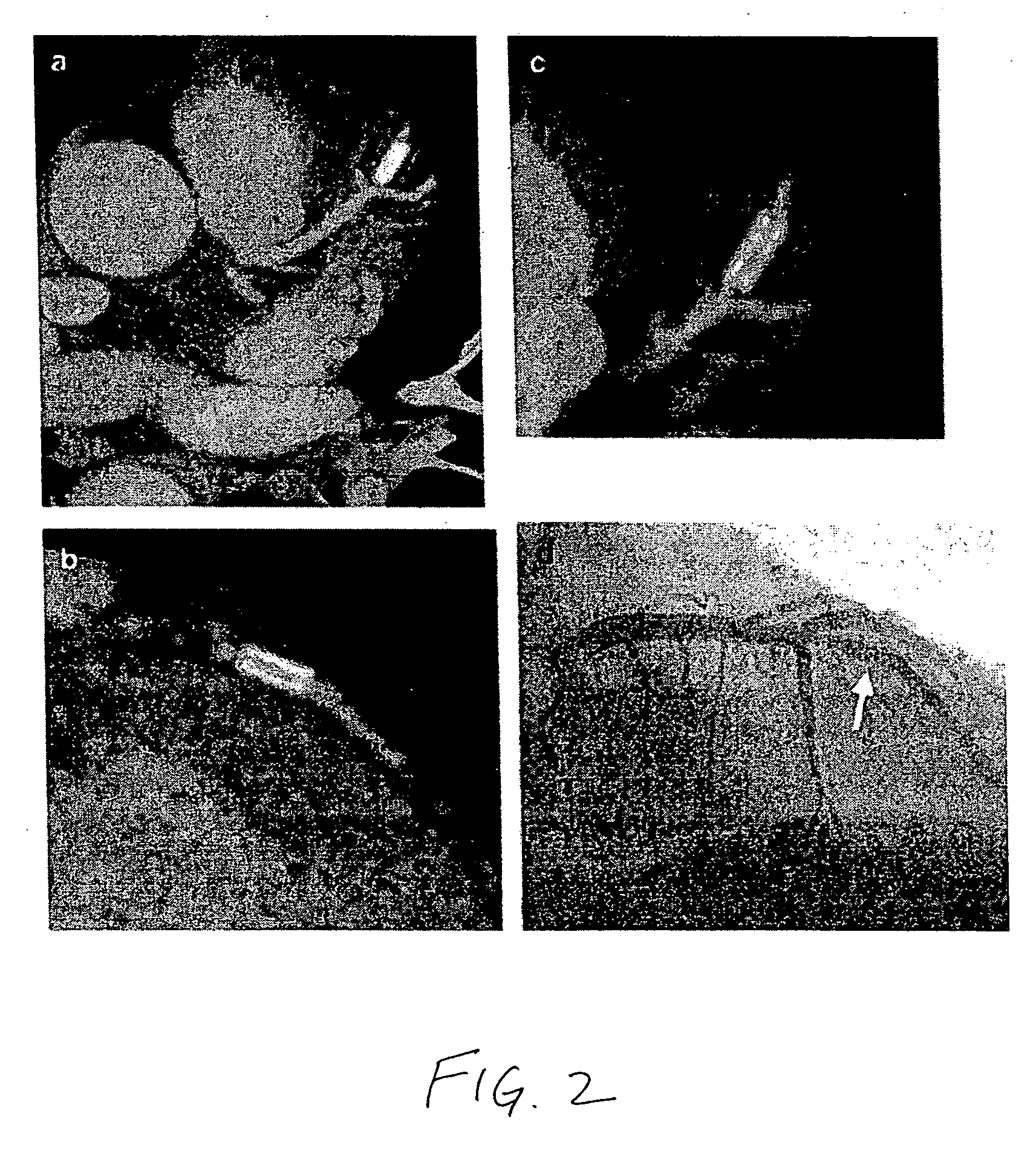
[0013]The present invention provides a medical device comprising radiopaque water-dispersible metallic nanoparticles, wherein the nanoparticles are released from the medical device upon implantation of the device.

[0014]The term “metallic nanoparticle” refers to a particle having a diameter in the range of about 1 nm to 1000 nm that comprises a metallic material, an alloy, or other mixture of metallic materials. The metallic material may be any metal having sufficient radiopacity for visualization under x-ray fluoroscopy, including iodine, barium, tantalum, tungsten, rhenium, osmium, iridium, noble metals, platinum, gold, and bismuth. Oxides and compounds of the metals listed, such as bismuth subcarbonate and bismuth oxychloride, may also be used. Salts of the metals listed, such as barium salts, iodine salts, or bismuth salts, may also be used.

[0015]The term “water-dispersible” refers to the ability of the material to form an essentially unaggregated dispersion of discrete particles...


PUM

PropertyMeasurementUnit
diameteraaaaaaaaaa
diameteraaaaaaaaaa
diameteraaaaaaaaaa

Abstract

A medical device comprising radiopaque water-dispersible metallic nanoparticles, wherein the nanoparticles are released from the medical device upon implantation of the device. The medical device of the present invention is sufficiently radiopaque for x-ray visualization during implantation, but loses its radiopacity after implantation to allow for subsequent visualization using more sensitive imaging modalities such as CT or MRI.
The nanoparticles are formed of a metallic material and have surface modifications that impart water-dispersibility to the nanoparticles. The nanoparticles may be any of the various types of radiopaque water-dispersible metallic nanoparticles that are known in the art. The nanoparticles may be adapted to facilitate clearance through renal filtration or biliary excretion. The nanoparticles may be adapted to reduce tissue accumulation and have reduced toxicity in the human body. The nanoparticles may be applied directly onto the medical device, e.g., as a coating, or be carried on the surface of or within a carrier coating on the medical device, or be dispersed within the pores of a porous layer or porous surface on the medical device. The medical device itself may be biodegradable and may have the nanoparticles embedded within the medical device itself or applied as or within a coating on the biodegradable medical device. The nanoparticles may be released by diffusion through the carrier coating, disruption of hydrogen bonds between the nanoparticles and the carrier coating, degradation of the nanoparticle coating, degradation of the carrier coating, diffusion of the nanoparticles from the medical device, or degradation of the medical device carrying the nanoparticles.

Description

TECHNICAL FIELD[0001]The present invention relates to implantable medical devices having a radiopaque coating.BACKGROUND[0002]Many medical devices are implanted inside the body with the aid of x-ray fluoroscopy, which provides real-time visualization of the implantation procedure. For example, vascular stents are typically implanted by a catheterization procedure using x-ray fluoroscopy to guide the stent through the vasculature and position it in the target artery. Thus, the stent and / or stent deployment system must be sufficiently radiopaque (not transparent to x-rays) for visualization under x-ray fluoroscopy.[0003]Weeks or months after the implantation of a stent, a subsequent visualization of the stented artery is often necessary in order to diagnose possible reocclusion (restenosis) of the artery. Typically, the diagnosis of restenosis is made by repeating an invasive catheterization procedure in order to obtain an x-ray fluoroscopic image (angiogram) of the stented artery.[00...

Claims


Application Information

Patent Timeline
Patent Type & Authority Applications(United States)
IPC IPC(8): A61K49/04A61F2/82
CPCA61L27/306A61L27/446A61L29/106A61L2400/12A61L31/088A61L31/128A61L31/18A61L29/18
Inventor CLARKE, JOHN T.O'BRIEN, BARRY
Owner BOSTON SCI SCIMED INC
Why Eureka
Try Eureka