Novel Method for the Preparation of Embryoid Bodies (Ebs) and Uses Thereof

a technology of embryoid bodies and embryos, which is applied in the field of embryoid body preparation, can solve the problems of insufficient amount of embryoid bodies, insufficient number of embryoid bodies, and repeated tests in multiple cell lines, etc., and achieves the effects of improving the yield of embryoid bodies, high density, and operating expense for large-scale production

Active Publication Date: 2008-01-24
EVOTECH INT GMBH
View PDF6 Cites 11 Cited by
  • Summary
  • Abstract
  • Description
  • Claims
  • Application Information

AI Technical Summary

Benefits of technology

[0036] In accordance with the present invention, it has surprisingly been found that embryonic stem (ES) cell aggregates, so called embryoid bodies (EBs) can be generated in large amounts and high density, which in turn can be induced to differentiate into particular cell types and tissue such as cardiomycytes, neurones, endothelial cells and the like. The present invention is based on the observation that agitation of a certain amount and concentration of ES cells in an appropriate container is superior to, for example, the stirring culture method for the preparation of ES cell aggregates, in particular embryoid bodies. Thus, in contrast to methods described in the prior art a culture system could be established that allows the generation of EBs in high density, which has no negative influence on the differentiation capacity towards different cell types such as cardiomyocytes, neurons, endothelial cells and liver cells. Moreover, compared with other methods, the method of the present invention does neither need sophisticated equipment like, e.g., fermenters of stirred bioreactors, nor time-consuming and laborious cell preparations like the encapsulation method described in international application WO03 / 004626. Thus, the advantage of the present invention is to generate large amounts of high-quality EBs with a simple and cheap method, which makes the method suitable for large-scale testing procedures like high throughput screening in drug discovery.
[0037] With the method of the invention the yield of embryoid bodies can be considerably be improved compared to conventional methods, since the cell aggregates can be cultured in large volumes and higher density than in the previous methods. The method of the invention also allows generating tissue in sufficient amounts for therapeutic uses, wherein the target tissue can be purified according to standard methods such as described in WO02 / 051987. Accordingly, the method of the present invention provides several advantages over the prior art methods for the preparation of embryoid bodies.
[0038] First, the embryoid bodies, and thus any desired precursors and cell types are provided in large amounts and high density and allow compound screening on industrial scale.
[0039] Second, the method of the invention is quite easy to perform contrary to, for example, the hanging drop method. In accordance with this, the method of the invention is much more reliable and reproducible than the classical methods.
[0040] Third, the operating expense for large-scale production of embryoid bodies in accordance with the method of the present invention is also quite low compared to other fermenter cultures.
[0041] Fourth, compared to cultures in spinner flasks, in the method of the invention the ES cells are much less exposed to shear stress, whereby the capability of the cells to differentiate in an appropriate manner is not negatively influenced.

Problems solved by technology

Employing conventional cell lines for these studies has the disadvantage that individual cell lines may not be fully representative of the complex biology of an intact organism.
Moreover, even repeating the tests in multiple cell lines does not reproduce or account for the complex interactions among cells and tissue that occur in an organism.
However, all of those methods are cumbersome and / or do not provide sufficient amounts of embryoid bodies suitable for example for high throughput screening (HTS) assays.

Method used


Image

  • Novel Method for the Preparation of Embryoid Bodies (Ebs) and Uses Thereof
  • Novel Method for the Preparation of Embryoid Bodies (Ebs) and Uses Thereof
  • Novel Method for the Preparation of Embryoid Bodies (Ebs) and Uses Thereof

Examples


example 1

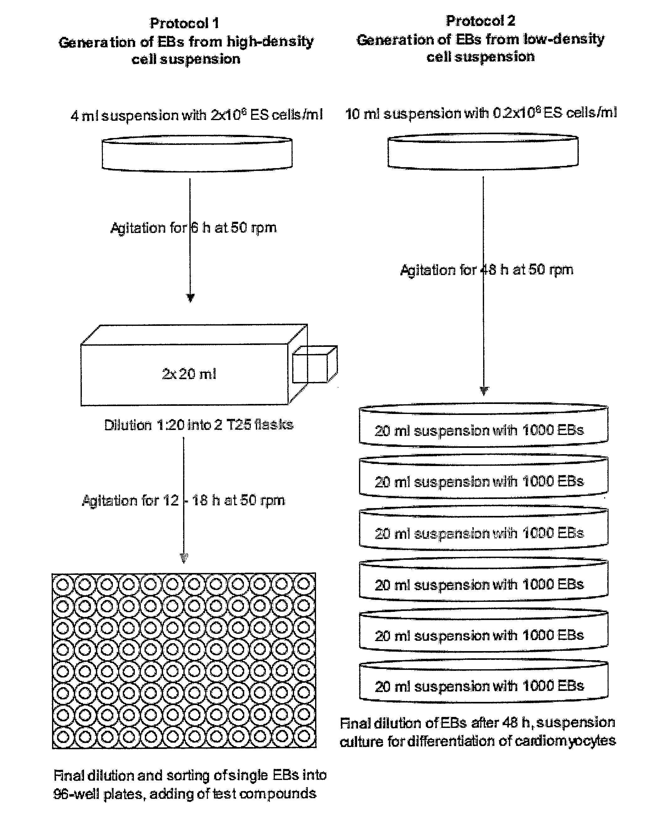
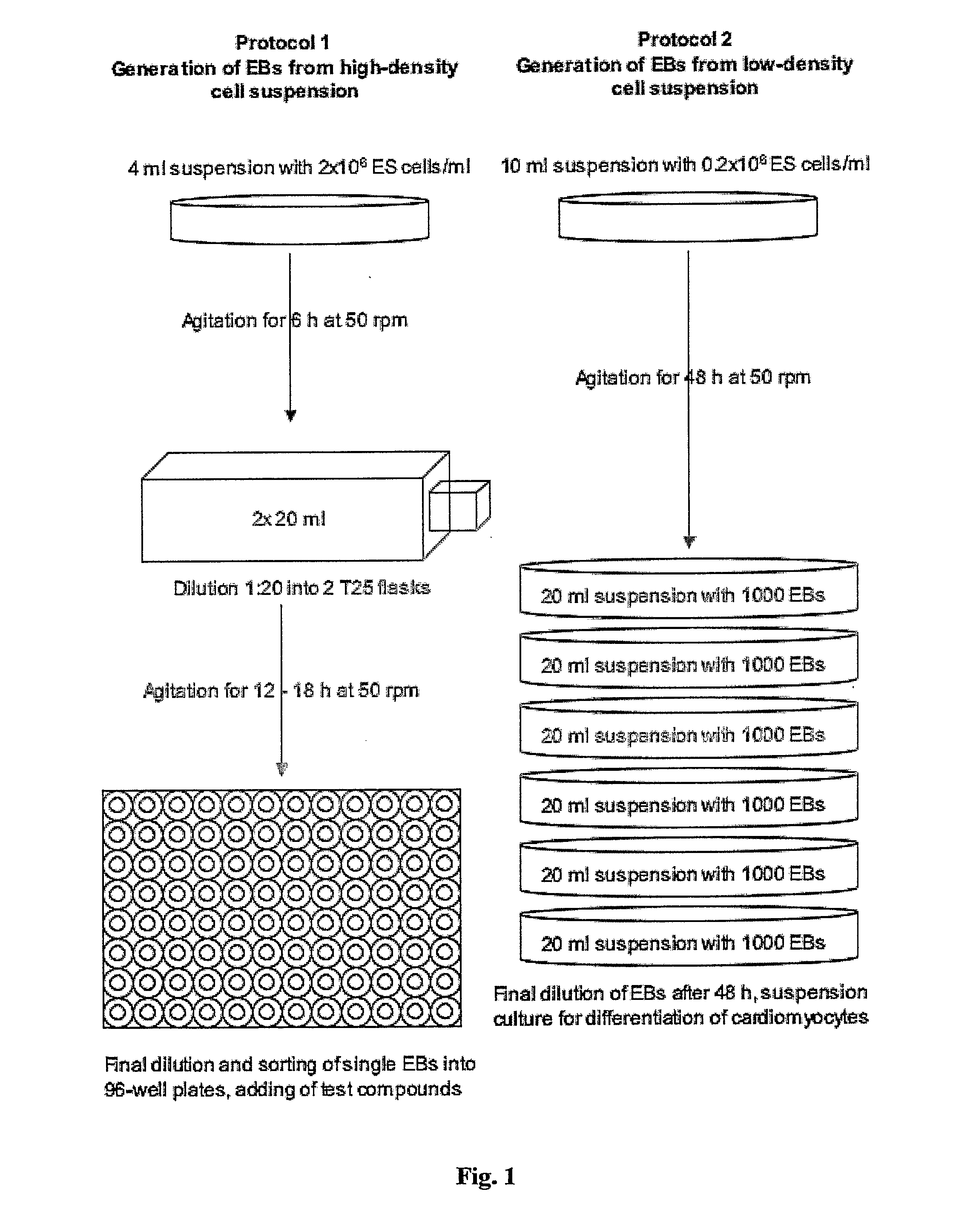
Generation of “Embryoid Bodies” (EBs) from High Density Cell Suspensions and Differentiation of Cardiac Cells (FIG. 1, Protocol 1)

[0169] Mouse embryonic stem cells (ES cells, clone D3, ATCC CRL 1934) were stably transfected with the pαMHC-GFP vector containing the gene of the green fluorescent protein under control of the cardiac a-myosin heavy chain (α-MHC) promotor. To obtain this vector, a 5.5 kb fragment containing the promotor region of the mouse (α-myosin heavy chain gene (Genbank 471441) was introduced into the polylinker of the pEGFP-1 vector (Clontech Laboratories).

[0170] ES cells were cultured on 10 cm petri dishes (Falcon, Becton Dickinson) at a density of 1.4×10⁶ in DMEM (Gibco, Invitrogen) supplemented with 15% FCS (Gibco, invitrogen, batch controlled) and 1×10³ U / ml LIF (Chemicon) on a layer of feeder cells (inactivated mouse embryonic fibroblasts, prepared according standard protocols; see also description of the invention above). Cells were incubated at 37° C., 7% ...

example 2

Generation of “Embryoid Bodies” (EBs) from Low Density Cell Suspensions and Differentiation of Cardiac Cells (FIG. 1, Protocol 2)

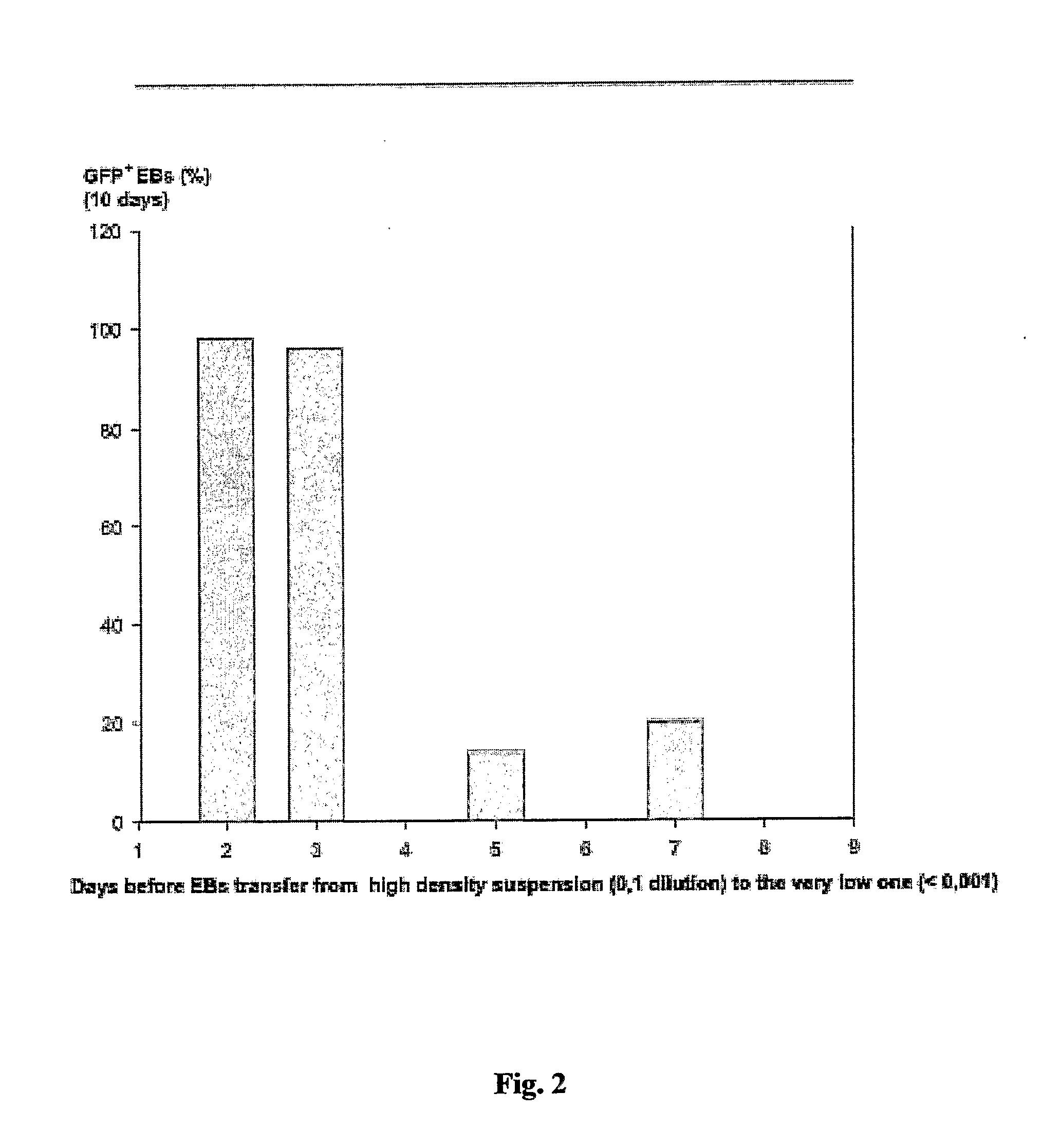
[0173] ES cells were cultured as describe in Example 1. To generate EBs, ES cells were cultured in suspension at a density of 0.2×10⁶ cells / ml in a 10 cm petri dish (Greiner, Darmstadt, Germany) in 10 ml IMDM with 20% FCS (Invitrogen, Karlsruhe, Germany) at 37° C., 5% CO2, 95% humidity on a rocking table (GFL 3006, GFL, Braunschweig, Germany) at 50 rpm for 48 hours. On day 2, 100-2000 EBs were transferred to 10 cm bacterial dishes (Greiner) into a volume of 10 ml IMDM 20% FCS and incubated at 37° C., 5% CO2, 95% humidity, either with or without agitation. On day 5 and 7, or every other day for high density suspensions such as 2000 EBs / 10 ml, the medium was replaced by 10 ml of fresh medium. On day 14, fluorescent areas representing cardiac cells were detected by fluorescence mircoscopy using a Zeiss Axiovert 200 M with a 10× Achroplan objective, a HQ-filt...

example 3

Effect of Embryotoxic Compounds on Differentiation Capacity of EBs towards Cardiomyocytes

[0174] EBs were generated as described in Example 1. At day 1, 30 EBs were transferred to each well of a bacteriological 6-well plate (Greiner) into a volume of 3 ml IMDM 20% FCS, and test compounds with known embryotoxic potential were added at different concentrations as indicated in the figures (solvent: DMSO, final concentration of DMSO: 0.1%). Test compounds were chosen from a list of compounds recommended for a validation study on in vitro embryotoxicity tests by the European Center for the Validation of Alternative Methods (ECVAM) (see Brown, NA 2002; ATLA 30, 177-198). Each compound concentration was tested in triplicate in three individual experiments. EBs were cultured at 37° C., 5% CO2, 95% humidity. On day 5, cultures were fed with 2 ml of fresh medium, and fresh test compounds were added. On day 14, EBs on each plate were counted, lysed in lysis buffer (20 mM Tris-HCl / 0.5% TritonX ...


PUM

PropertyMeasurementUnit
humidityaaaaaaaaaa
timeaaaaaaaaaa
timeaaaaaaaaaa

Abstract

Provided are means and methods for producing embryoid bodies (EBs) from multi- or pluripotent cells. In particular, a method of generating embryoid bodies (EBs) is described comprising agitation of a liquid suspension culture of multi- or pluripotent cells in a container until generation of cell aggregates, optionally diluting the suspension, and further agitation of the suspension until formation of EBs. Furthermore, the present invention relates to the use of the novel culturing method and EBs obtained thereby for a variety of applications including genomics, diagnostic assays, teratogenic / embryotoxicological and pharmacological assays as well as for the provision of tissue grafts.

Description

FIELD OF THE INVENTION [0001] The present invention relates to a method for producing embryoid bodies (EBs) from multi- or pluripotent cells. In particular, the instant invention concerns a method of generating embryoid bodies (EBs) comprising agitation of a liquid suspension culture of multi- or pluripotent cells in a container until generation of cell aggregates, optionally diluting the suspension, and further agitation of the suspension until formation of EBs. Furthermore, the present invention relates to the use of the method and EBs so obtained for a variety of applications including, but not limited to “loss of function” assays with ES cells containing homozygous mutations of specific genes, “gain of function” assays with embryonic stem (ES) cells overexpressing exogenous genes, developmental analysis of teratogenic / embryotoxic compounds in vitro, pharmacological assays and the establishment of model systems for pathological cell functions, and application of differentiation a...

Claims


Application Information

Patent Timeline
Patent Type & Authority Applications(United States)
IPC IPC(8): A01N1/00A61K48/00A61P43/00C12N5/00C12N5/10C12N5/16C12Q1/02C12N5/073C12N5/077
CPCC12N5/0603C12N5/0657C12N2502/13C12N2506/02C12N2510/00C12N2503/02A61P9/00A61P9/04A61P43/00
Inventor KOLOSSOV, EUGENKETTENHOFEN, RALFKOPP, ISABELLABOHLEN, HERIBERTSCHWENGBERG, SILKE
Owner EVOTECH INT GMBH
Why Eureka
Try Eureka