System and method for co-registering multi-channel images of a tissue micro array

a multi-channel image and tissue technology, applied in the field of tissue micro array processing and imaging, can solve the problems of inconsistent and inconclusive, tma fails to incorporate differences between serial images, and efforts are less than optimal

Active Publication Date: 2008-02-07
LEICA MICROSYSTEMS CMS GMBH
View PDF35 Cites 128 Cited by
  • Summary
  • Abstract
  • Description
  • Claims
  • Application Information

AI Technical Summary

Problems solved by technology

Such efforts have proved to be less than optimal because of the inability to automate the analysis of the TMA after each serial stain is applied.
Although systems exist that attempt to combine the serial images, these combined images result in inconsistent and inconclusive results because the serial images are merely co-registered based on the mechanical placement of the TMA on the imaging microscope.
These combined images that are co-registered based solely on the mechanical location of the TMA fail to incorporate differences between the serial images including, but not limited to, mechanical misplacement, tissue distortion, autofluorescence, differing levels of focus when each image is taken, and anomalies associated with the curvature of the cells in the tissue sample.
Simultaneous labeling of both techniques is not used because of the auto-fluorescence characteristics of the H&E dyes.
It is not possible to simultaneously image H&E dyes with molecular biomarker using current imaging techniques because of the auto-fluorescence characteristics of the H&E dyes.
Chemical interactions of H&E dyes with the antibodies to which the molecular biomarkers are attached significantly limit the simultaneous imaging of H&E with biomarkers.
As noted, although serial sections of H&E and fluorescent images have previously been serially compared, registration between such serial sections is poor because of tissue distortion and the optical and chemical effects of the stains.

Method used


Image

  • System and method for co-registering multi-channel images of a tissue micro array
  • System and method for co-registering multi-channel images of a tissue micro array
  • System and method for co-registering multi-channel images of a tissue micro array

Examples


Embodiment Construction

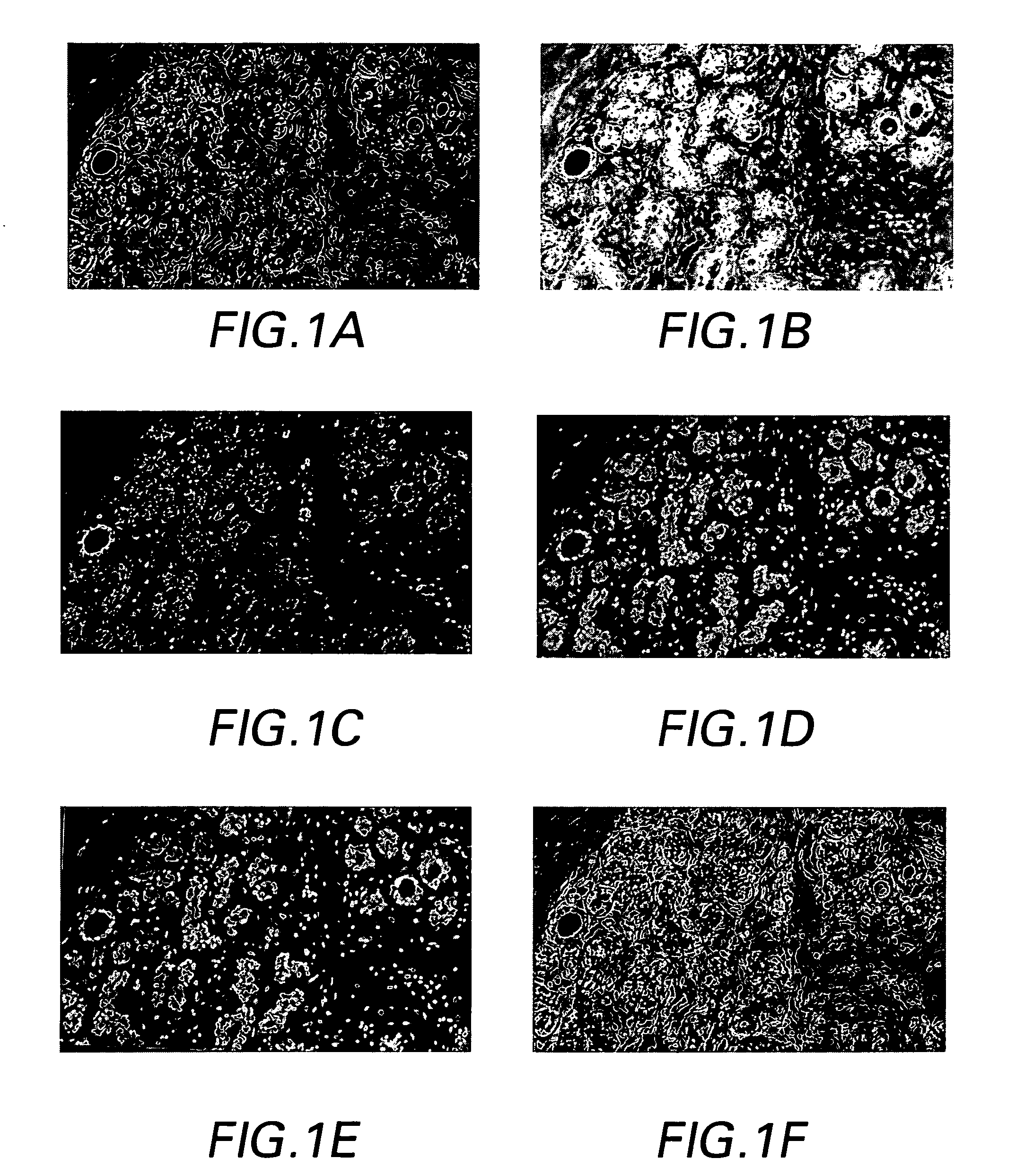
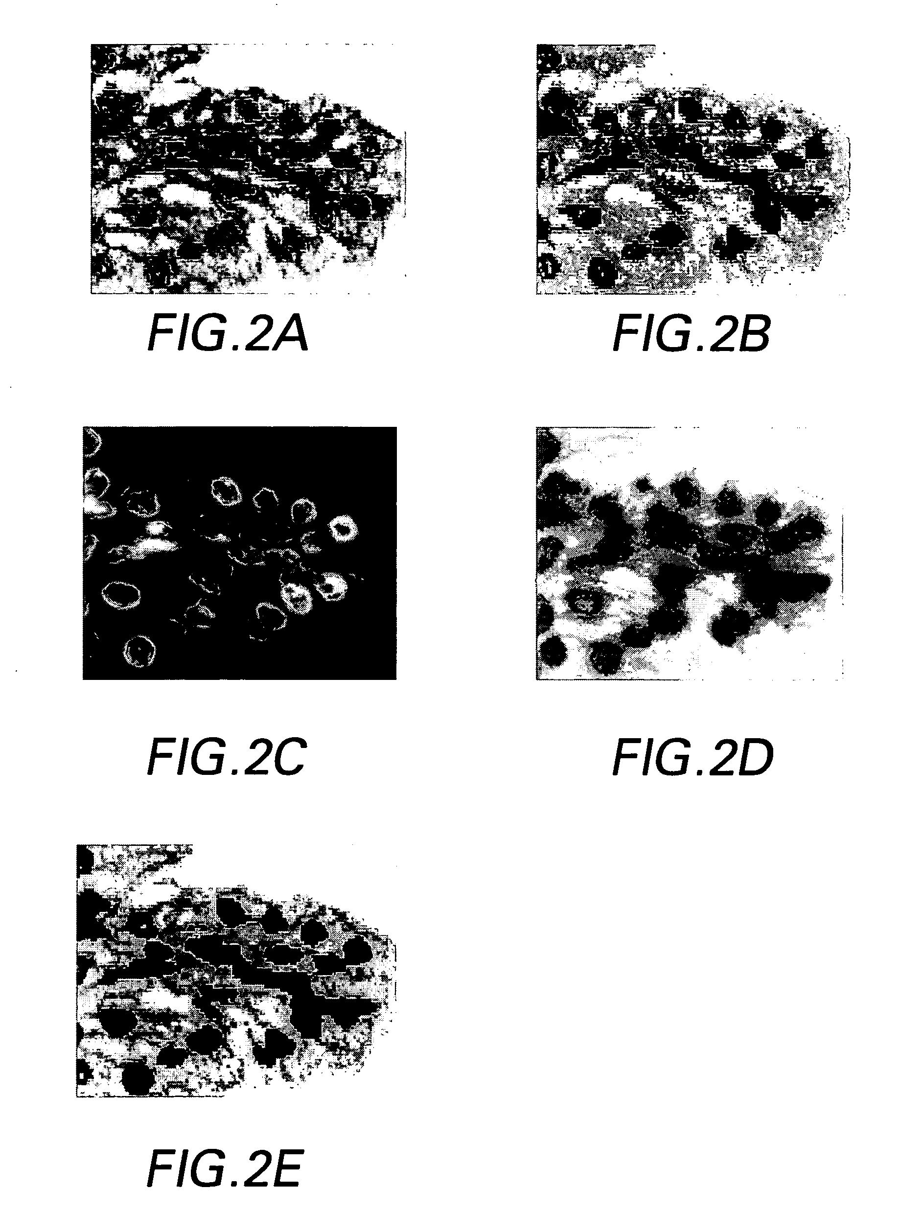
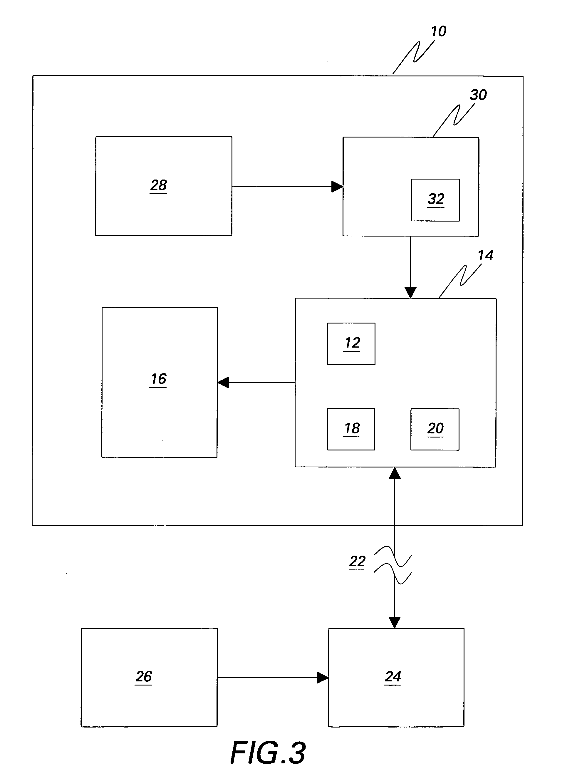
[0030]The preferred methods and preferred embodiments of system of the invention allow both molecular and morphological markers to be imaged from the same tissue sample using sequential imaging and co-registration techniques. Generally, the tissue, which is fixed or otherwise provided on a substrate such as, but not limited to, a TMA, a slide, a well, or a grid, is labeled with molecular biomarkers, and imaged through a fluorescent microscope. Then the tissue is re-labeled with one or more morphological stains such as H&E dyes, and imaged again. The methods are not limited to two images and can be adapted to co-register more than two images as needed. The images are overlaid using both hardware and software registration techniques, and the information is merged, whereby the technical effect is to co-register or otherwise produce multi-channel images. Every pixel in the multi-channel image represents both molecular and H&E information. This multi-channel registered image can be used ...


PUM

PropertyMeasurementUnit
fluorescentaaaaaaaaaa
morphological structuresaaaaaaaaaa
joint entropyaaaaaaaaaa

Abstract

A system and methods for co-registering multi-channel images of a tissue micro array, comprising the steps of, providing a biological material on a substrate; applying one or more molecular probes, adapted to provide fluorescent molecular markers, to the biological material; obtaining a first digital image of the biological material and the fluorescent molecular markers; applying a morphological stain to the biological material; obtaining a second digital image of the biological material, computing information common to the first and second images; and co-registering the second image with the first image based on one or more registration metrics.

Description

BACKGROUND[0001]The invention relates generally to tissue micro array processing and imaging.[0002]Tissue micro arrays (TMA) are used for many analytic and diagnostic purposes, one of which is to diagnose diseased tissue at the molecular level. Whatever the purpose, tissue micro arrays, on which tissue samples are fixed, are typically stained with a morphological stain or biomarker and then analyzed manually with a microscope, or an image may be taken of the TMA to retain the image for subsequent analysis or comparison. After the first stain is applied and imaged, one or more serial or successive stains or biomarkers may be applied and the TMA is analyzed again. The two or more serial images may then be compared. It has been an ongoing goal to automate this system while maintaining the quality and consistency of the analysis and resulting conclusions or data. Such efforts have proved to be less than optimal because of the inability to automate the analysis of the TMA after each seri...

Claims


Application Information

Patent Timeline
Patent Type & Authority Applications(United States)
IPC IPC(8): G01N1/30G06F19/00G06T7/40
CPCG01N1/30G06T2207/30072G06T2207/30024G06T7/0034G06T7/35G06V20/695G06V10/761
Inventor CLINE, HARVEY ELLISCAN, ALI
Owner LEICA MICROSYSTEMS CMS GMBH
Why Eureka
Try Eureka