Respiratory Volume/Flow Gating, Monitoring, and Spirometry System for Mri

a spirometry system and spirometry technology, applied in the field of medical imaging, diagnostics, therapy and intervention, can solve the problems of incompatibility of conventional methods of air flow measurement with the magnetic field environment of mr systems, inability to accurately track the volume changes in the lungs, and many limitations, and achieve the effect of high precision and accuracy

Inactive Publication Date: 2008-03-27
THE CHILDRENS HOSPITAL OF PHILADELPHIA
View PDF15 Cites 44 Cited by
  • Summary
  • Abstract
  • Description
  • Claims
  • Application Information

AI Technical Summary

Benefits of technology

[0006] The preferred embodiments of the invention eliminate the aforementioned deficiencies of the bellows method and provide additional utility. These benefits are provided because the preferred embodiments measure actual calibrated respiratory volume and flow (preferably through a mask covering the nose and mouth) with high precision and accuracy using microphone technology. In addition, the software allows for gating image acquisition, respiratory motion suppression, monitoring, pulmonary diagnostic spirometry, and data storage of respiratory data for real time and subsequent analysis.

Problems solved by technology

Conventional methods for air flow measurement are not compatible with the magnetic field environment of MR systems.
The Applicants discovered that the pressure signal resulting from conventional respiratory gating signals corresponds approximately to the respiratory volume in breath timing, but is subject to many limitations.
In particular, the respiratory signal: a) does not provide flow rate; b) is not calibrated while representing volume; c) can exhibit significant time lags with the actual volume changes in the patient; d) is not sensitive to airway obstructive elements that may register as breaths due to torso expansion and not actually involving airflow; and e) may be distorted in patients with thoracic or abdominal deformity.
The Applicants have also discovered that the current systems do not accurately track volume changes in the lungs because the waveform shape is dependent on the placement of the belt, thoracically or abdominally, or in between.
Further, the sensitivity of the bellows system signal is affected by the tightness with which the belt is initially placed around the patient.
Other methods of respiratory plethysmography (volume measurement / tracking) that measure chest motion (e.g., inductive, impedance, piezo electric) suffer from the same deficiencies, and require metallic cables and components on the patient.

Method used


Image

  • Respiratory Volume/Flow Gating, Monitoring, and Spirometry System for Mri
  • Respiratory Volume/Flow Gating, Monitoring, and Spirometry System for Mri
  • Respiratory Volume/Flow Gating, Monitoring, and Spirometry System for Mri

Examples


Embodiment Construction

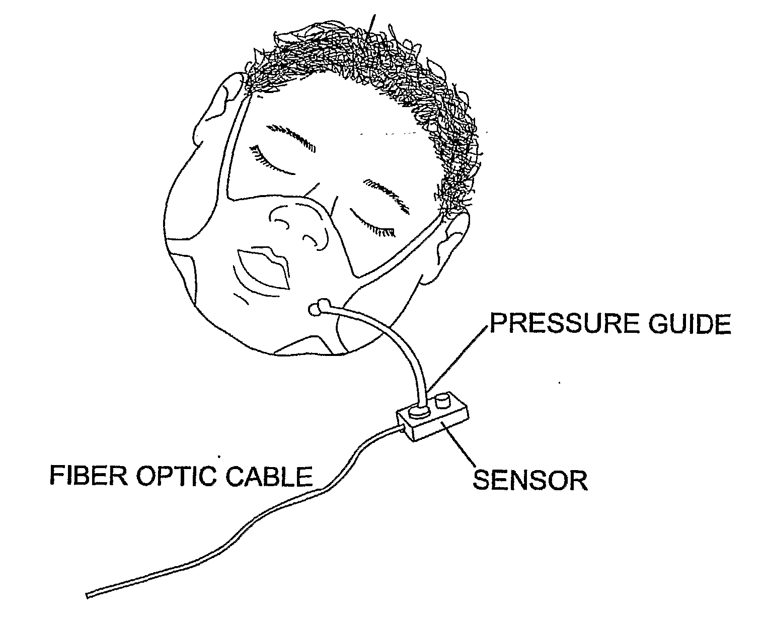
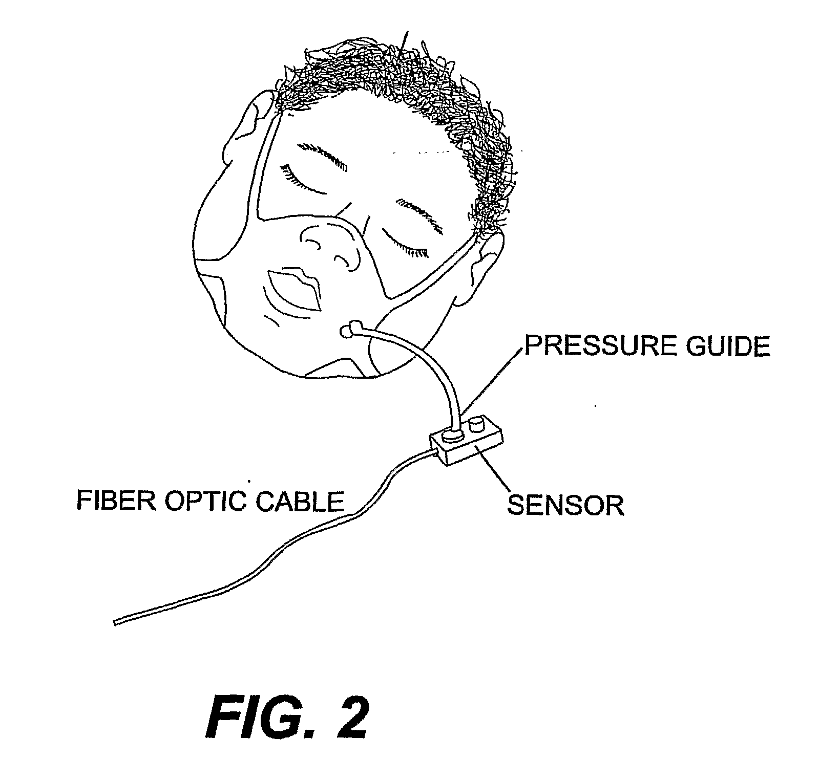
[0020] In accordance with an example of the preferred embodiments, a patient in an MRI scanner wears a mask preferably covering the nose and mouth, and held on the face with straps. The mask is preferably a tightly fitted low profile plastic mask or other modified mask in communication with a pneumotach assembly, including a pressure sense chamber and an air pressure sensor. A port on the mask allows the patient to inhale and exhale room air or other gas mixtures as required. While not being limited to a particular theory, the port is fitted with the pressure sense chamber. The pressure sense chamber includes a laminarizing element, a resistive element and a conduit communicating with the air pressure sensor. The proximal end of the pressure sense chamber includes a laminarizing element having a baffle. The baffle serves to limit turbulence and flow eddies in the pressure sense chamber. The distal end of the pressure sense chamber includes a non-metallic, hydrophobic, porous screen ...


PUM


Abstract

A respiratory volume/flow monitoring system, including a pneumotach that measures micromovements of a membrane in a MRI environment, gates (triggers) a MRI machine to start image acquisition at specific lung volumes or airflow rates during the breathing cycle or other breathing maneuvers of a patient. The system provides breathing motion artifact suppression for any imaging test susceptible to breathing motion artifact, allows respiratory monitoring of the subject during an MRI procedure, and provides the capability to perform spirometric testing while the subject is in the MRI environment. Embodiments of the invention are also applicable for patients undergoing CT imaging or any other imaging modality that allows external triggering.

Description

FIELD OF INVENTION [0001] The inventive subject matter relates to medical imaging, diagnostics, therapy and intervention. More particularly, this invention relates to devices and methods for monitoring, imaging and diagnosing a patient's breathing in a magnetic resonance imaging (MRI) environment. BACKGROUND OF THE INVENTION [0002] Conventional respiratory gating technology for MRI testing consists of tracking the respiratory wave form via a single sensor on a belt encircling the torso of the patient. This sensor is typically a rubber bellows attached on either end to an inelastic belt. As the chest and / or abdomen expands and contracts during breathing, air pressure in the bellows falls and rises as it stretches and contracts. Conventional methods for air flow measurement are not compatible with the magnetic field environment of MR systems. These devices generally involve metallic components, wires, and the close proximity of transducers and computers. [0003] Applicants developed th...

Claims


Application Information

Patent Timeline
Patent Type & Authority Applications(United States)
IPC IPC(8): A61B5/08
CPCA61B5/055A61B6/541A61B5/7285A61B5/0873G01R33/5673
Inventor MCDONOUGH, JOSEPH M.ARENS, RAANANSIN, SANGHUN
Owner THE CHILDRENS HOSPITAL OF PHILADELPHIA
Why Eureka
Try Eureka