Manganese-enhanced magnetic resonance imaging of neurons using electrical stimulation

Inactive Publication Date: 2008-04-03
UNIV KANSAS MEDICAL CENT
View PDF1 Cites 7 Cited by
  • Summary
  • Abstract
  • Description
  • Claims
  • Application Information

AI Technical Summary

Benefits of technology

[0010]The present invention is directed to a method for improving imaging of neurons in areas such as the spinal cord and the brain. In a preferred embodiment, electrical stimulation is used to improve

Problems solved by technology

Unfortunately, in these previous studies, manganese labeling of the cortical spinal tract weake

Method used


Image

  • Manganese-enhanced magnetic resonance imaging of neurons using electrical stimulation
  • Manganese-enhanced magnetic resonance imaging of neurons using electrical stimulation
  • Manganese-enhanced magnetic resonance imaging of neurons using electrical stimulation

Examples


Example

EXAMPLE 1

Electrical Stimulation Improves Cortical Spinal Tract Tracing of Spinal Cord Using Manganese Enhanced MRI

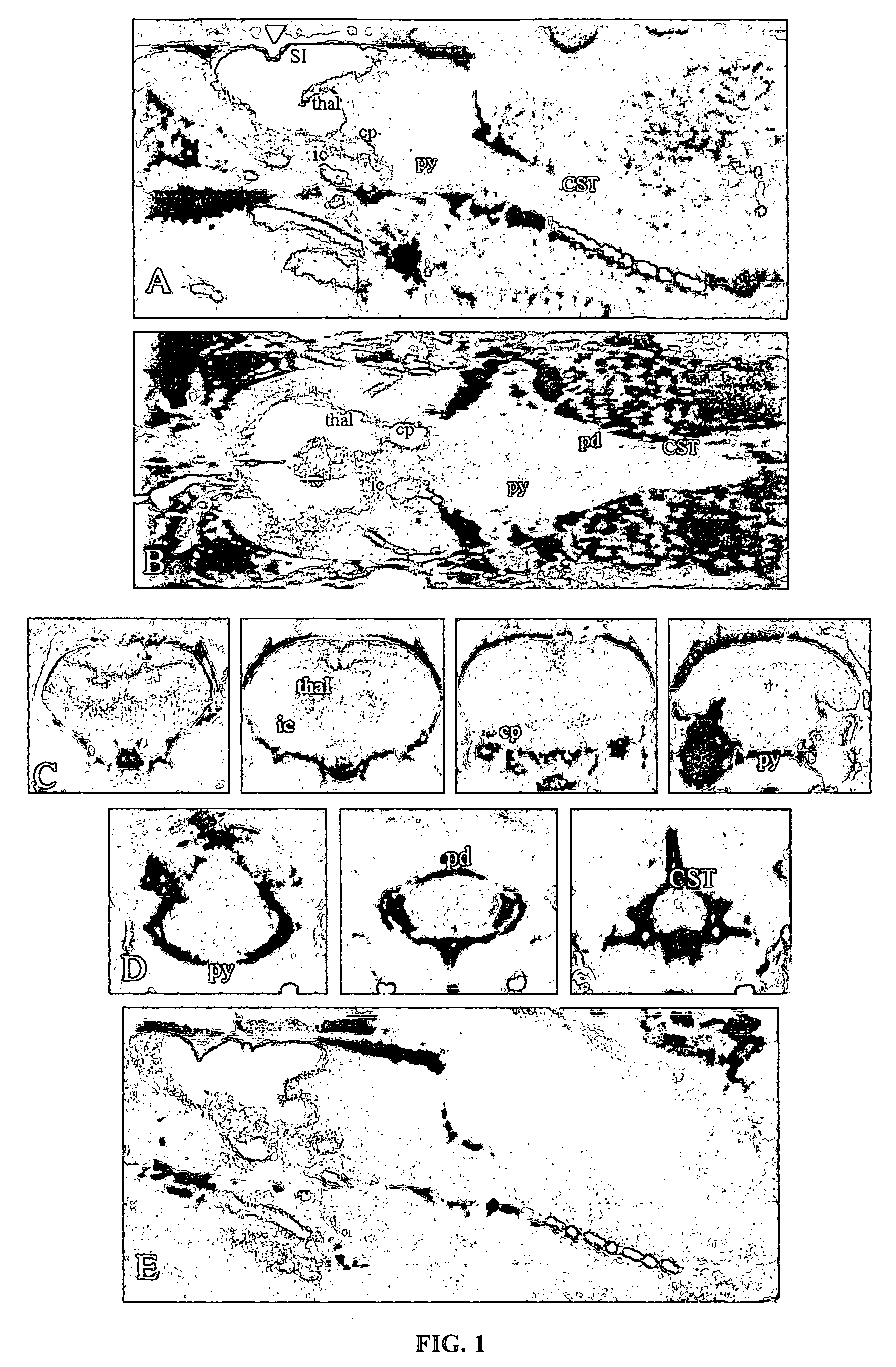
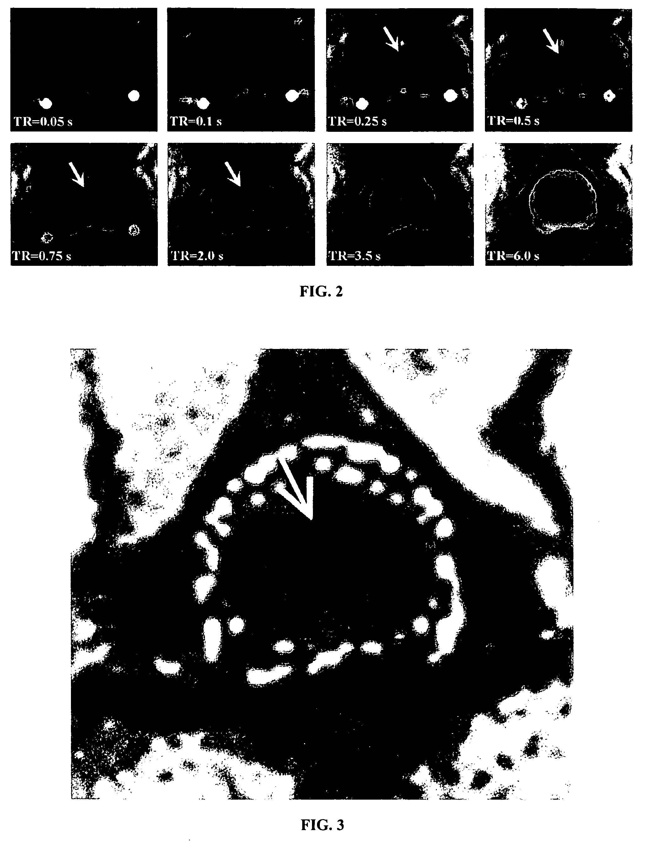
[0036]In this example, electrical stimulation was applied to the rat cortex in order to improve visualization of the rat spinal cord using manganese-enhanced MRI. The experiments were conducted on twelve Sprague-Dawley rats weighing between 300 and 350 g under a protocol approved by the University of Kansas Medical Center Institutional Animal Care and Use Committee. Six rats were studied without electrical stimulation in the cortex and served as the control group. The remaining six rats formed the stimulation group. These rats received electrical stimulation of motor cortex to test the merits of this procedure as a means of enhancing the delineation of the cortical spinal tract in the spinal cord using manganese-enhanced magnetic resonance imaging.

[0037]Surgical Procedures

[0038]Rats were anesthetized using ketamine hydrochloride delivered intramuscularly. The advantage o...

Example

EXAMPLE 2

Electrical Stimulation Improves Imaging of Injured Spinal Cord Using Manganese-Enhanced MRI

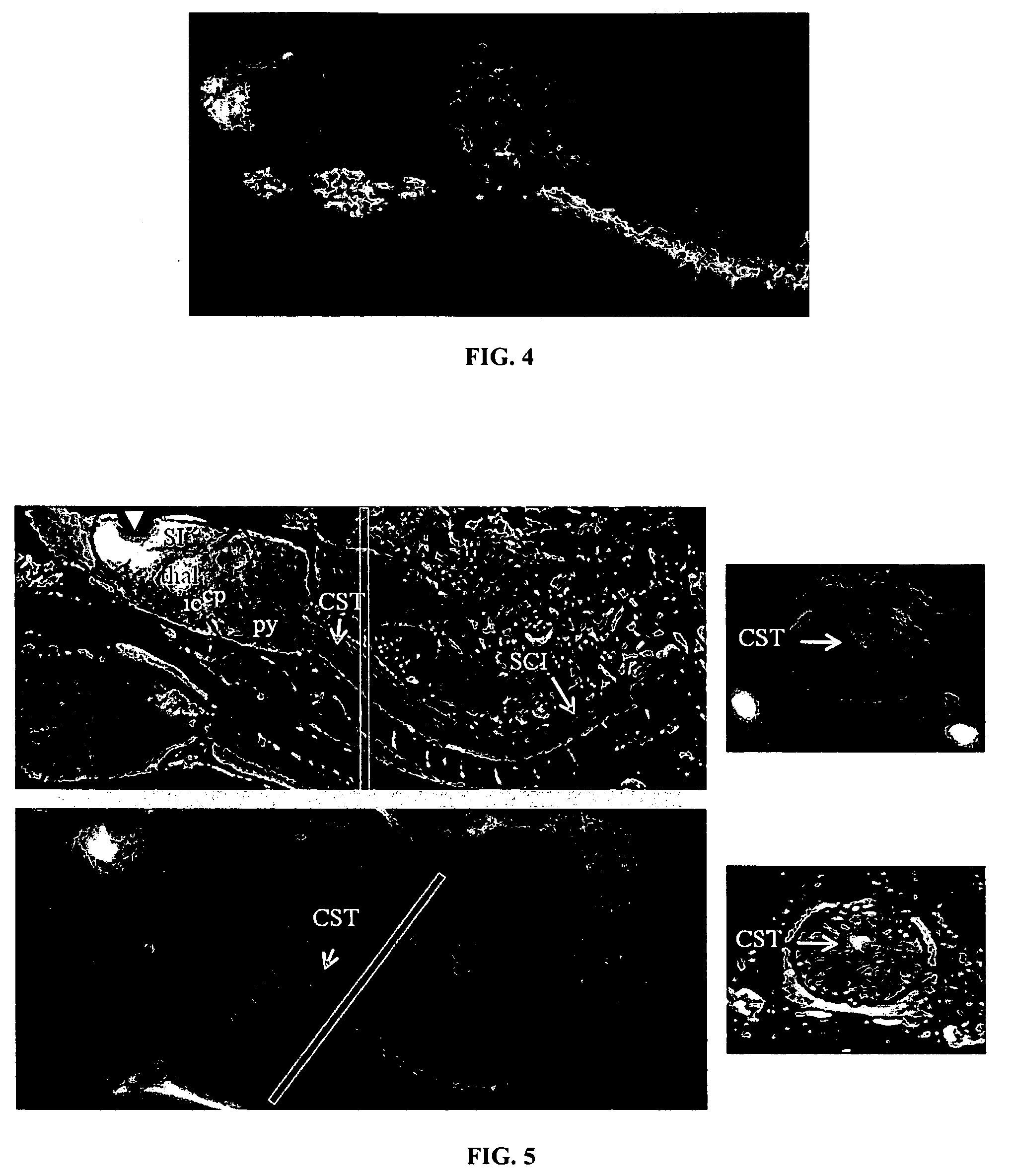
[0055]Understanding the temporal changes in CNS tissue, such as reorganization of the cortical spinal tract, is an important and growing focus of spinal cord injury (“SCI”) research. In the example, manganese enhanced MRI with electrical stimulation was expanded to include tracing intact neuronal fibers, not only in the cortical spinal tract but also fibers in other tracts, that project through the site of injury.

[0056]The experiments were conducted on one Sprague-Dawley rat (about 300 g) under a protocol approved by the University of Kansas Medical Center Institutional Animal Care and Use Committee. Established procedures described previously for the surgery, SCI, manganese delivery and manganese enhanced MRI scans were followed. See Bilgen, A new device for experimental modeling of central nervous system injuries, Neurorehabil. Neural. Repair, 19-226 (2005); Bilgen et al., Ex vivo m...


PUM


Abstract

A method for improving uptake of contrast agents, such as manganese-based contrast agents, is neuronal imaging of areas such as the spinal cord and cortical spinal tract with magnetic resonance imaging are provided. Electrical stimulation is applied to the subject in order to increase uptake of the contrast agent in to the neuron, resulting in an improved image.

Description

STATEMENT REGARDING FEDERALLY SPONSORED RESEARCH OR DEVELOPMENT[0001]The present invention was supported in part by NIH Grant NS051825 and the government may have certain rights in the invention.CROSS-REFERENCE TO RELATED APPLICATIONS[0002]Not applicable.BACKGROUND OF THE INVENTION[0003]1. Field of the Invention[0004]The present invention relates to methods for enhancing neuroimaging using contrast agents, such those involving magnetic resonance imaging (“MRI”). More specifically, improved methods of imaging the neuronal tracts, such as those in the cortical spinal tract and spinal cord using manganese-enhanced MRI are disclosed.[0005]2. Description of Related Art[0006]Various neuroimaging techniques which employ contrast agents are known in the art. Such techniques include computer tomography (“CT”), computer axial tomography (“CAT”), positron emission tomography (“PET”), single photon emission computed tomography (“SPECT”), and MRI. In particular, MRI is well established as a medi...

Claims


Application Information

Patent Timeline
IPC IPC(8): A61K49/10
CPCA61K49/103A61K49/06
Inventor BILGEN, MEHMETBROOKS, WILLIAM M.CHENEY, PAUL D.
Owner UNIV KANSAS MEDICAL CENT
Why Eureka
Try Eureka