Compositions and methods for the treatment and prevention of fibrotic, inflammatory, and neovascularization conditions of the eye

a technology of fibrosis or scarring, which is applied in the field of ocular disorders, can solve the problems of insufficient treatment options, complex search for effective treatments, and unclear exact pathogenesis of amd, and achieve the effects of reducing or preventing aberrant fibrogenesis, fibrosis or scarring, and modulating surgical and traumatic wound healing responses of the ey

Inactive Publication Date: 2008-09-04
LPATH
View PDF99 Cites 45 Cited by
  • Summary
  • Abstract
  • Description
  • Claims
  • Application Information

AI Technical Summary

Benefits of technology

[0072]In accordance with the present invention, methods are provided for treating ocular diseases or conditions through administration of a pharmaceutical composition comprising an immune-derived moiety (e.g, an antibody) reactive against a bioactive lipid, in order to decrease the effective concentration so that the bioactive lipid is inhibited in whole or in part from eliciting its undesired effects. In some embodiments, the immune-derived moiety is a monoclonal antibody or fragment, variant or derivative thereof. In some embodiments, the immune-derived moiety is reactive against a lysolipid, such as S1P or LPA. Methods are also provided for decreasing or preventing aberrant fibrogenesis, fibr

Problems solved by technology

Multiple theories exist but, the exact etiology and pathogenesis of AMD are still not well understood.
The multiple cellular and cytokine interactions which are associated with exudative AMD greatly complicate the search for effective treatments.
While CNV and edema are manageable in part by anti-VEGF therapeutics, potential treatments to mitigate scar formation and inflammation have not been adequately addressed [Bylsma and Guymer (2005), Clin Exp Optom, vol 88: 322-34 and Pauleikhoff (2005), Retina, vol 25: 1065-84].
The results in a larger patient population may be less robust.
Tissue damage can result from either component.
These other aspects of the pathologic process are not addressed by current anti-VEGF treatments.
There is no effective treatment for the atrophic (dry) form of AMD.
Secondary to RPE dysfunction, macular rods and cones degenerate leading to the irreversible loss of vision.
However, new vessels when uncontrolled, are commonly defecti

Method used


Image

  • Compositions and methods for the treatment and prevention of fibrotic, inflammatory, and neovascularization conditions of the eye
  • Compositions and methods for the treatment and prevention of fibrotic, inflammatory, and neovascularization conditions of the eye
  • Compositions and methods for the treatment and prevention of fibrotic, inflammatory, and neovascularization conditions of the eye

Examples


example 1

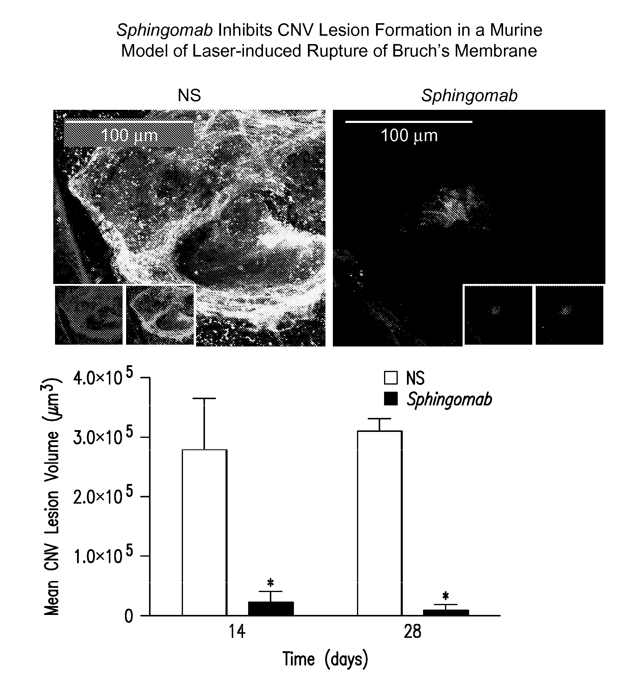
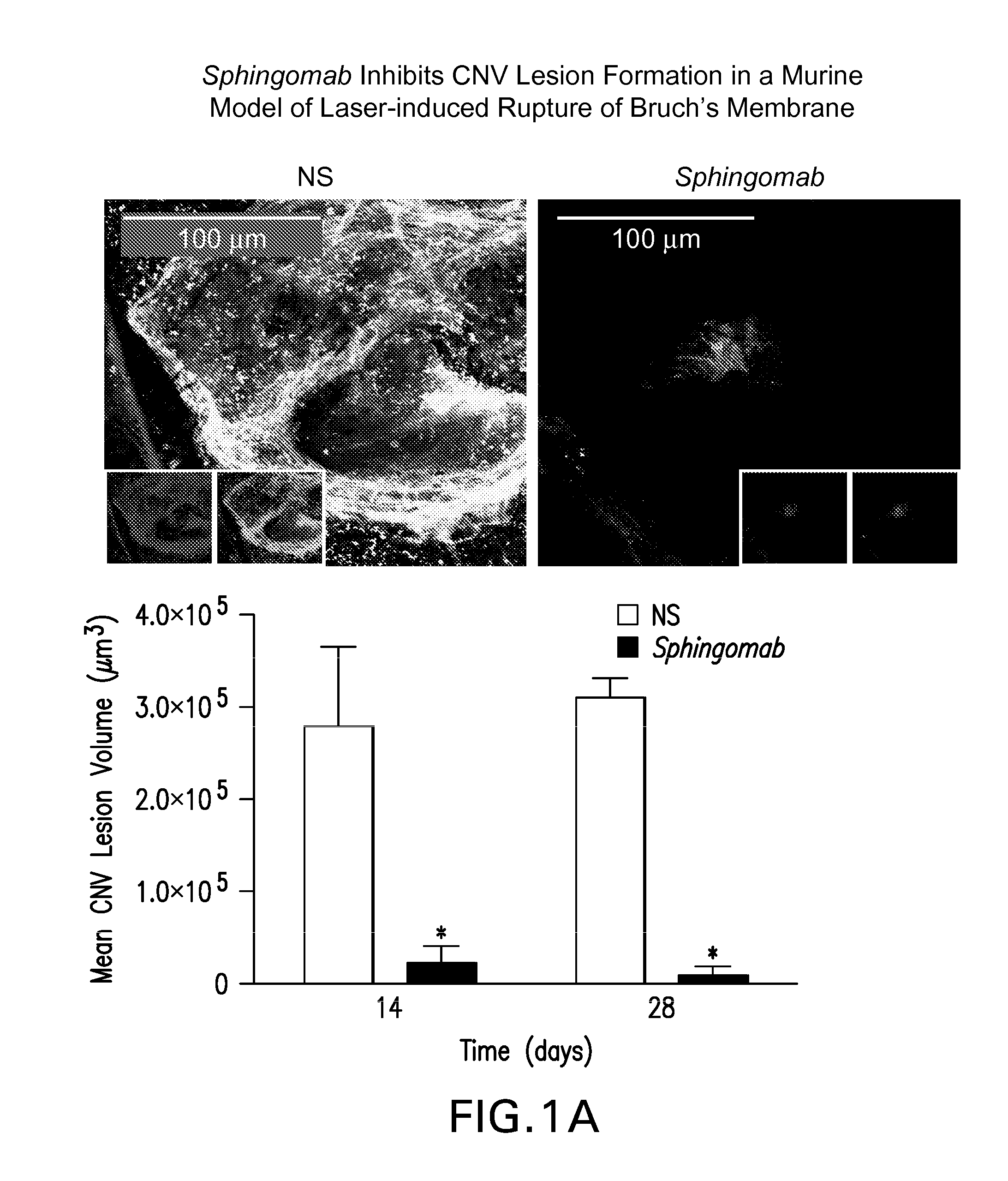
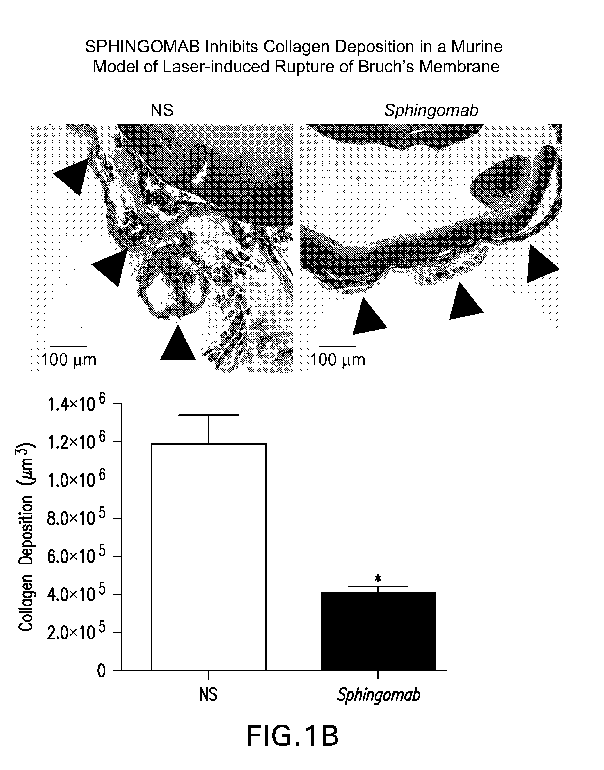
SPHINGOMAB Significantly Reduced CNV and Scar Formation in a Murine Model of CNV

[0156]Female C57BL6 / J mice were subjected to laser-induced rupture of Bruch's membrane and administered either 0.5 μg of Sphingomab or an isotype-matched non-specific (NS) antibody diluted in 2 μl of physiological saline. Mice were sacrificed 14 and 28 days after laser rupture.

[0157]To induce CNV lesions, the pupils were dilated with ophthalmic tropicamide (0.5%) and phenylephrine (2.5%). A coverslip was placed on the eye. An Oculight GL 532 nm (Iridex Corporation, Mountain View, Calif.) coupled to a slit lamp set to deliver a 100 msec pulse at 150 mW with a 50 μm spot size was used to rupture Bruch's membrane in three quadrants of the right eye located approximately 50 μm from the optic disc at relative 9, 12 and 3 o'clock positions. The left eye served as an uninjured control in all cases. Any lesion not associated with a vapor bubble or lesions that became confluent were excluded from analysis.

[0158]T...

example 2

SPHINGOMAB Inhibits Neovascularization Through Multiple Mechanisms Including Inhibition of Endothelial Cell Migration and Tube Formation

[0161]S1P promotes the migration of human umbilical vein endothelial cells (HUVECs) and, in Matrigel and other assays, the formation of de novo BV formation in vitro [112]; SPHINGOMAB can neutralize these effects of S1P Experiments were performed as described by Visentin et al. (Cancer Cell 2006 March; 9(3):225-38). Data in FIG. 2A suggest that HUVECs seeded onto GF-reduced Matrigel formed multiple capillary-like structures in the presence of S1P and failed to form capillary-like structures in the absence of S1P or when co-incubated with SPHINGOMAB and S1P. Data in FIG. 2B demonstrate the potent ability of 0.1-1 μM S1P to stimulate HUVEC migration 2-2.5 fold over non-treated HUVECs, or HUVECs co-incubated with SPHINGOMAB in a Matrigel chemoinvasion assay. Combined, these studies demonstrate that SPHINGOMAB can efficiently mitigate the pro-angiogenic...

example 3

SPHINGOMAB Inhibits Neovascularization Through Multiple Mechanisms Including Mitigation of the Effects of S1P, VEGF and bFGF In Vivo

[0162]Based on in vivo studies showing that S1P increased endothelial capillary growth into subcutaneously implanted Matrigel plugs[54], we speculated that SPHINGOMAB could reduce de novo BV formation in vivo. To investigate this, we employed the in vivo Matrigel Plug assay for neovascularization. In one set of experiments, Matrigel was supplemented with either 1 μM S1P, 0.5 μg / mL bFGF or 1 g / mL VEGF and then injected I.P. into mice (n=4). After 10 days, the mice were heparinized and injected with the fluorescent lectin, Isolectin B4-FITC, which binds to adhesion molecules expressed by vascular EC that form the growing BVs. The plugs were then excised, frozen in OCT, sectioned and viewed for FITC-stained BVs. Data in FIG. 3A suggest that S1P is a more potent stimulator of neovascularization in vivo than bFGF or VEGF[Lee, et al., (1999), Biochem Biophys ...


PUM

PropertyMeasurementUnit
Volumeaaaaaaaaaa
Volumeaaaaaaaaaa
Volumeaaaaaaaaaa

Abstract

The present invention relates to compositions and methods for prevention and treatment of ocular diseases and conditions characterized by aberrant fibrogenesis or scarring, inflammation and/or aberrant neovascularization or angiogenesis. The compositions and methods of the invention utilize immune-derived moieties that are specifically reactive against bioactive lipids and which are capable of decreasing the effective concentration of said bioactive lipid. In some embodiments, the immune-derived moiety is a monoclonal antibody that is reactive against sphingosine-1-phosphate (S1P) or lysophosphatidic acid (LPA).

Description

RELATED APPLICATIONS[0001]This application is a continuation-in-part of U.S. patent application Ser. No. 11 / 588,973, filed 27 Oct., 2006, which claims priority to the following: U.S. provisional patent application Ser. No. 60 / 854,971 [attorney docket no LPT-3010-PV, entitled “Compositions and Methods for Binding Sphingosine-1-Phosphate”] and U.S. provisional patent application Ser. No. 60 / 855,003 [attorney docket no LPT-3020-PV, entitled “Humanized Antibodies to Sphingosine-1-Phosphate in the Treatment of Ocular Disorders”], both filed on Oct. 27, 2006; U.S. provisional patent application Ser. No. 60 / 835,569 [attorney docket no LPT-3100-PV2], filed 12 Aug., 2006; and U.S. patent application Ser. No. 11 / 261,935, filed 28 Oct., 2005. This application claims the benefit of, and incorporates by reference for all purposes all of the aforementioned applications, each in its entirety:TECHNICAL FIELD[0002]The present invention relates to methods of treatments for ocular disorders using immu...

Claims


Application Information

Patent Timeline
IPC IPC(8): A61K39/395A61P27/02
CPCA61K2039/505C07K16/18C07K2316/96C07K2317/92C07K2317/56C07K2317/565C07K2317/24A61P27/02C07K2317/76
Inventor SABBADINI, ROGER A.GARLAND, WILLIAM A.STOLLER, GLENN L.
Owner LPATH
Why Eureka
Try Eureka