Systems and Methods for Reducing Intraocular Pressure

a technology of intraocular pressure and systemic pressure, applied in the field of systems and methods for reducing intraocular pressure, can solve the problems of increased intraocular pressure, obstruction of drainage system, blindness if not properly treated, etc., and achieve the effect of preventing cellular adhesion and reducing scarring over the devi

Inactive Publication Date: 2008-09-11
BECTON DICKINSON & CO
View PDF6 Cites 32 Cited by
  • Summary
  • Abstract
  • Description
  • Claims
  • Application Information

AI Technical Summary

Benefits of technology

[0015]In other embodiments, the systems of the present invention may include an implant that can be placed across the cornea to drain the anterior chamber of the eye. The implant may include a head, a foot, a tubular conduit between the foot and the head that has an interior channel in fluid communication with the anterior chamber, and a filter that can be impacted within the anterior chamber to regulate outflow of aqueous humor and to restrict incursion or minimize ingress of microorganisms or obstruct their passage.
[0016]In yet other embodiments, the systems of the present invention may include a transcorneal shunt and may further include a delivery device for implanting the shunt in this transcorneal position. In certain embodiments, the transcorneal shunt to be implanted with the delivery device may have a head, a foot, a substantially cylindrical body between the head and the foot having a channel therethrough, and a filter positioned within the channel to regulate the flow rate of aqueous humor through the channel and further to restrict the ingress of microorganisms. In certain embodiments, the delivery device may include a tip dimensionally adapted for holding the shunt and for positioning the shunt for insertion through the external surface of the cornea, and may further include a plunger slidable from a proximal position to a distal position wherein sliding the plunger dislodges the shunt and urges it through the external surface of the cornea into a transcorneal position.
[0018]The shunt according to the present invention is intended to solve certain of the abovementioned problems that have persisted within the opthalmological arts for treatment of elevated IOP. First, the shunt, its delivery device and the methods for their use are adapted for positioning of a drainage system across the clear cornea, thereby avoiding the difficulties that accompany subconjunctival or subscleral drainage. Second, the outflow of aqueous humor is consistently regulated by a filtration system without implicating mechanisms of wound healing, so that a predictable outflow rate can be calculated to avoid the dangers of hypotony on one hand and inadequate drainage on the other. Third, the filter provides a tortuous path to inhibit bacterial ingress; in addition, the slit opening in the head is shaped and sized to resist bacterial invasion; furthermore, the head itself is fabricated from a material that resists cellular adhesion, including the adhesion of microorganisms. Fourth, the device is made of materials well tolerated by the cornea. The head and the foot resist cellular adhesion and discourage scarring over the device, while the body is made of materials that encourage cellular adhesion, thereby to affix the device securely in the transcorneal position. These and other objects, features and advantages of the present invention will become more evident from the following discussion and drawings, wherein like numbers represent like components.

Problems solved by technology

Obstruction to the drainage system may be a cause of elevated intraocular pressure.
Persistence of elevated IOP produces the condition known as glaucoma, wherein an elevated IOP may damage the optic nerve and affect vision, leading eventually to blindness if not properly treated.
Medical management may fail, however, because of poor patient compliance, high cost, or any one of a number of well-recognized complications and side effects.
Several known limitations accompany such procedures, however.
First, normal wound healing tends to interfere with the patency of the fistula and with the dimension of the drainage pocket, so that these operations may have an unacceptable rate of failure.
Such treatment increases the incidence of a second sort of problem associated with these procedures: excessive or overly rapid outflow of aqueous humor.
It is well known that removal of too much aqueous humor too quickly can reduce intraocular pressure precipitously to dangerously low levels, a condition called hypotony, potentially causing a number of sight threatening complications.
Those treatments that inhibit wound healing therefore increase the risks associated with excessive aqueous humor drainage.
A third kind of problem accompanies this type of conventional drainage procedure: an increased risk of infection.
Drainage of aqueous humor into a scleral or subconjunctival bleb poses a risk for infection by providing a fluid milieu that microorganisms can invade.
Furthermore, if an infection becomes established in the fluid-filled pocket, the microorganisms can travel retrograde through the drainage channel to enter the anterior chamber of the eye and infect it as well, a much more serious condition.
The problems described above that affect soft tissue surgery also affect implantation surgery, however.
Indeed, artificial materials may overstimulate local wound healing, leading to excessive scar tissue formation.
In addition, infection remains a risk.
Finally, the eye, like most tissues of the body, has limited tolerance for the long-standing presence of artificial materials.
A locally positioned implant may irritate the surrounding tissues.
Even if properly positioned initially, however, the implant can be displaced by local tissue motion or can be extruded by constrictive wound healing processes.
As mentioned earlier, however, these devices face the problems of regulating aqueous outflow, resisting infection and avoiding local tissue irritation and trauma.
The first problem, regulating aqueous outflow, arises because the drainage rate of this fluid depends substantially on the mechanical characteristics of the implant until there has been sufficient wound healing to restrict fluid outflow biologically.
Effective balancing of biological and mechanical resistance to aqueous humor outflow remains a problem for implant-based drainage procedures.
Each of these mechanisms, though, may become a liability once wound healing has been established.
Restrictive elements within the implant, when combined with the restriction effected by wound healing, may inordinately reduce the rate of aqueous humor outflow, possibly to non-therapeutic levels.
The second problem, the possibility of intraocular infection, arises because the presence of an implant provides a conduit through which bacteria can gain entry to the interior of the anterior chamber.
These mechanisms have limitations, however: even when effective in resisting the transit of microorganisms, they have hydraulic effects on fluid outflow that may also impair effective drainage.
Finally, the problem of local tissue tolerance arises with certain prior art devices because these foreign bodies may incite tissue reactions culminating in local inflammation or extrusion, and may further be perceptible or uncomfortable for the patient: these reactions to the presence of the implant may make its use clinically unsuitable.
These problems, as previously discussed, affect transcorneal devices as well.
Finally, the problem of positional stability has not been solved satisfactorily.

Method used


Image

  • Systems and Methods for Reducing Intraocular Pressure
  • Systems and Methods for Reducing Intraocular Pressure
  • Systems and Methods for Reducing Intraocular Pressure

Examples


Embodiment Construction

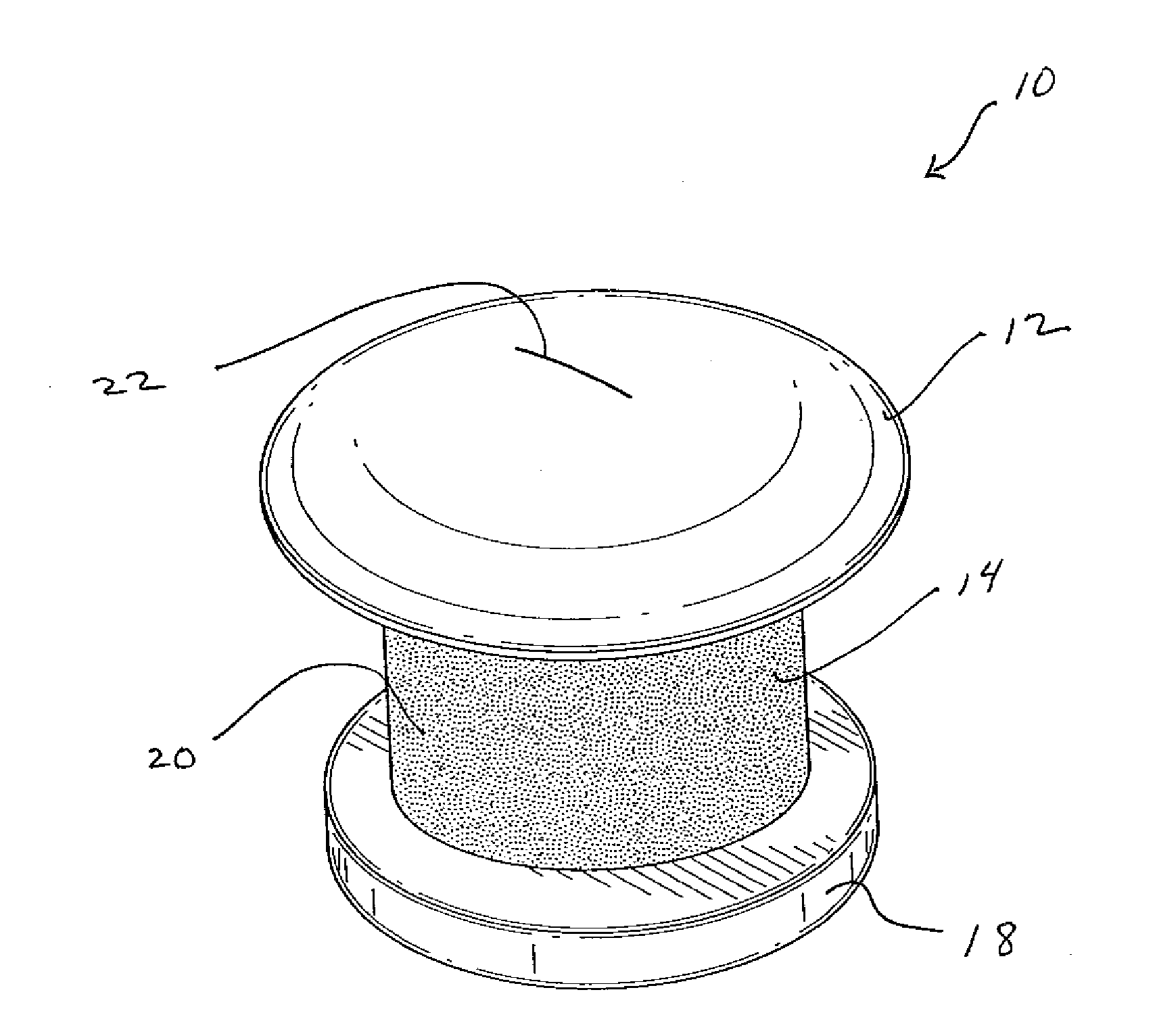
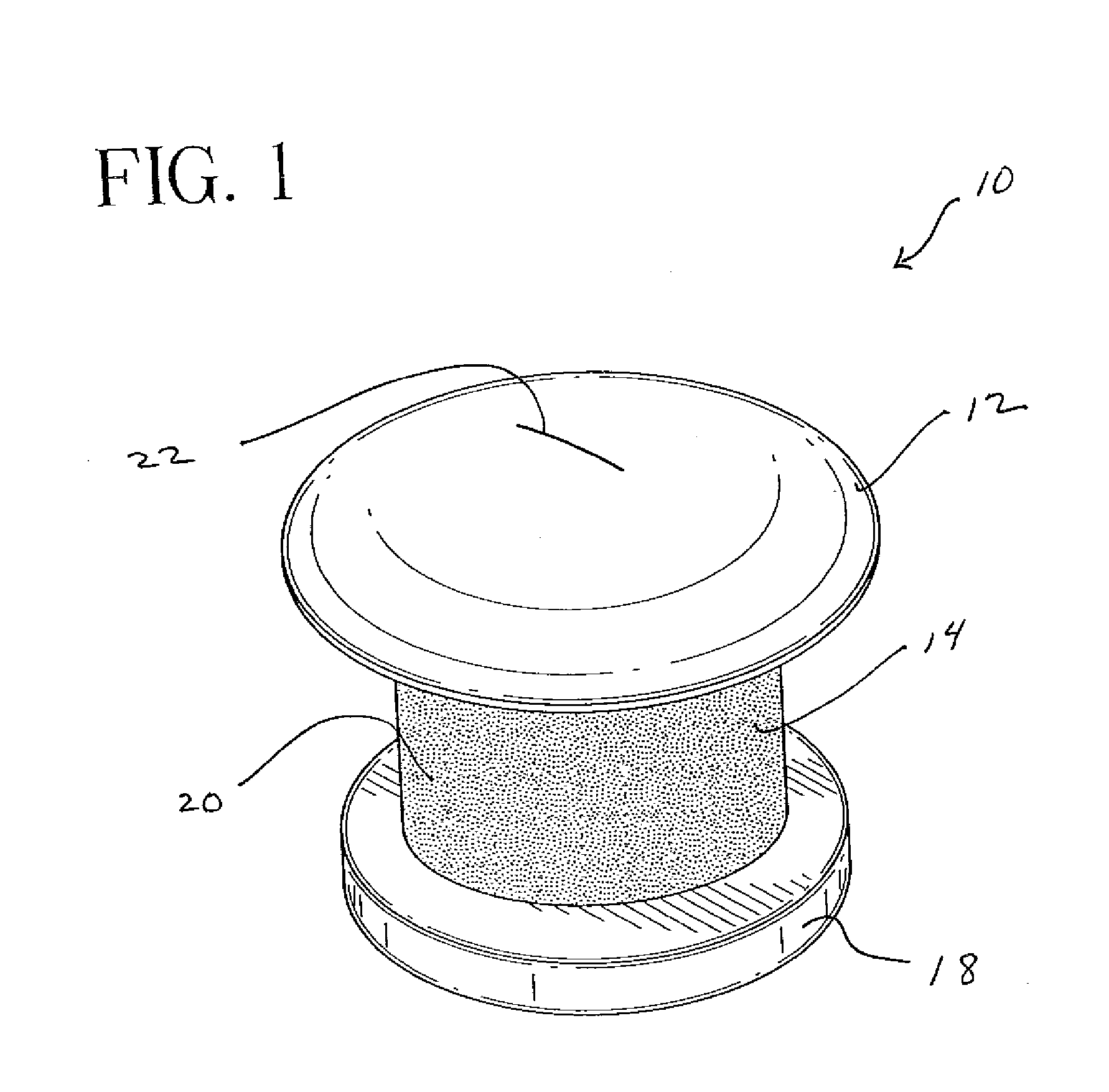
[0026]With reference to FIG. 1, a perspective view of a shunt 10 according to the present invention may be seen. In a representative embodiment, the shunt 10 may be approximately one millimeter long with an outer diameter of approximately 0.5 mm. While the shunt 10 illustrated in this and the following figures is shown as a cylindrical structure, it is understood that other shapes of tubular conduits may be suitable as well. For example, the shunt 10 may assume a more oval shape or a more lenticular shape. FIG. 1 shows the shunt 10 from its top or external aspect. The shunt 10 dimensionally adapted for transcorneal positioning. The head 12 will be located on the external or epithelial surface of the cornea when the shunt 10 is in position. As shown in this figure, the head 12 may be dome-shaped to provide a continuous transition surface from the device to the cornea. This shape may also be well tolerated by the patient's eyelid. While this shape seems particularly advantageous, othe...


PUM


Abstract

The present invention provides systems and methods for reducing intraocular pressure, thereby to treat glaucoma and other disorders. The systems of the present invention include a shunt insertable across the clear cornea and a delivery device for inserting the shunt in the transcorneal position. The shunt has a body with a head at one end and a foot at the opposite end, and a channel therethrough permitting the passage of aqueous humor from the anterior chamber to the external surface of the cornea. A removable filter is positioned within the channel to regulate aqueous humor outflow and to resist the incursion of microorganisms.

Description

RELATED APPLICATION[0001]This patent application is a continuation of, and claims priority under 35 U.S.C. §120 to, U.S. patent application Ser. No. 10 / 182,833 filed Dec. 27, 2002, which is the national stage of International Application No. PCT / US01 / 00350, filed Jan. 5, 2001, which claims the benefit of U.S. provisional patent application Ser. No. 60 / 175,658, filed Jan. 12, 2000, the entire content of each being incorporated herein by reference. International Application No. PCT / US01 / 00350 was published under PCT Article 21(2) in English.BACKGROUND[0002]1. Field of the Invention[0003]The invention relates generally to systems and methods for reducing intraocular pressure. In one embodiment, the invention relates to implantable devices for drainage of aqueous humor to relieve high intraocular pressures characteristic of glaucoma.[0004]2. Description of Related Art[0005]The eyeball is a substantially spherical structure whose shape and tone is maintained by endogenous fluid materials...

Claims


Application Information

Patent Timeline
Patent Type & Authority Applications(United States)
IPC IPC(8): A61F9/007A61M27/00A61F9/00
CPCA61M27/00A61F9/00781
Inventor COTE, DANAMULHEM, MARGARETPIERCE, ROBERTWANDEL, THADDEUSSTOY, VLADIMIR
Owner BECTON DICKINSON & CO
Why Eureka
Try Eureka