

Method For Imaging And Differential Analysis Of Cells
a cell and imaging technology, applied in the field of cell imaging and differential analysis, can solve the problems of significant local tissue damage and inflammation, conventional flow cytometric methods do not provide direct morphologic evidence of cell death, and it is difficult to achieve statistically valid comparisons
- Summary
- Abstract
- Description
- Claims
- Application Information
AI Technical Summary
Problems solved by technology
Method used
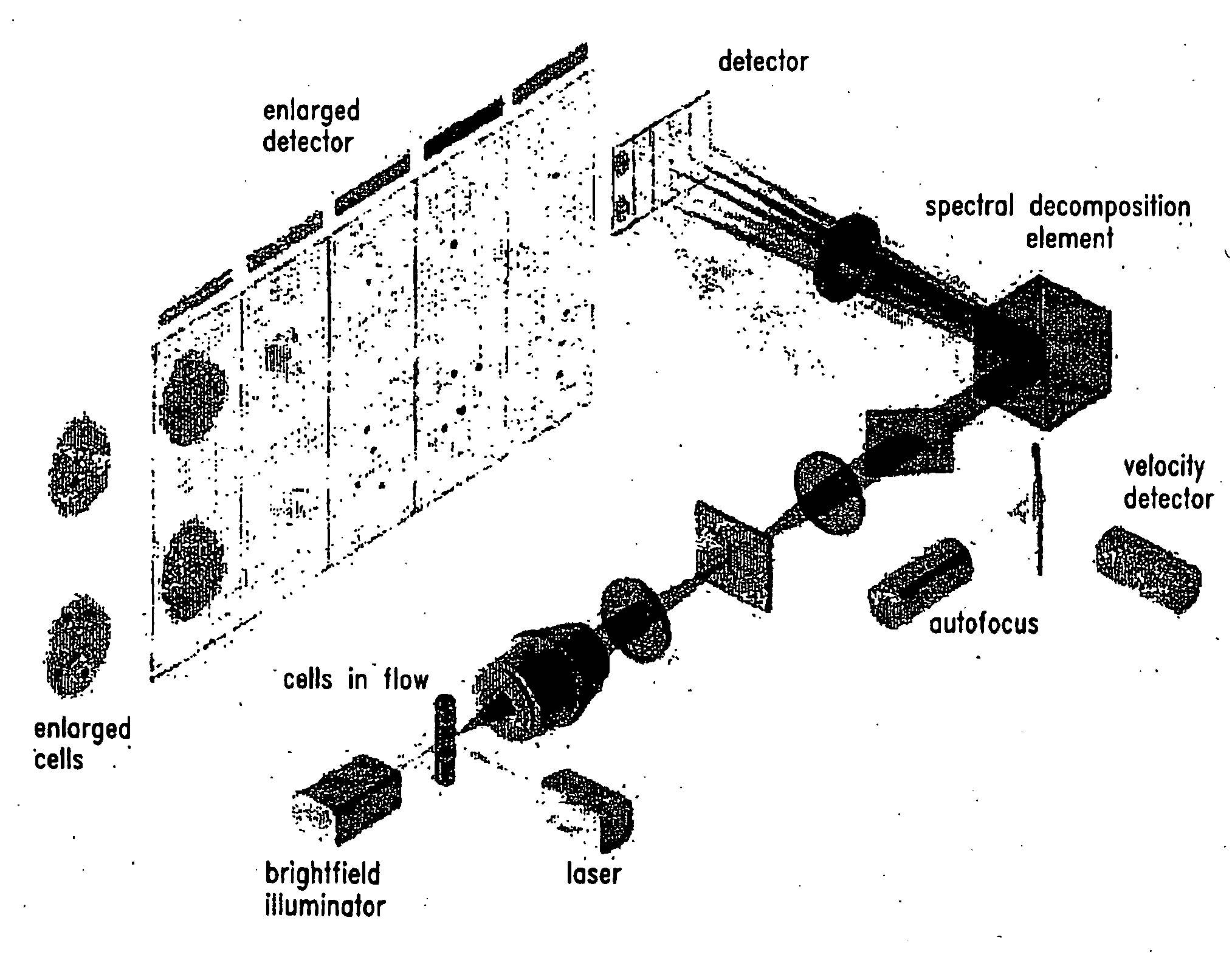
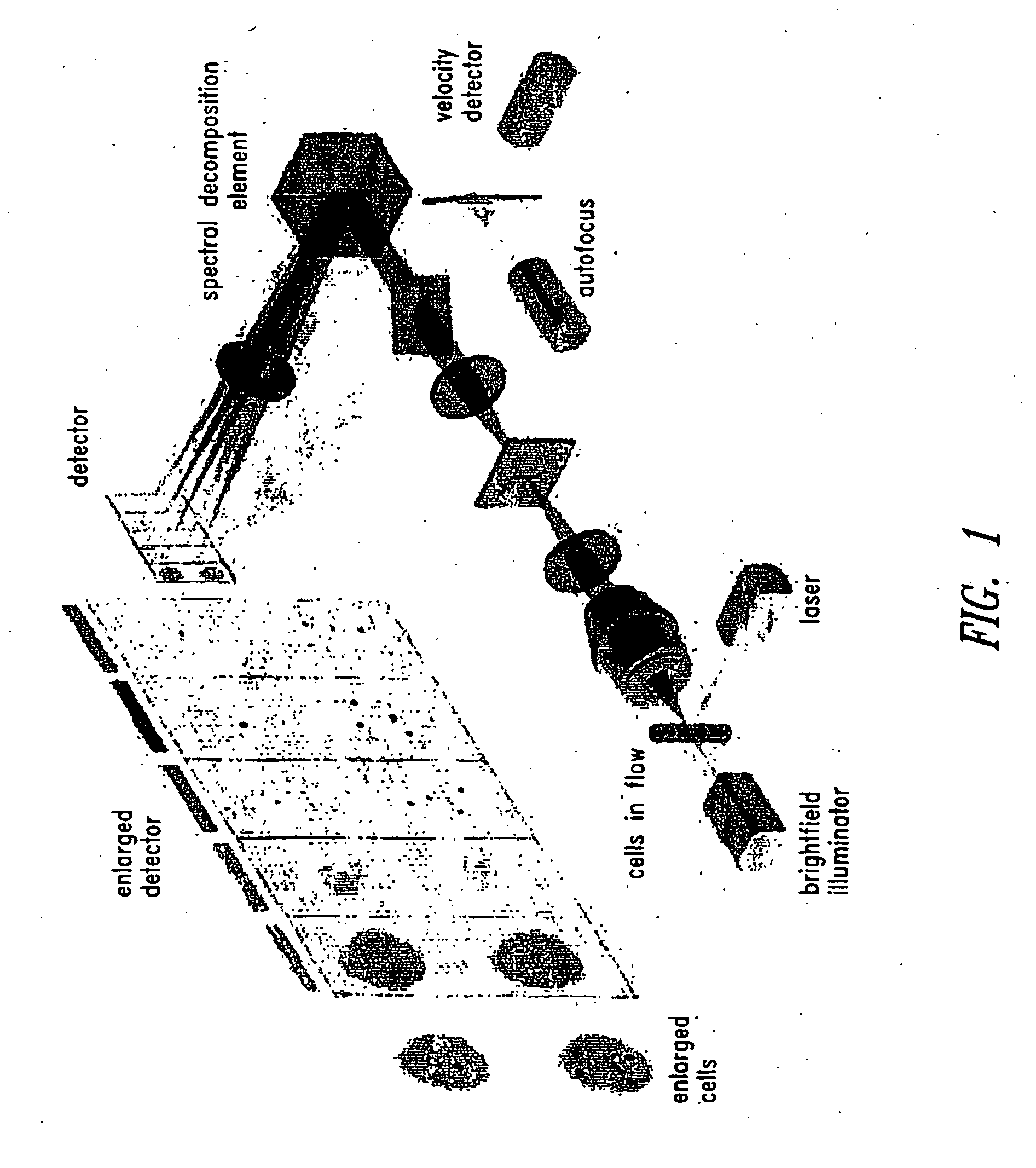
Image

Examples
example 1
Induction of Apoptosis
[0031]Human acute T leukemic Jurkat cell line was obtained from ATCC (Rockville, Md.; catalog number CRL-1990) and maintained in RPMI 1640 (Gibco, Grand Island, N.Y.) containing 5% fetal bovine serum, 1 mM sodium pyruvate (Mediatech, Herndon, Va.), 100 μM nonessential amino acids, 100 U / ml penicillin, 100 μg / ml streptomycin, and 2 mM L-glutamine (BioWhittaker, Walkersville, Md.) in 5% CO2 atmosphere at 37° C. The density of exponentially growing cells was less than 3×10⁵ cells per ml at the time of all treatments. To induce apoptosis, cells were treated for 18 hours with 1 μM camptothecin (CPT, Sigma), a DNA topoisomerase I inhibitor.
example 2
Induction of Necrosis
[0032]Human acute T leukemic Jurkat cell line was obtained from ATCC (Rockville, Md.; catalog number CRL-1990) and maintained in RPMI 1640 (Gibco, Grand Island, N.Y.) containing 5% fetal bovine serum, 1 mM sodium pyruvate (Mediatech, Herndon, Va.), 100 μM nonessential amino acids, 100 U / ml penicillin, 100 μg / ml streptomycin, and 2 mM L-glutamine (BioWhittaker, Walkersville, Md.) in 5% CO2 atmosphere at 37° C. The density of exponentially growing cells was less than 3×10⁵ cells per ml at the time of all treatments. To induce necrosis, cells were treated for 1 hour with 0.3% hydrogen peroxide (Sigma, St. Louis, Mo.).
example 3
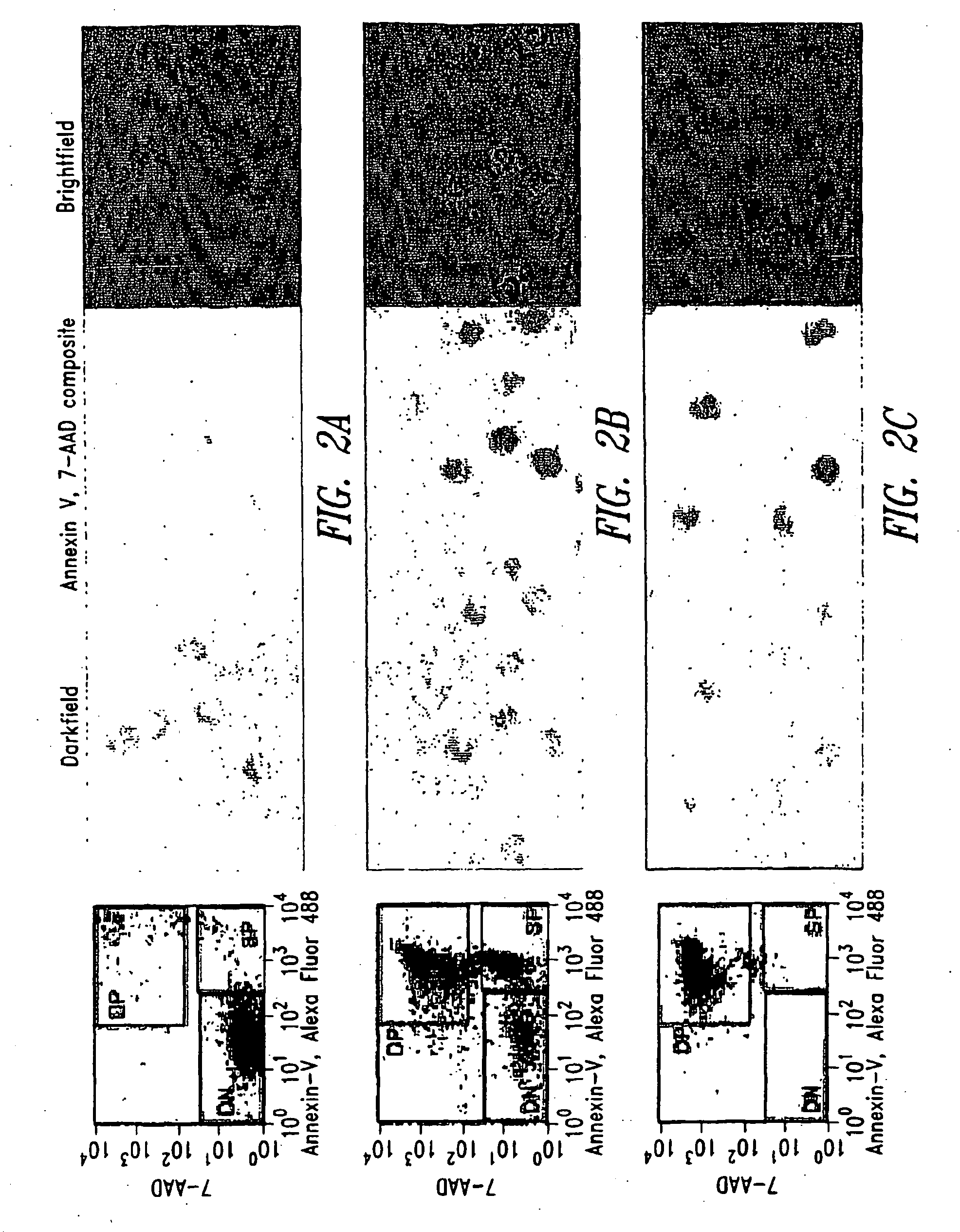
Staining to Identify Apoptotic Cells and Necrotic Cells
[0033]Control (untreated) cell, CPT treated (apoptotic) cells, and peroxide treated (necrotic) cells were independently counted and washed once in phosphate buffered saline (PBS, Fair Lawn, N.J.). Each cell group was resuspended at 107 cells / ml in annexin V Binding Buffer (BD Pharmingen, San Diego, Calif.) containing Alexa Fluor 488 annexin V (Molecular Probes, Eugene, Oreg.) and 10 μM 7-aminoactinomycin D (7-AAD, Molecular Probes) for 10 minutes at room temperature. Necrotic cells were additionally stained with phycoerythrin (PE)-labeled anti-HLA-A, B, C (clone G46-2.6, BD Pharmingen; anti-HLA class I). Each cell group was washed in annexin V Binding Buffer, fixed in 2% paraformaldehyde (Sigma), and analyzed as either single populations or as a mixture by flow cytometry and immunofluorescence microscopy.
PUM
 Abstract
Description
Claims
Application Information