
Use of an in vitro hemodynamic endothelial/smooth muscle cell co-culture model to identify new therapeutic targets for vascular disease

Active Publication Date: 2009-02-26
HEMOSHEAR LLC +1
View PDF40 Cites 26 Cited by
  • Summary
  • Abstract
  • Description
  • Claims
  • Application Information

AI Technical Summary

Benefits of technology

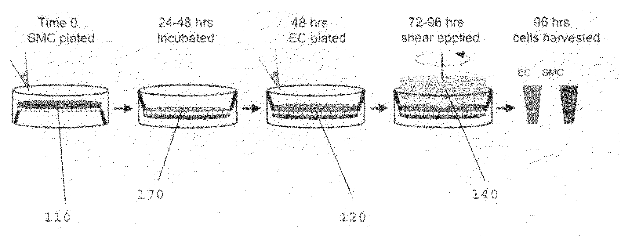
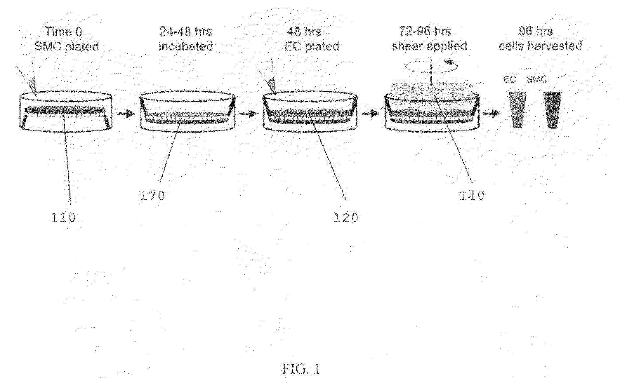
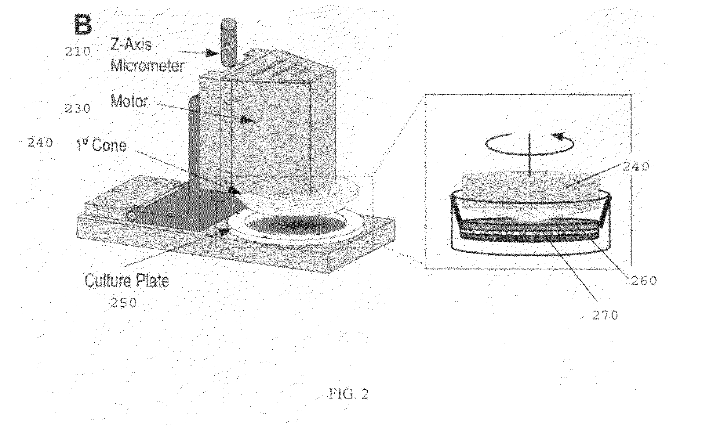
"The invention is a biomechanical model that can replicate the hemodynamic forces of the human circulation using a cone-shaped device. This model can be used to study the interactions between different cell types in the vascular system and to test the effects of drugs and other compounds on these interactions. The model can also be used to isolate individual cell types for further analysis. The device can be used to investigate the development of atherosclerosis and to test the compatibility and behavior of cells from vascular stent material under hemodynamic conditions."

Problems solved by technology

Additionally, few studies have shown the intricate interactions and cross-communications of ECs and SMCs in the presence of any type of flow, and no known studies to date have examined how in vivo-derived human hemodynamic forces on the endothelium regulate SMC phenotypic switching, as it is classically defined by the literature.

Method used


Image

  • Use of an in vitro hemodynamic endothelial/smooth muscle cell co-culture model to identify new therapeutic targets for vascular disease
  • Use of an in vitro hemodynamic endothelial/smooth muscle cell co-culture model to identify new therapeutic targets for vascular disease
  • Use of an in vitro hemodynamic endothelial/smooth muscle cell co-culture model to identify new therapeutic targets for vascular disease

Examples


example

[0070]The following is an example of a method of using the present invention, and is not intended to limit the scope of the invention to the exact method described in this example.

[0071]Human Cell Isolation and Culture.

[0072]Primary human ECs and SMCs were isolated from umbilical cords, expanded, and used as cell sources. Human ECs were isolated from the umbilical vein (human umbilical vein ECs) as previously described, followed by isolation of SMCs from the vein using a similar method as previously described.

[0073]ECs were used for experimentation at passage 2 and SMCs were for experimentation used up to passage 10, both of which have been established to retain the basal EC / SMC phenotype based on the retention of specific EC and SMC markers. Cell types were separately cultured and passaged using medium 199 (M199; BioWhitaker) supplemented with 10% FBS (GIBCO), 2 mM L-glutamine (BioWhitaker), growth factors [10 μg / ml heparin, (Sigma), 5 μg / ml endothelial cell growth supplement (Sigm...


PUM

PropertyMeasurementUnit
timeaaaaaaaaaa
pore diameteraaaaaaaaaa
thicknessaaaaaaaaaa

Abstract

An in vitro biomechanical model used to applied hemodynamic (i.e., blood flow) patterns modeled after the human circulation to human / animal cells in culture. This model replicates hemodynamic flow patterns that are measured directly from the human circulation using non-invasive magnetic resonance imaging and translated to the motor that controls the rotation of the cone. The cone is submerged in fluid (i.e., cell culture media) and brought into close proximity to the surface of the cells that are grown on the plate surface. The rotation of the cone transduces momentum on the fluid and creates time-varying shear stresses on the plate or cellular surface. This model most closely mimics the physiological hemodynamic forces imparted on endothelial cells (cell lining blood vessels) in vivo and overcomes previous flow devices limited in applying more simplified nonphysiological flow patterns. Another aspect of this invention is directed to incorporate a transwell co-cultured dish. This permits two to three or more different cell types to be physically separated within the culture dish environment, while the inner cellular surface is exposed to the simulated hemodynamic flow patterns. Other significant modifications include custom in-flow and out-flow tubing to supply media, drugs, etc. separately and independently to both the inner and outer chambers of the coculture model. External components are used to control for physiological temperature and gas concentration. The physical separation of adherent cells by the artificial transwell membrane and the bottom of the Petri dish permits each cell layer, or surface to be separately isolated for an array of biological analyses (i.e., protein, gene, etc.).

Description

CROSS REFERENCE TO RELATED APPLICATIONS[0001]This application claims priority under 35 U.S.C. § 119(e) from U.S. Provisional Patent Application No. 60 / 879,710, filed on Jan. 10, 2007, which is incorporated herein by reference.STATEMENT REGARDING SPONSORED RESEARCH OR DEVELOPMENT[0002]Not Applicable.REFERENCE TO SEQUENCE LISTING[0003]Not Applicable.BACKGROUND OF THE INVENTION[0004]1. Field of the Invention[0005]This invention relates generally to devices and methods for in vitro analysis of fluid flow (e.g., hemodynamics) on cells (e.g., endothelial cells). More specifically, this invention relates to a method of using a device that permits more than one different cell types to be physically separated within the culture dish environment, while the inner cellular surface is exposed to the simulated hemodynamic flow patterns.[0006]2. Description of Related Art[0007]Atherosclerosis is a vascular inflammatory disease characterized by lesion formation and luminal narrowing of the arteries...

Claims


Application Information

Patent Timeline
Patent Type & Authority Applications(United States)
IPC IPC(8): C12Q1/02C12N5/00C12M1/00
CPCC12N5/0691C12N2521/00C12N2502/28
Inventor BLACKMAN, BRETT R.WAMHOFF, BRIAN R.
Owner HEMOSHEAR LLC
Learn More