Methods of Detection of Changes in Cells
a cell and cell technology, applied in the field of cell and cell change detection, can solve the problems of incongruous data sets and difficult to determine the effect of the experiment on the outcom
- Summary
- Abstract
- Description
- Claims
- Application Information
AI Technical Summary
Benefits of technology
Problems solved by technology
Method used
Image
Examples
example 1
[0119]Current techniques for studying cell-protein interactions and other cell interactions are time consuming and labor intensive because they can involve many steps including radioisotope or fluorescence labeling, washing, blocking, and detecting. See e.g. Table 2. Current techniques for studying cell-protein interactions and other cell interactions can also use extensive amounts of expensive reagents. The instant invention provides compositions and methods for determining cell interactions that are faster than conventional methods and that require the use of fewer reagents than conventional methods.
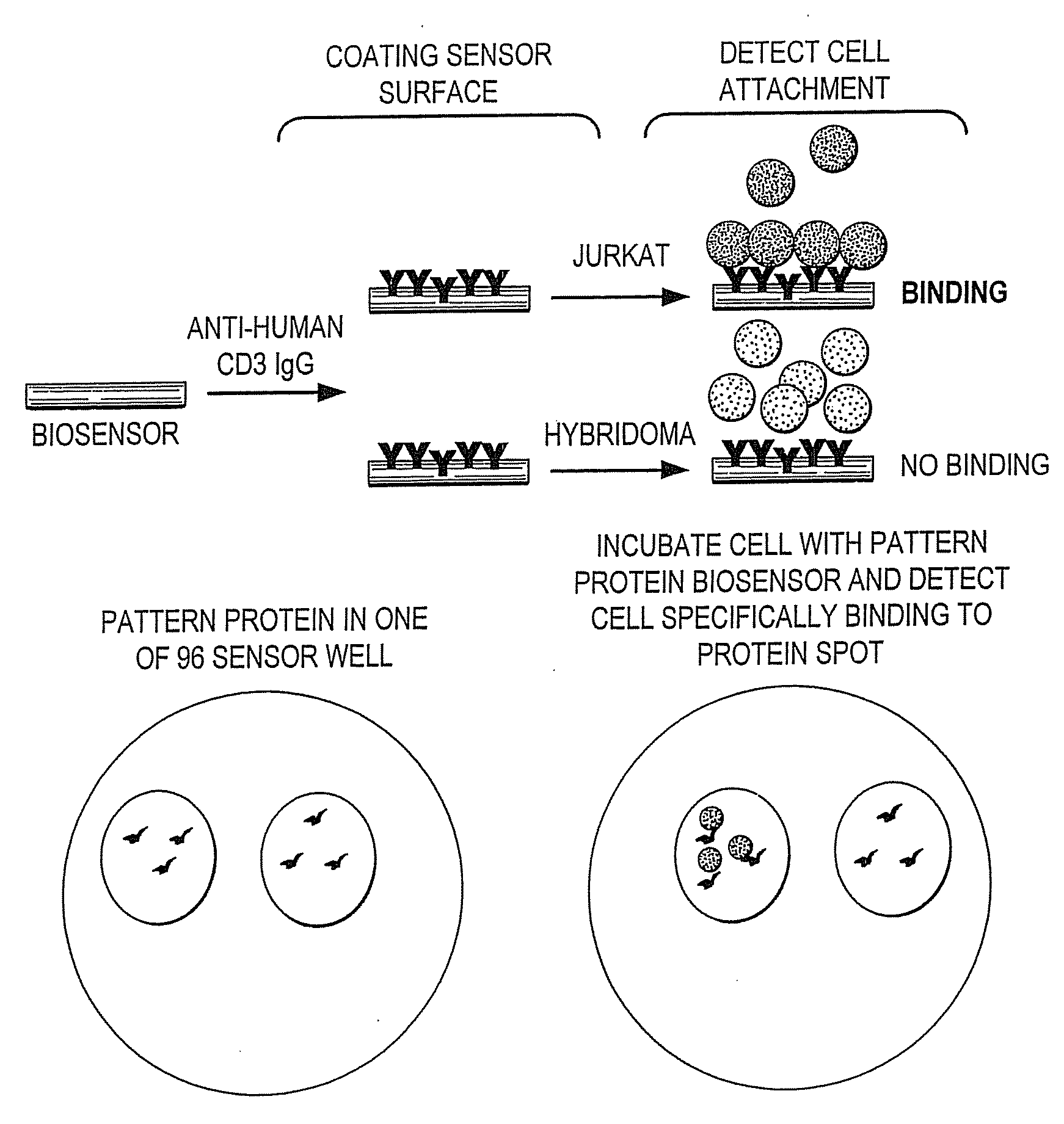
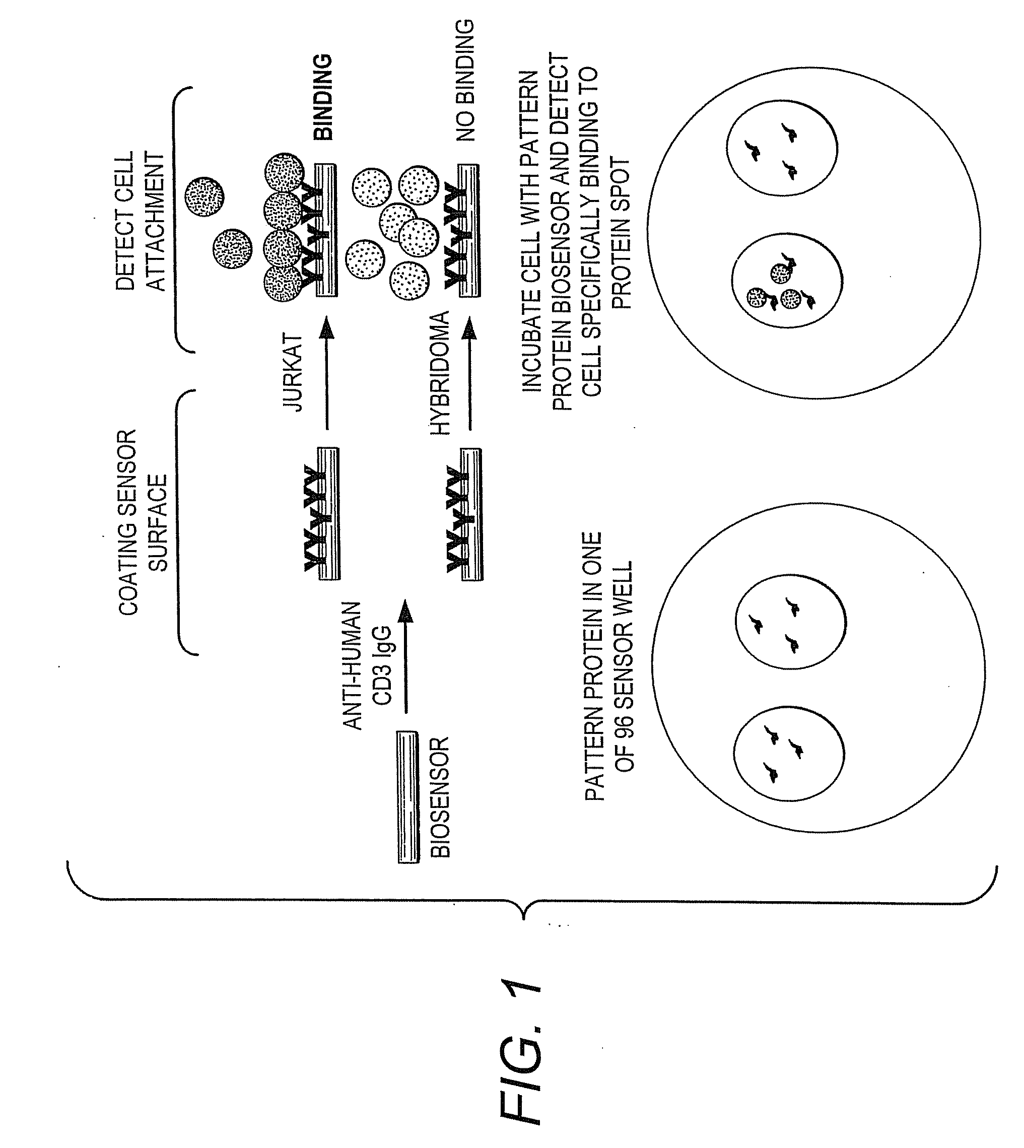
[0120]The instant methods and compositions provide a label-free, simple, high throughput assay to identify cell specificity for specific binding substances, including proteins, cell migration, cell chemotaxis, specific binding substance-cell interactions, cell-extracellular matrix interactions, and cell-cell interactions.
[0121]In one embodiment of the invention the methods and composit...
example 2
Development of a Wash-Free CBA Protocol
Plating of CHO and HEK Cells Directly into Starvation Medium
[0130]The first established protocol for cell based assays using biosensor technology relies primarily on two important steps referred to as cell washing and starvation.
[0131]These steps are important in reducing assay time as cell harvesting is currently carried out by trypsin treatment of large scale cultures. Cell harvesting is currently carried out by trypsin treatment of large scale cultures. Trypsinization of cell monolayers is effective and reproducible and can be easily adapted to most cell lines. In practicality, trypsin mediated cell harvesting is a two step process whereby supplemented tissue culture media is washed off tissue culture flaks in a divalent cation-free buffer (Hank's buffered saline solution, HBSS) followed by trypsinization of cellular junctions in the presence of EDTA. The absence of Ca++ weakens and / or breaks down protein-protein interaction events that medi...
example 3
Collagen Coating and Storage of Colorimetric Resonant Reflectance Biosensors: Performance Evaluation Using mP-M4 Cells
[0145]Conditions were evaluated for coating a colorimetric resonant reflectance biosensor with the extracellular matrix (ECM) protein collagen (COLL) and storage of the resulting coated biosensor. Collagen I has been identified as a culturing substrate that mediates the attachment of muscle cells, hepatocytes, spinal ganglion, embryonic lung cells, Schwann cells, among other primary cells. Collagen is a specific ECM ligand for 14 Chem-1 cells. These cells over express muscarinic GPCRs M4 and respond to carbachol in a dose dependent manner when plated on freshly deposited collagen coated colorimetric resonant reflectance biosensor.
[0146]Three different colorimetric resonant reflectance 384-well biosensor plates were coated with different concentrations of collagen in couplet rows. The plates were stored dry or stored in glycerol or freshly made. The freshly made colla...
PUM
| Property | Measurement | Unit |
|---|---|---|
| grating period | aaaaa | aaaaa |
| grating depth | aaaaa | aaaaa |
| height | aaaaa | aaaaa |
Abstract
Description
Claims
Application Information
 - R&D
- Intellectual Property
- Life Sciences
- Materials
- Tech Scout
- Unparalleled Data Quality
- Higher Quality Content
- 60% Fewer Hallucinations
Browse by: Latest US Patents, China's latest patents, Technical Efficacy Thesaurus, Application Domain, Technology Topic, Popular Technical Reports.
© 2025 PatSnap. All rights reserved.Legal|Privacy policy|Modern Slavery Act Transparency Statement|Sitemap|About US| Contact US: help@patsnap.com


