Bioartificial Renal Tubule

a technology of artificial kidney and kidney, which is applied in the field of bioartificial kidney, can solve the problems of deterioration of artificial kidney tubule device, renal tubule device serious deterioration, study suspension, etc., and achieves simple and sustainable treatment, improved symptoms, and efficient and selective reabsorption.

Inactive Publication Date: 2009-08-20
TOKAI UNIV
View PDF4 Cites 14 Cited by
  • Summary
  • Abstract
  • Description
  • Claims
  • Application Information

AI Technical Summary

Benefits of technology

[0021]The bioartificial renal tubule according to the present invention aims to faithfully reproduce the reabsorption function of a kidney and is an artificial renal tubule device which is prepared by attaching a confluent monolayer of renal tubular epithelial cells to the inner surface of an artificial membrane so as to efficiently and selectively reabsorb a useful substance, an electrolyte, and water.
[0022]The following problem can be prevented over a long period in such a manner that a MEK inhibitor is used to maintain the contact inhibition of the renal tubular epithelial cells: a problem that other renal tubular epithelial cells overlap a renal tubular epithelial cells and therefore active transport is prevented or a problem that the renal tubular epithelial cells are stratified and therefore lumens of the artificial membrane are narrowed so that the flow of primitive urine is interrupted and therefore the reabsorption of useful substances. This allows the function of the renal tubular epithelial cells, which are attached to the inner surface of the artificial membrane in the form of a confluent monolayer, to be maintained.
[0023]The bioartificial renal tubule according to the present invention is a device which forms an artificial kidney together with a bioartificial glomerulus. The bioartificial renal tubule is useful in providing a safe, simple sustainable treatment system that prevents a disadvantage due to the continuous use of an anticoagulant agent such as heparin when blood needs to be continuously filtered to improve symptoms, such as chronic or acute cardiac failure, chronic or acute renal failure, and multiple organ failure, causing overhydration or accumulation of metabolites.

Problems solved by technology

However, the study was suspended in 1989.
The inventors have also found that a renal tubule device seriously deteriorates in performance because cells used are stratified one to two weeks later after the cells form a confluent monolayer depending on the type of the cells.
For this while, the stratification of cells used causes the deterioration of the artificial renal tubule device.
When the artificial renal tubule devices need to be used to repeatedly treat a chronic patient, the cells are stratified during cultivation on treatment intervals and therefore the artificial renal tubule devices are deteriorated in performance.

Method used


Image

  • Bioartificial Renal Tubule
  • Bioartificial Renal Tubule
  • Bioartificial Renal Tubule

Examples


example 1

Method

[0076]On each transwell filter unit, 2×10⁶ Lewis-lung cancer porcine kidney (LLC-PK1) cells, which were cryopreserved porcine proximal renal tubular epithelial cell lines, were seeded. The LLC-PK1 cells were cultured in (1) a high-glucose-DMEM (DMEM-HG) medium, (2) a DMEM-HG medium containing 1% dimethyl sulfoxide (DMSO), or (3) a DMEM-HG medium, containing 50 μM U0126 and 1% DMSO, placed in outside of inserts (lumens were filled with the DMEM-HG medium). The LLC-PK1 cells on 0 day, the first day, the second day, the third day, and the sixth day after seeding were colored with trypan blue and then measured for number for each group. The LLC-PK1 cells were gently washed with a sterilized PBS solution containing no magnesium or calcium and were then trypsin-treated with 1 ml of a trypsin-EDTA solution at 37° C. for 15 minutes. The treated cells (1 ml) were added to 4 ml of DMEM, were colored with trypan blue, and were then measured for number with a hemocytometer. Measurement wa...

example 2

Method

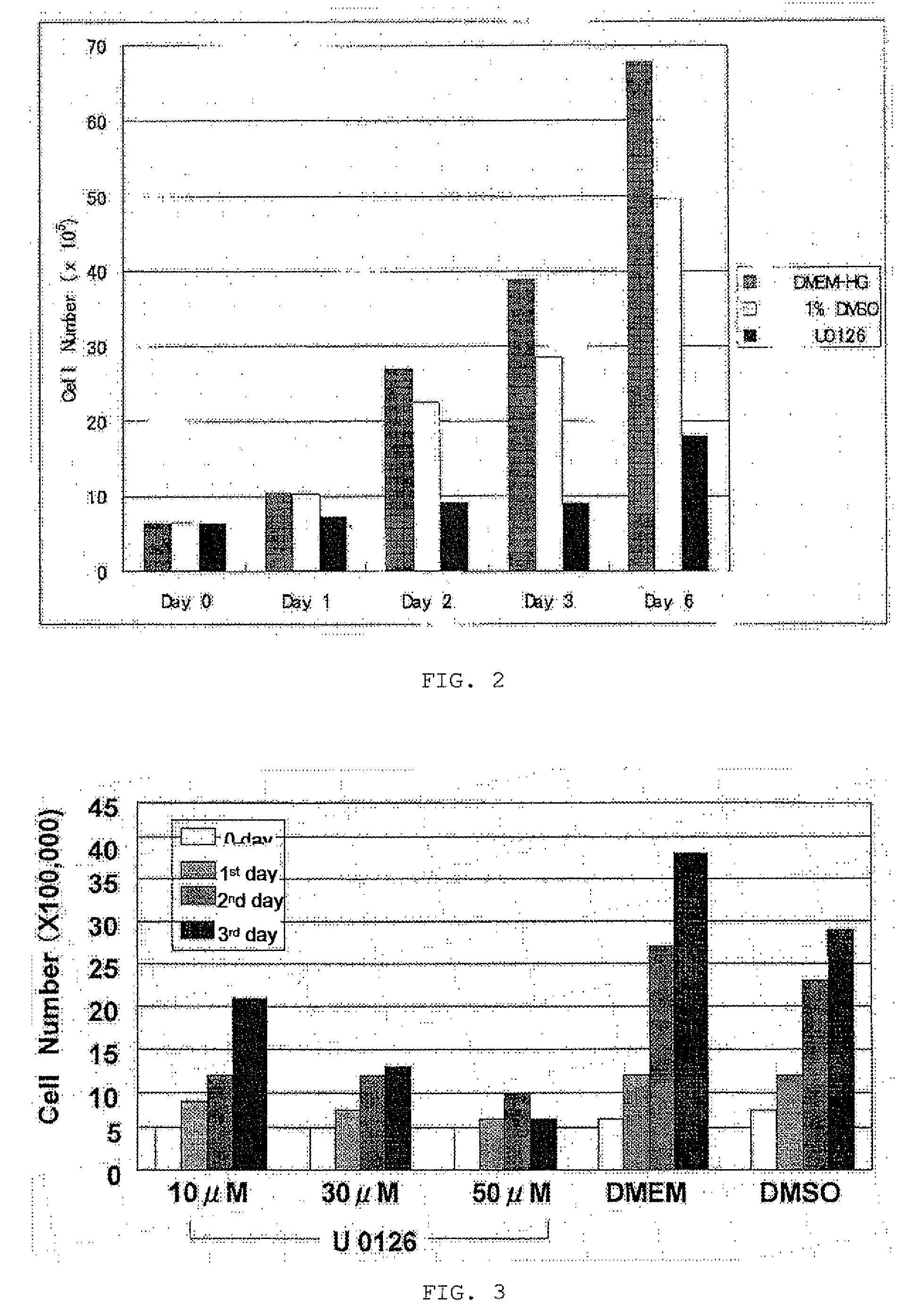
[0079]On each 6-well plate, 5×10⁵ LLC-PK1 cells were seeded. Broths used to culture the cells were replaced with the following three media 24 hours later after seeding: (1) a high-glucose DMEM (DMEM-HG) medium containing 10 μM U0126, which is a MEK inhibitor, and 1% DMSO; (2) a DMEM-HG medium containing 30 μM U0126 and 1% DMSO 1%; and (3) a DMEM-HG medium containing 50 μM U0126 and 1% DMSO. Furthermore, two groups of the cells cultured in (4) a DMEM-HG medium and (5) a DMEM-HG medium containing 1% DMSO were checked. The cells on 0 day, the first day, the second day, and the third day after seeding were colored with trypan blue and then measured for number.

[0080]Results

[0081]FIG. 3 shows the change in number of the LLC-PK1 cells grown in the media (1) to (5) from 0 day to the third day. In the 1% DMSO-containing medium group, the increase of the cells is suppressed to some extent as compared to in the DMEM-HG medium group, but the cells clearly increase in number with day. The ...

example 3

Method

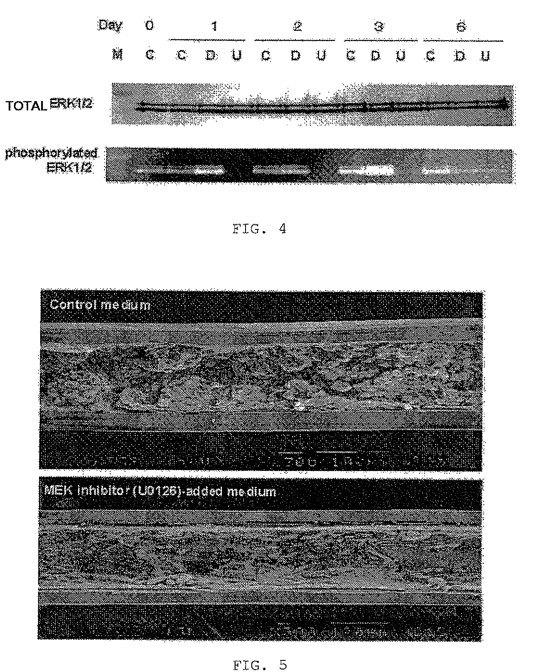
[0082]In each of 6-well plates, 5×10⁵ LLC-PK1 cells were seeded. The cells were divided into three groups on the following day (0 day). That is, the cells of each group were cultured in a corresponding one of the following three media for three days: (1) a DMEM-HG medium (C) used as a control, (2) a DMEM-HG medium containing 1% DMSO (D), and (3) a DMEM-HG medium containing 50 μM U0126 and 1% DMSO (U). Then the media (D) and (U) were replaced with a DMEM-HG medium. The cells of each group were further cultured for three days. The cells were subjected to Western blot analysis for ERK1 / 2 on 0 day, the first day, the second day, the third day, and the sixth day.

[0083]The cultured LLC-PK1 cells were used in the form of a cytolytic solution for Western blot analysis. The sampled cells of each group were rinsed with ice-chilled PBS and were then centrifuged at 4° C. for five minutes at 1500 rpm. The centrifugate was collected and then maintained at −80° C.

[0084]Proteins were taken fr...


PUM

PropertyMeasurementUnit
sizeaaaaaaaaaa
areaaaaaaaaaaa
inner diameteraaaaaaaaaa

Abstract

A bioartificial renal tubule is provided that forms an artificial kidney together with a bioartificial glomerulus suitable for continuous hemofiltration. The bioartificial renal tubule includes an artificial membrane having an inner surface coated with renal tubular epithelial cells and a vessel containing the artificial membrane. The cells are prevented by the use of a MEK inhibitor from being stratified and therefore form a confluent monolayer on the artificial membrane. The renal tubular epithelial cells are characterized in that the contact inhibition thereof is maintained by the use of the MEK inhibitor. The MEK inhibitor is preferably U0126. The attachment of cells capable of reproducing the function of a kidney allows dialysis to be continuously performed for 24 hours with high efficiency and also allows the ability of a renal tubule to reabsorb useful substances to be achieved.

Description

TECHNICAL FIELD[0001]The present invention relates to a bioartificial renal tubule, that is, an artificial renal tubule containing a confluent monolayer of renal tubular epithelial cells and also relates to a method of maintaining of a monolayer of the cells.BACKGROUND ART[0002]Aebischer et al. were the first to develop a bioartificial renal tubule using an artificial membrane and renal tubular epithelial cells and released a basic study on the bioartificial renal tubule in 1987 (Non-patent Documents 1 to 3). However, the study was suspended in 1989. In 1998, Humes et al. at University of Michigan reported that a bloartificial renal tubule prepared by grafting proximal renal tubular epithelial cells onto the inner surface of a polysulfone hollow fiber was capable of inhibiting the leakage of inulin and had various metabolic functions due to almost complete adhesion of the cells (Non-patent Document 4). Humes et al. succeeded in extracorporeal circulation experiments on renal failure...

Claims


Application Information

Patent Timeline
Patent Type & Authority Applications(United States)
IPC IPC(8): C12N11/12C12N11/00C12N11/04
CPCA61M1/3472C12M25/10C12M29/16A61M1/3489C12N2501/727C12N2533/30C12N5/0686
Inventor SAITO, AKIRAYOKOYAMA, TUN AUNG
Owner TOKAI UNIV
Why Eureka
Try Eureka