Imaging Catheter With Integrated Contrast Agent Injector

Inactive Publication Date: 2009-09-17
BOSTON SCI SCIMED INC
View PDF22 Cites 51 Cited by
  • Summary
  • Abstract
  • Description
  • Claims
  • Application Information

AI Technical Summary

Benefits of technology

[0009]The imaging catheter with the integrated contrast agent injector advantageously reduces the volume of contrast agent that needs to be injected into the patient by injecting the contrast agent locally near the region to be imaged. The imaging catheter also provides easier synchronization between the injection of contrast agent and imaging. This is because the exit port of the contrast lumen is located at a short known distance from the imager, making it easier to estimate the time for the injected contrast agent to reach the region being imaged by the imager. In addition, the imaging catheter p

Problems solved by technology

Another problem with these methods is the difficulty of synchronizing the injection of contrast agent with imaging.
This is due in part to the difficulty of estimating the time its takes the injected contrast agent to reach the region of interest, which can be as long as two minutes.
Such methods canno

Method used


Image

  • Imaging Catheter With Integrated Contrast Agent Injector
  • Imaging Catheter With Integrated Contrast Agent Injector
  • Imaging Catheter With Integrated Contrast Agent Injector

Examples


Embodiment Construction

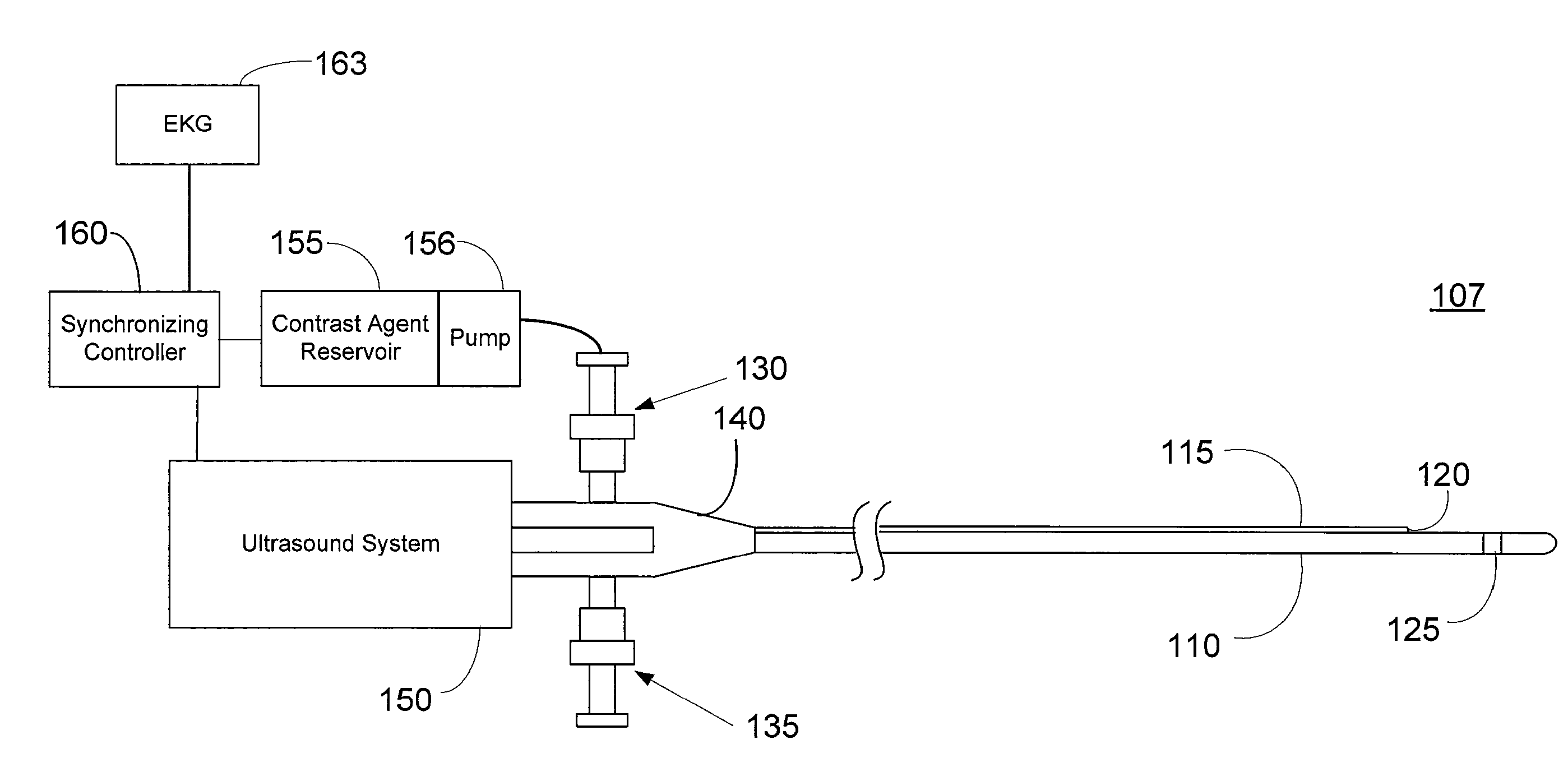
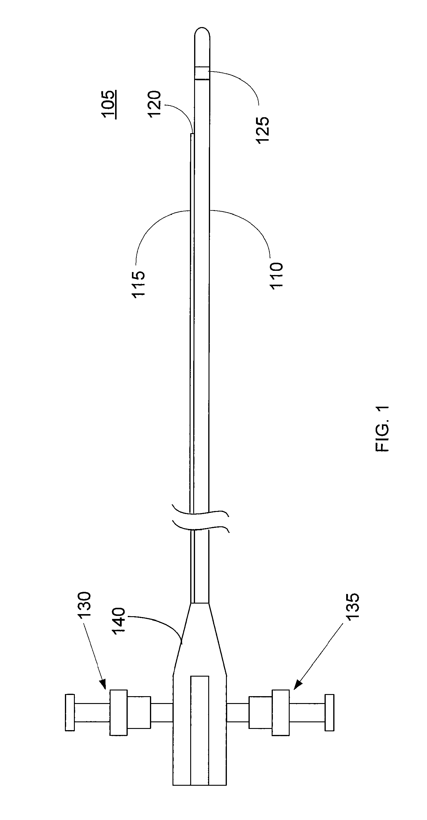
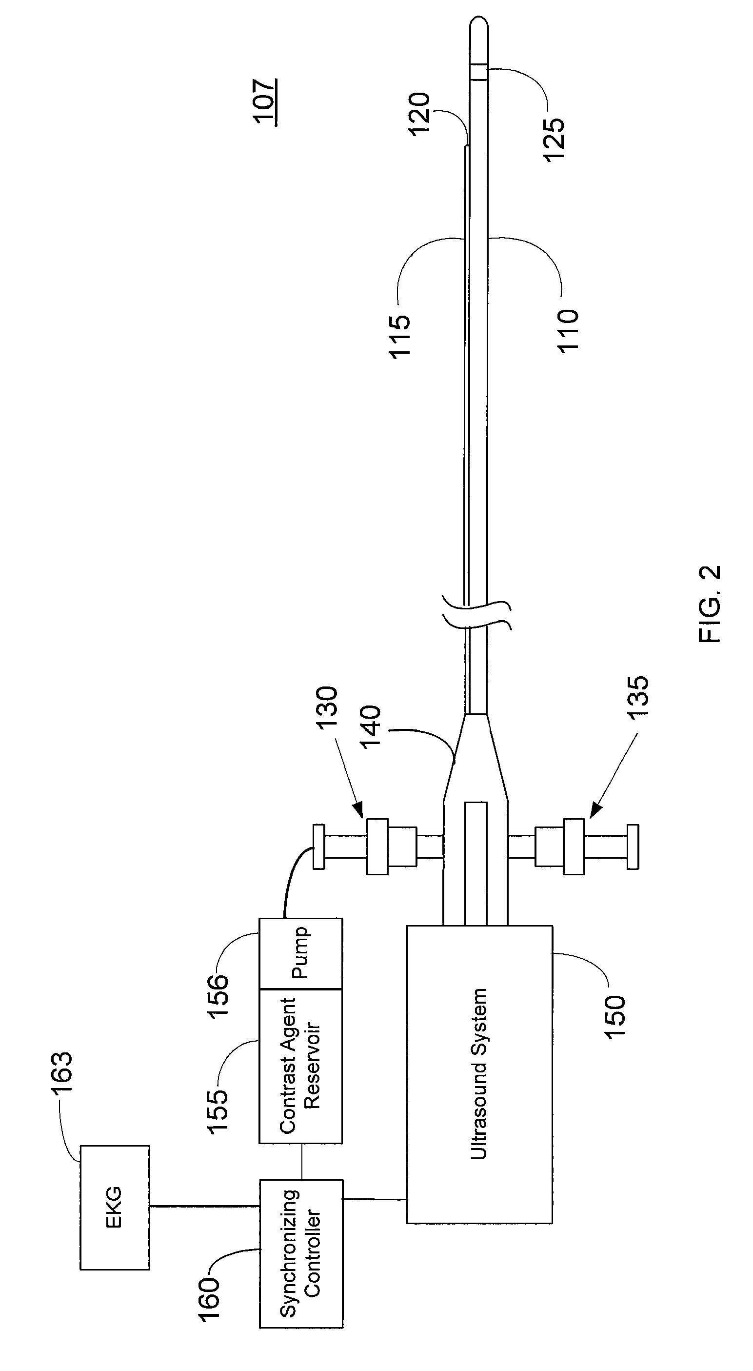
[0021]FIG. 1 shows an imaging catheter 105 apparatus with an integrated contrast agent injector according to an embodiment of the present invention. The catheter 105 comprises an elongated catheter sheath 110 and a contrast agent lumen 115 that extends along the catheter sheath 110. In a preferred embodiment, the catheter sheath 110 and contrast agent lumen 115 are adapted to be inserted into a patient's blood vessel, e.g., coronary artery, for contrast enhanced imaging within the blood vessel. The catheter may be adapted to be inserted in other passages in the body for imaging, e.g., the esophagus or urethra. The catheter sheath 110 and contrast lumen 115 may be made of a variety of polymeric materials, such as polytetrafluoroethylene (PTFE), polyethylene, PEEK, PEBAX or the like. The contrast lumen 115 includes an exit port 120 at its distal end for injecting contrast agent into the blood vessel.

[0022]The catheter 105 further comprises an ultrasound imager 125. The ultrasound imag...


PUM


Abstract

Described herein are systems and methods that integrate the injection of contrast agents with imaging catheters. In an embodiment, an imaging catheter comprises a catheter sheath and an imager, e.g., ultrasound transducer. The imaging catheter further comprises a contrast lumen having one or more exit ports for injecting contrast agent into the patient. The contrast lumen extends along the catheter sheath and may be external to or integrated into the catheter sheath. Preferably, the exit port of the contrast lumen is positioned along the catheter sheath at a relatively short known distance from the imager. The catheter may include multiple contrast lumens for injecting different types of contrast agents. In an embodiment, a synchronizing controller is provided to automatically synchronize the injection of contrast agent with imaging. In another embodiment, drug-filled microbubbles in combination with ultrasound imaging are used to deliver a controlled drug dose to a specific treatment site.

Description

FIELD OF THE INVENTION[0001]The present invention relates to imaging catheters, and more particularly to imaging catheters with integrated contrast agent injectors.BACKGROUND INFORMATION[0002]Intravascular ultrasound (IVUS) is an established tool for obtaining ultrasound images within the vascular system. IVUS is a method by which a catheter-based transducer is used to obtain images of the lumen and vascular wall of larger vessels. It is an established interventional cardiology tool for gaining insight into the size, structure, and composition of atherosclerotic plaque located within coronary arteries.[0003]Ultrasound contrast imaging employs a contrast agent to enhance the detection and imaging of organs, blood vessels, or tissues within the body. Contrast agents may be comprised of echogenic microbubbles or other substances having ultrasound scattering properties that are distinct from those of the surrounding media (tissue and blood), thereby enhancing the visibility of organs, b...

Claims


Application Information

Patent Timeline
IPC IPC(8): A61B8/14A61N7/00
CPCA61B8/06A61B8/12A61B8/481A61B8/543A61M2025/0039A61M5/172A61M37/0092A61M2025/0037A61M5/007
Inventor KNIGHT, JON M.TEO, TAT-JINROMLEY, RICHARDLI, WENGUANG
Owner BOSTON SCI SCIMED INC
Why Eureka
Try Eureka