Process for the identification of novel enzyme interacting compounds

a technology of enzymes and complexes, applied in biochemistry apparatuses, instruments, peptide/protein ingredients, etc., can solve the problems of inefficient capture of kinases or rapid elution, kinases are not preferentially captured, and it is difficult to develop selective atp-competitive inhibitors, etc., to achieve rapid and focused identification and quantification

Inactive Publication Date: 2009-09-24
CELLZOME LTD
View PDF11 Cites 14 Cited by
  • Summary
  • Abstract
  • Description
  • Claims
  • Application Information

AI Technical Summary

Problems solved by technology

A major challenge in this field is the development of PDE isotype specific inhibitors in order to avoid cross-reactivity that is responsible for side effects (Card et al., 2004, “Structural basis for the activity of drugs that inhibit phosphodiesterases”, Structure 12, 2233-2247).
The ATP binding pocket of different kinases is structurally similar and therefore it is considered difficult to develop selective ATP-competitive inhibitors.
One disadvantages of immobilized ATP is that the affinity for kinases is rather low leading to inefficient capturing of kinases or rapid elution due to high off-rates.
Another disadvantage is that kinases are not preferentially captured, but also other classes of ATP-binding proteins which can be expressed at much higher levels in the cell.
The more abundant ATP-binding proteins can cause inefficient capturing due to competition or can lead to problems during the mass spectrometry analysis of the bound proteins if the analytical depth is not sufficient.
This approach is only successful if the structure-affinity-relationship (SAR) is not destroyed through the chemical modification of the drug but fails if the SAR is impaired.
In addition, it is difficult to identify targets mediating unwanted side effects because the SAR of the cognate drug target and the side-effect-target can be different and the latter SAR is usually not known.
A disadvantage of this method is that the kinases need to be cloned and only a fraction of the phage-tagged kinases are folded in the correct native state.
Furthermore, such protein preparations do not reflect at all the natural situation in a cell.
However, it remains unclear what structural and / or catalytic properties are shared by these sulfonate-targeted enzymes.
Another limitation of this approach is the difficulty to distinguish specific interactions with enzymes and non-specific interactions caused by the intrinsic reactivity of the probes.
One major limitation of this approach is that only proteins phosphorylated on tyrosine can be captured.

Method used


Image

  • Process for the identification of novel enzyme interacting compounds
  • Process for the identification of novel enzyme interacting compounds
  • Process for the identification of novel enzyme interacting compounds

Examples


example 1

Preparation of Kinobeads

[0222]This example illustrates the preparation of kinobeads with 4 different ligands. These kinobeads were later used in example 2 and example 3.

[0223]Broad spectrum capturing ligands were covalently immobilized on a solid support through covalent linkage using suitable functional groups (e.g. amino or carboxyl or groups). Compounds that do not contain a suitable functional group were modified in order to introduce such a group. The necessary chemical methods are known in the medicinal chemistry literature and illustrated below.

1. Selection and Synthesis of Ligands

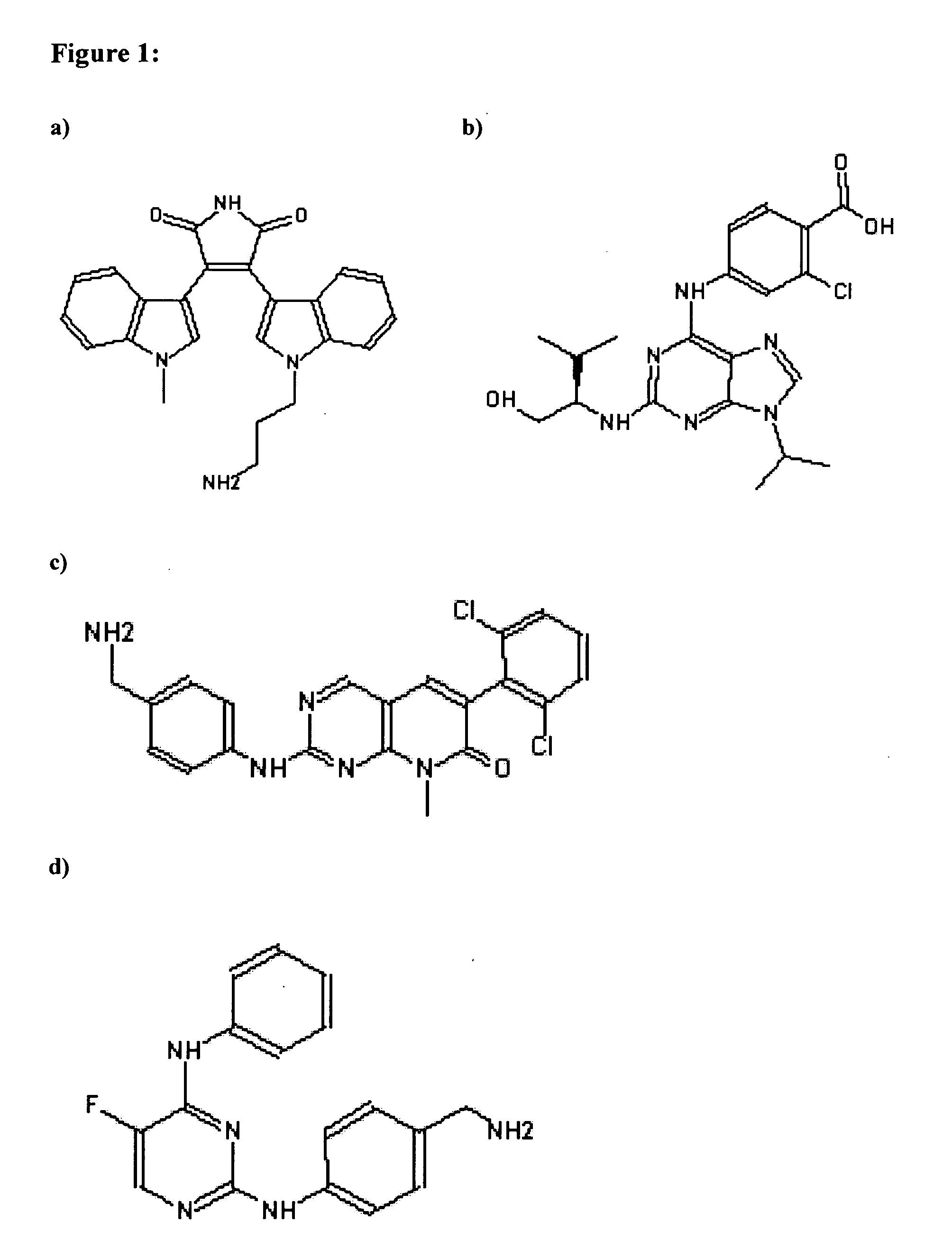
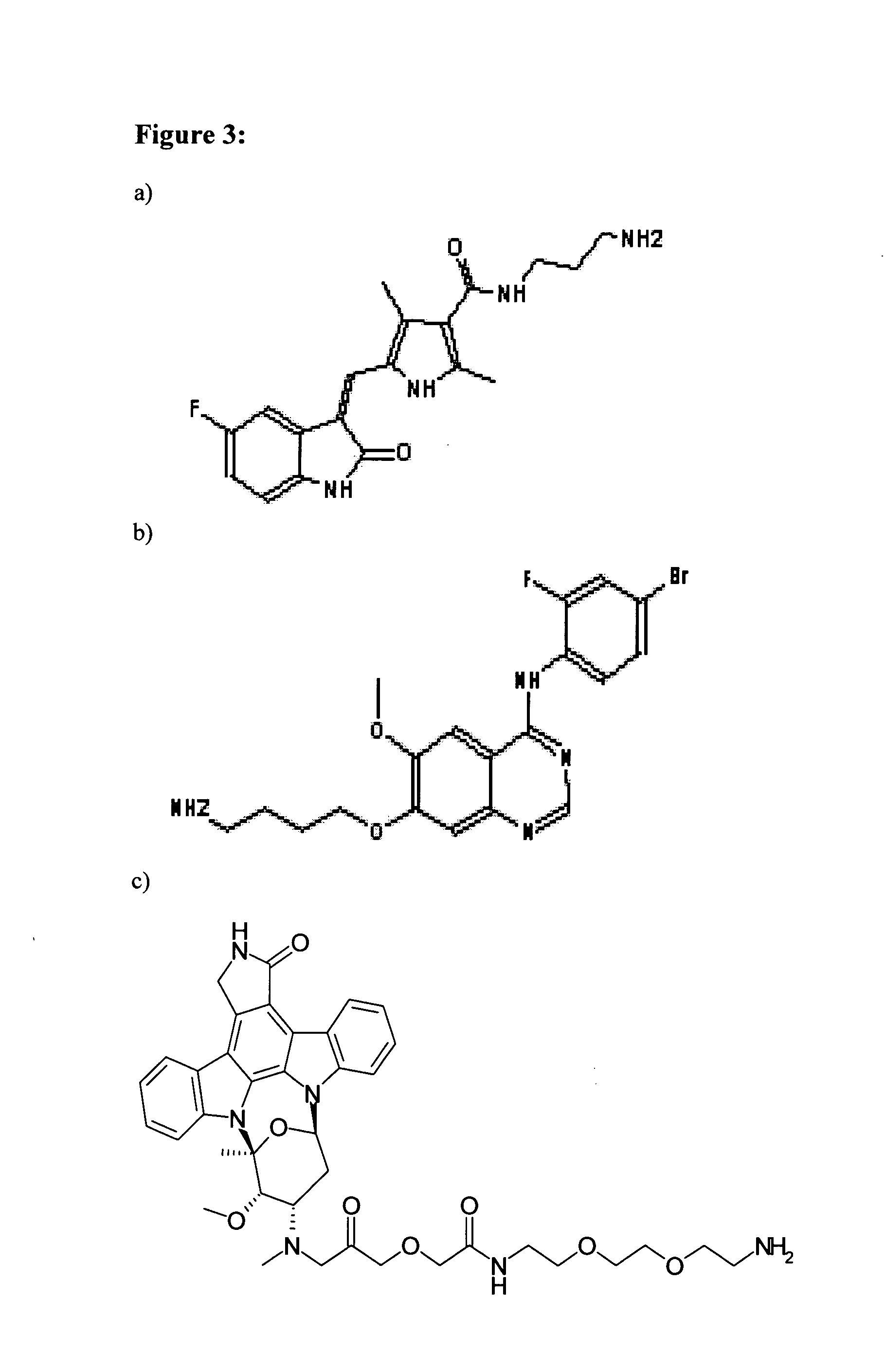
[0224]The following four broad specificity ligands (Kinobead ligands 1 to 4; FIG. 1) were covalently coupled to beads in separate reactions as described below and then the four types of beads were mixed and used for the drug pulldown experiments.

[0225]Kinobead ligand 1: Bisindolylmaleimide VIII-Acetate (Chemical Formula: C24H22N4O2. CH3COOH; MW 398.5; CAS number 138516-31-1; Alexis Biochemicals, AXX...

example 2

Signalokinome

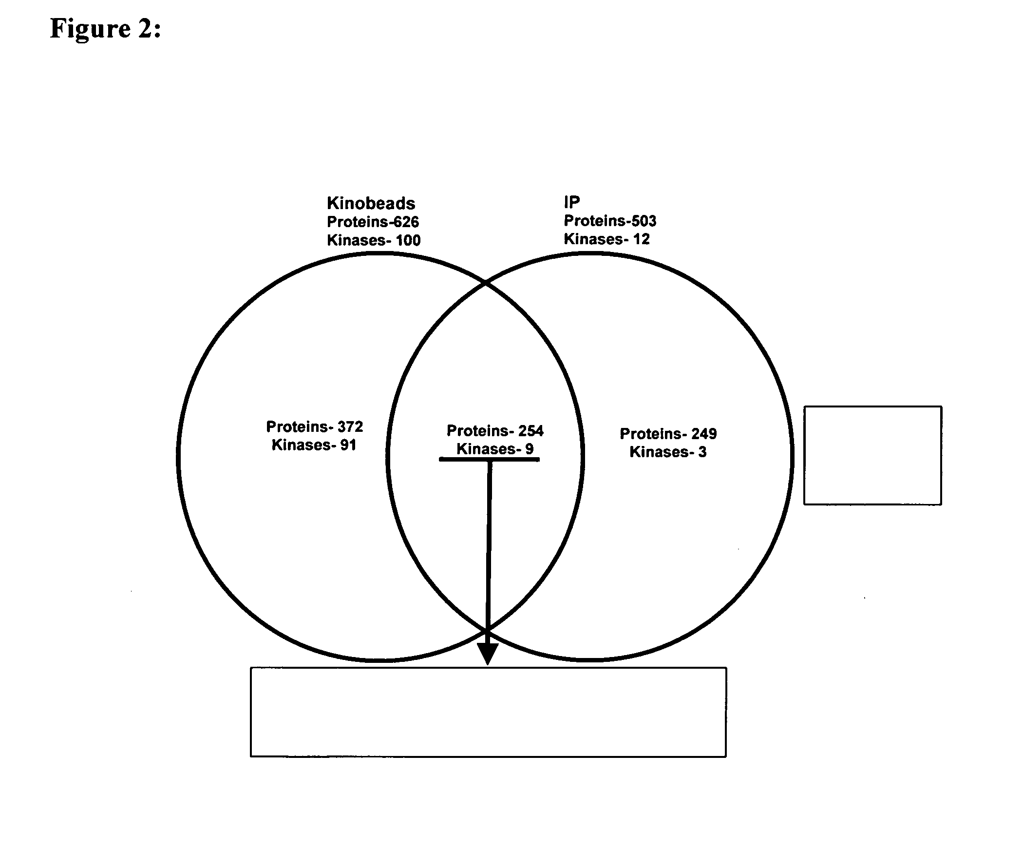
[0271]This example illustrates the treatment of cells with compounds (see particularly the second aspect of the invention). HeLa cells were treated with epidermal growth factor (EGF), a cell lysate was prepared and analysed using Kinobeads (experimental protocol in section 2.1) and mass spectrometry. The preparation of Kinobeads is described in Example 1.

[0272]In parallel the cell lysate was subjected to immunoprecipitation with an anti-phosphosphotyrosine antibody (experimental protocol in section 2.2) and analysed by mass spectrometry.

[0273]The result (FIG. 2) shows that kinobeads identify significantly more kinases (Table 8) compared to the immunoprecipitation procedure (Table 6).

1. Preparation of the Biological Sample (Cell Lysate)

1.1 Cell Culture and Treatment of Cells

[0274]Cell culture. HeLa cells (American Type Culture Collection-No CCL-2) were grown in MEM medium (without L-Arginine and without L-Glutamine; Promocell C-75280), 10% dialyzed Fetal Bovine Serum (Gi...

example 3

Screening Assay using Test Compounds for Protein Elution

[0345]This example illustrates competitive elution of proteins bound to kinobeads with non-modified test compounds (see particularly the fourth aspect of the invention). The kinobeads (as described in example 1) were contacted with mouse brains lysate, bound proteins were eluted with various test compounds and the released proteins were analysed by mass spectrometry.

1. Preparation of the Biological Sample (Tissue Lysate)

1.1 Preparation of Lysates

[0346]A mouse brain lysate was prepared by mechanical disruption in lysis buffer (5 ml buffer per mouse brain) under gentle conditions that maintain the structure and function of proteins. The following steps were performed:[0347]Thaw the tissue quickly at room temperature or 37° C., then transfer tissue to a glass bottle containing the 1× lysis buffer (use a vial big enough to be used with Polytron PT 3100 homogenizer)[0348]Lyse the organ / tissue with 4×10 sec pulses at 5000-7000 rpm at...


PUM

PropertyMeasurementUnit
ionic strengthaaaaaaaaaa
molecular weightaaaaaaaaaa

Abstract

The present invention relates to methods for the characterization of enzymes or of enzyme-compound complexes, wherein the enzyme is obtained from a protein preparation with the help of at least one broad spectrum ligand immobilized on a solid support and wherein the enzyme is characterized by mass spectrometry. These methods are useful for the screening of non-immobilized compound libraries, selectivity profiling of lead compounds and mechanism of action studies in living cells.

Description

CROSS-REFERENCE TO RELATED APPLICATIONS[0001]This application is a continuation-in-part of International Application No. PCT / EP2006 / 062984, filed Jun. 7, 2006, which claims priority to U.S. Provisional Application Nos. 60 / 711,399, filed Aug. 25, 2005, and 60 / 782,170, filed Mar. 14, 2006, and European Application No. 05012722.4, filed Jun. 14, 2005, each of which is hereby incorporated by reference.REFERENCE TO TABLES SUBMITTED ON COMPACT DISC[0002]Supplementary Tables 1 and 2 are submitted on duplicate compact discs, which are hereby incorporated by reference. Each disc contains the files Supplemental_Table—1—1.txt, 10,779 kB and Supplemental_Table—1—2.txt, 10,817 kB, both created Dec. 13, 2007, and Supplemental_Table—2.txt, 3,513 kB, created Dec. 12, 2007. Supplementary Table 1 is found in files Supplemental_Table—1—1.txt and Supplemental_Table—1—2.txt, and Supplementary Table 2 is found in Supplemental_Table—2.txt. LENGTHY TABLESThe patent application contains a lengthy table sect...

Claims


Application Information

Patent Timeline
Patent Type & Authority Applications(United States)
IPC IPC(8): A61K38/43G01N33/573
CPCC12Q1/485G01N33/6848
Inventor DREWES, GERARDKUESTER, BERNHARDKRUSE, ULRICHHOPF, CARSTENEBERHARD, DIRKBANTSCHEFF, MARCUSREADER, VALERIERAIDA, MANFREDMIDDLEMISS, DAVID
Owner CELLZOME LTD
Why Eureka
Try Eureka