

DNA virus detection by DNA chip
a technology of dna virus and chip, applied in the direction of microbiological testing/measurement, biochemistry apparatus and processes, etc., can solve the problems of high cost of chemicals and disposable items, loss of samples, and considerable time required for gene amplification and purification, so as to facilitate detection, facilitate detection, and reduce interference with the detection of amplified materials
- Summary
- Abstract
- Description
- Claims
- Application Information
AI Technical Summary
Benefits of technology
Problems solved by technology
Method used
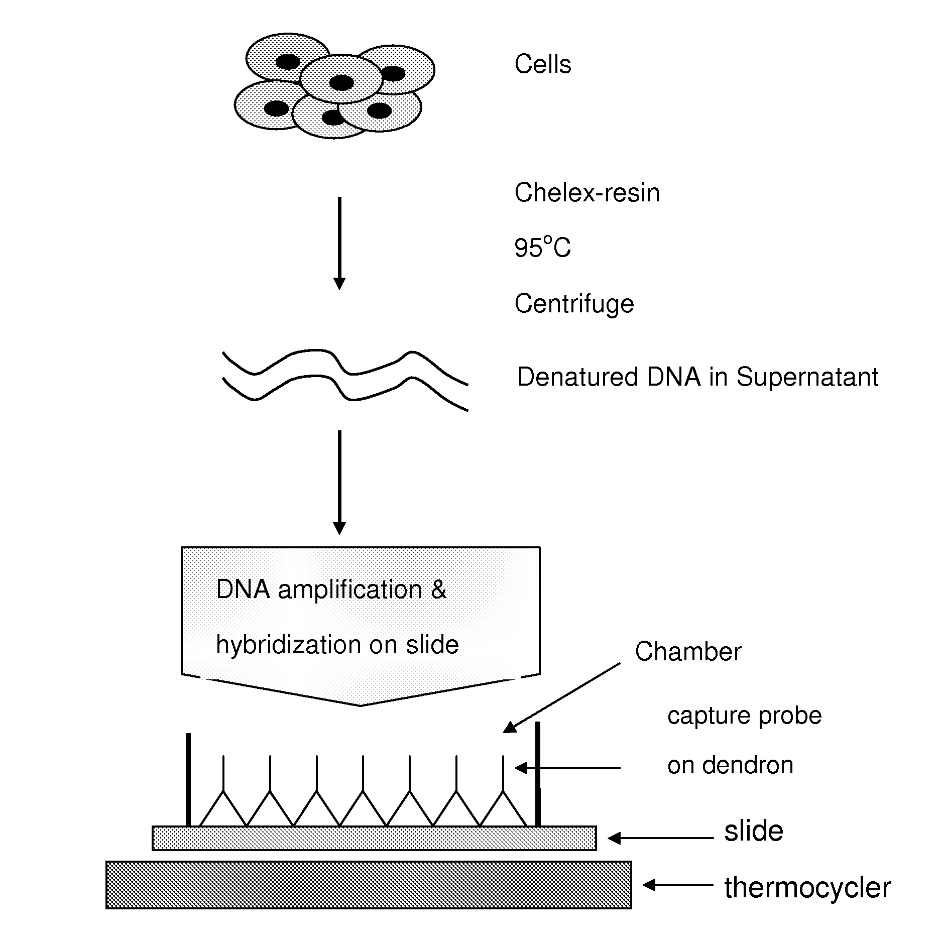
Image

Examples
example 1
Verification of Capture Probes for HPV Assay
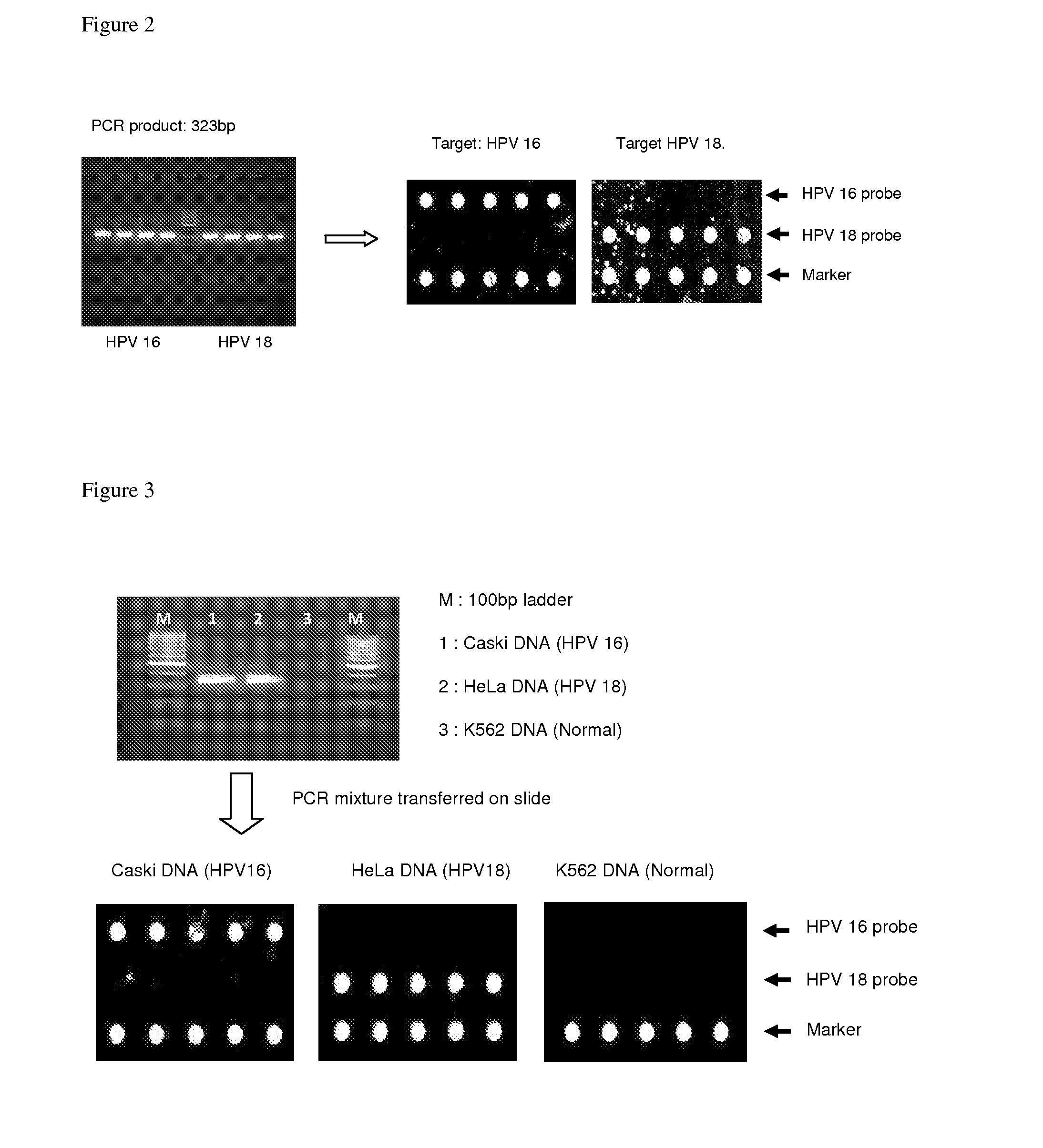
[0149]The following capture probes and primers for HPV were synthesized:
Forward primer (GP4F):5′ Cy3-GATGGTGATATGGTWSATACTGGMTWTGG 3′Reverse primer (GP4R):5′ Cy3-GMRTCAGAGGTAACMATAGARCCACTWGG 3′HPV 16 probe:5′ CTCTGGGTCTACTGCAAATTTAGCCAGTT 3′HPV 18 probe:5′ CACAGGTATGCCTGCTTCACCTG 3′Marker position probe:5′ TTTACACCTAGTGGCTCTATGGTGTCCTCT 3′
[0150]M denotes mixture of A and C; R that of A and G; W that of A and T; S that of C and G.
[0151]The primers and the capture probes were derived from the L1 region of HPV as described in Albrecht et al. J. Virol Methods 137:236-244, 2006 and a published Korean patent document (10-2006-0019042). The underlined sequence in the Marker position probe is complementary to the underlined sequence of the fluorescent reverse primer. Therefore, the marker position probe will always hybridize with the reverse primer and will be labeled positive in the final DNA slide. The capture probes were crosslinked to dendron...
example 2
Verification with the Cells Infected with HPV16 and HPV18
[0157]Human cervical carcinoma-derived cell line Caski containing HPV16 sequence (ATCC CC1-2), the HeLa S3 containing HPV18 (ATCC CRL-1550), and a leukemia cell line K562 which does not carry HPV sequence (ATCC KCLB-10243), were grown in appropriate culture medium.
[0158]DNA was extracted from the cell lines described above using G-DEXTM IIc Genomic DNA Extraction Kit (Cell / Tissue), INTRON, Co, Korea). PCR reaction mixture was assembled and PCR was carried out as described in Example 1 and transferred to the chamber assembled on the DNA slide. The slide was incubated in a PCR machine using the hybridization conditions as described in Example 1. The result showed that the HPV sequences in the infected cells can be identified correctly as shown in FIG. 3.
example 3
Cell Samples Used for PCR in a Tube or on the DNA Chip Surface Gave Similar Results
[0159]The HPV infected (Caski and HeLa cells) and non-infected (K562 cells) cells were grown. The cells were scrapped from the culture dish and suspended in PBS buffer. The cells (about 10,000 cells) were pelleted in a 1.5 ml Eppendorf tube, and to the pellet was added 50 μl of 5% Chelex-100 resin (Bio-Rad Laboratories). The content of the tube was heated at 95° C. for 5 min and the tube was centrifuged for 5 minutes. The supernatant was saved. Two sets of 30 μl PCR reaction mixtures were prepared with 2 μl each of the supernatant as described in Example 1.
[0160]One set of PCR mixture was transferred into a chamber assembled on DNA chip, and PCR and hybridization were carried out under the condition described in Example 1. PCR was carried out in the tubes with another set of reaction mixtures. After PCR the content was transferred into the chamber assembled on DNA slide for hybridization. The results ...
PUM
| Property | Measurement | Unit |
|---|---|---|
| Temperature | aaaaa | aaaaa |
| Fluorescence | aaaaa | aaaaa |
Abstract
Description
Claims
Application Information