Method and apparatus for positioning a tissue recovery instrument in confronting adjacency with a target tissue volume

a tissue recovery and target tissue technology, applied in medical science, surgery, vaccination/ovulation diagnostics, etc., can solve the problems of scarring at the location of the incision, high blood loss, and risk of false negative, so as to avoid thermal damage

Inactive Publication Date: 2009-11-26
INTACT MEDICAL
View PDF2 Cites 14 Cited by
  • Summary
  • Abstract
  • Description
  • Claims
  • Application Information

AI Technical Summary

Benefits of technology

[0023]The present invention is addressed to method and apparatus for accessing a target tissue volume with a tissue recovery instrument. With the method, upon determining the instrument entry location and attitude at the skin surface, a cold scalpel incision is made through the skin. That incision will have a length generally corresponding with the cross-sectional dimension of the cannular instrument tip and a depth effective to avoid thermal damage by electrosurgically excited precursor electrodes located forwardly of the instrument tip surface. To assure the proper initial positioning of these precursor electrodes prior to their electrical excitation, the incision is expanded with a pair of retractor components having mutually outwardly disposed tiss

Problems solved by technology

As with other similar procedures, such as those described above, open surgery may result in high levels of blood loss, scarring at the location of the incision and permanent disfigurement, due to the removal of relatively large amounts of tissue.
While a fine needle aspiration biopsy has the advantages of being a relatively simple and inexpensive office procedure, there are some drawbacks associated with its use.
With fine needle aspiration, there is a risk of false-negative results, which most often occur in cases involving extremely fibrotic tumor.
In addition, after the procedure has been performed there may be insufficient specimen material for diagnosis.
Rather, fragmented portions of tissue are withdrawn which do not allow for the same type of pathological investigation as the tissue removed during an open surgery biopsy.
This limitation also is observed with respect to large core needle biopsies.
These samples may provide some histological d

Method used


Image

  • Method and apparatus for positioning a tissue recovery instrument in confronting adjacency with a target tissue volume
  • Method and apparatus for positioning a tissue recovery instrument in confronting adjacency with a target tissue volume
  • Method and apparatus for positioning a tissue recovery instrument in confronting adjacency with a target tissue volume

Examples


Embodiment Construction

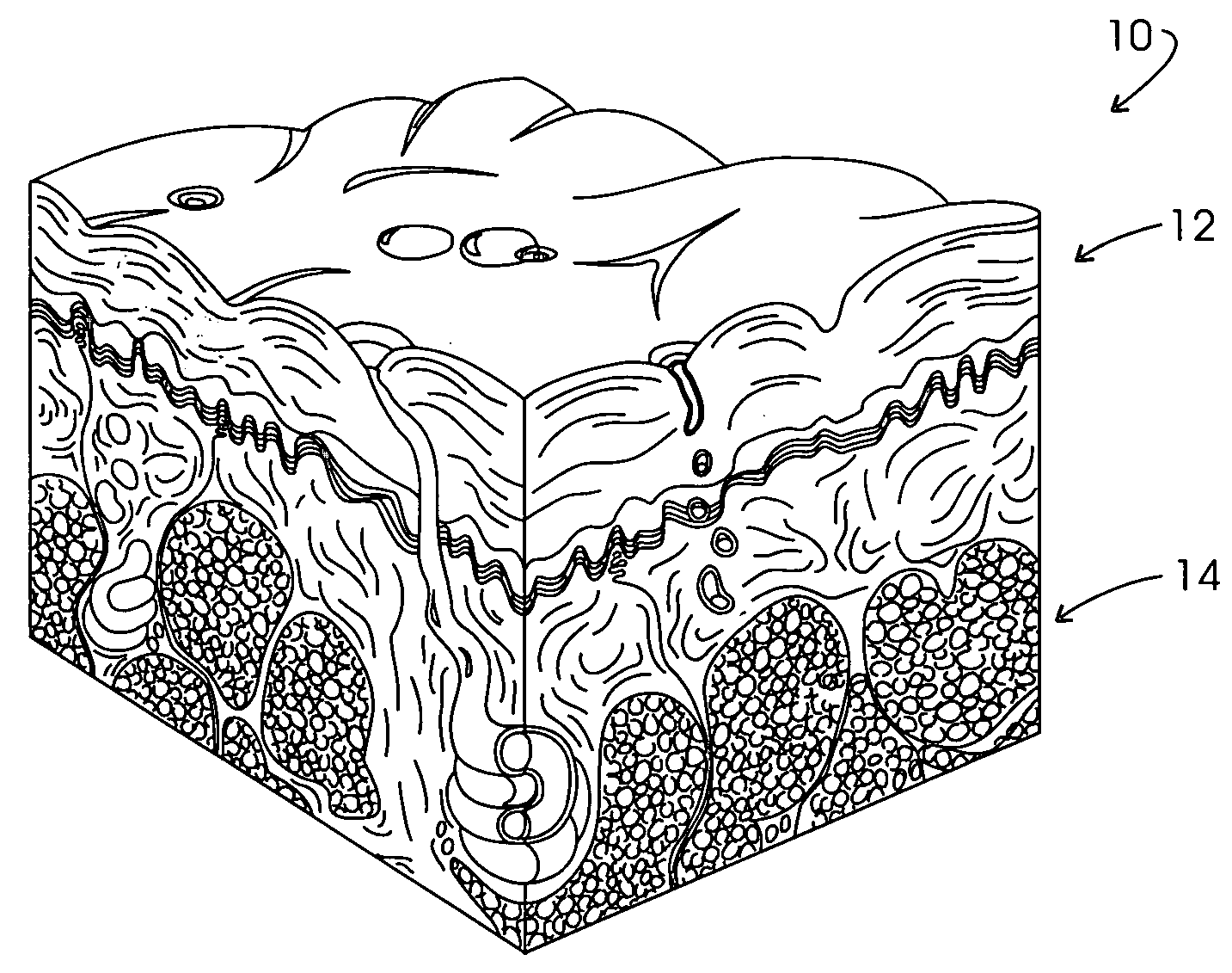
[0067]As a prelude to considering the method and apparatus involved with the initial subcutaneous positioning of the then un-energized tip of the tissue capture instrument, some insight into the mechanical structure of tissue involvement may be beneficial. The initial tissue to be encountered in the procedure is the skin, which is an anatomically and physiologically specialized boundary lamina ranging from about 1.5 mm to 4.0 mm in total thickness. Structurally, skin is complex and highly specialized, being formed as an intimate association between two distinct tissues: keratinized stratified, squamous, epithelium, superficially, the epidermis, and a deeper layer of moderately dense connective tissue, the dermis. This combination results in an integument providing a most effective barrier against a variety of externally encountered phenomena including thermal and mechanical excursions.

[0068]Referring to FIG. 1, a schematic representation of the organization of the skin is represente...


PUM


Abstract

A target tissue volume is accessed with a cannular instrument, the tip surface of which supports a precursor electrode assemblage which is electrosurgically excitable. The instrument tip initially is inserted through an incision made in the skin of a patient utilizing a pair of retractor components, the tips of which are located at a proper depth for positioning the precursor electrodes of the recovery instrument. The retractor components also are configured to define a guidance channel for receiving the tip of the recovery instrument. By stabilizing the instrument when the precursor electrodes are adjacent the tips of the retractor components and then slidably removing the retractor apparatus along the surface of the instrument, the tissue is “set” to assure proper precursor electrode positioning. The precursor electrodes are configured to exhibit an equivalent diameter having a value of at least about 90% of the diameter of the recovery instrument and are spaced forwardly of the tip surface of the instrument a distance for enhancing instrument maneuvering.

Description

CROSS-REFERENCE TO RELATED APPLICATIONS[0001]The present application is a Continuation-in-Part of application Ser. No. 09 / 904,396 filed Jul. 12, 2001 entitled “Minimally Invasive Intact Recovery Of Tissue” by Eggers, et al. which, in turn, is a Continuation-in-Part of application Ser. No. 09 / 472,673, filed Dec. 27, 1999, now U.S. Pat. No. 6,277,083 by Eggers, et al., issued Aug. 21, 2001 and entitled “Minimally Invasive Intact Recovery Of Tissue”.STATEMENT REGARDING FEDERALLY SPONSORED RESEARCH[0002]Not applicable.BACKGROUND OF THE INVENTION[0003]Developments of the diagnosis of tumorous cancer and its subsequent treatment continues to somewhat rapidly evolve. These developments particularly have been apparent in connection with cancer of the breast, perhaps in consequence of an estimation that one out of eight women will face cancer at some point in her life.[0004]Among the developments, techniques for detection with imagining devices have permitted the identification of suspect tu...

Claims


Application Information

Patent Timeline
IPC IPC(8): A61B1/32A61B18/14A61BA61B10/00A61B10/02A61B17/00A61B17/02A61B17/32A61B17/34
CPCA61B10/0266A61B17/02A61B17/3439A61B18/1442A61B2018/144A61B2017/008A61B2018/00333A61B2018/00601A61B2018/1407A61B18/1482
Inventor EGGERS, PHILIP E.EGGERS, ERIC A.EGGERS, ANDREW R.
Owner INTACT MEDICAL
Why Eureka
Try Eureka