
Rapid detection of replicating cells

a technology of replicating cells and detection time, applied in the field of replicating cell detection, can solve the problems of not substantially increasing the sensitivity or decreasing the time, bacteria that are very sensitive to antibiotics cannot grow, and the broth becomes visibly cloudy in less than 24 hours, so as to reduce the time needed to achieve results, analyze the entire membrane efficiently, and analyze large sample volumes efficiently

Inactive Publication Date: 2009-12-24
RAPID MICRO BIOSYSTEMS INC
View PDF74 Cites 29 Cited by
  • Summary
  • Abstract
  • Description
  • Claims
  • Application Information

AI Technical Summary

Benefits of technology

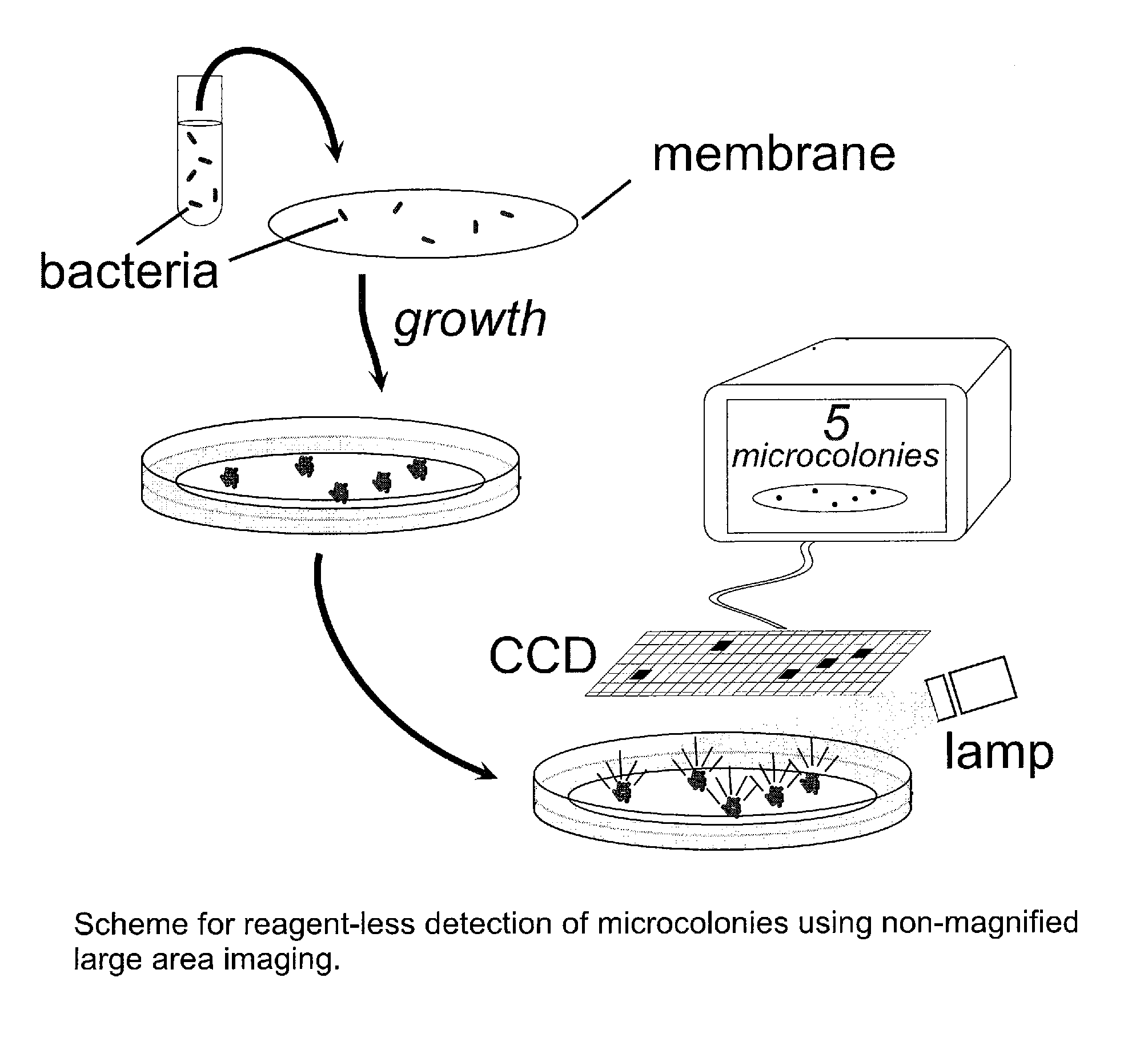
The invention is a method and instrument for detecting living cells by forming microcolonies of cells and detecting them using large area imaging. This allows for efficient, rapid, and sensitive enumeration of living cells without the need for labeling reagents. The method can be used in clinical and industrial microbiology to detect cellular microcolonies and determine antimicrobial resistance. The instrument features a photoelectric array detector and an illumination source capable of imaging a detection area with at least one dimension that is ≧1 cm. The instrument does not optically magnify more than 5 fold.

Problems solved by technology

For example, a single bacterial cell, which is much too small to see by eye (about one millionth of a meter), when placed in nutrient broth, can cause the broth to become visibly cloudy in less than 24 hours.
Most such methods automate colony counting but do not substantially increase the sensitivity or decrease the time to results compared to traditional enumeration by eye.
However, bacteria that are very sensitive to the antibiotic can not grow unless they are far from the edge of the filter.
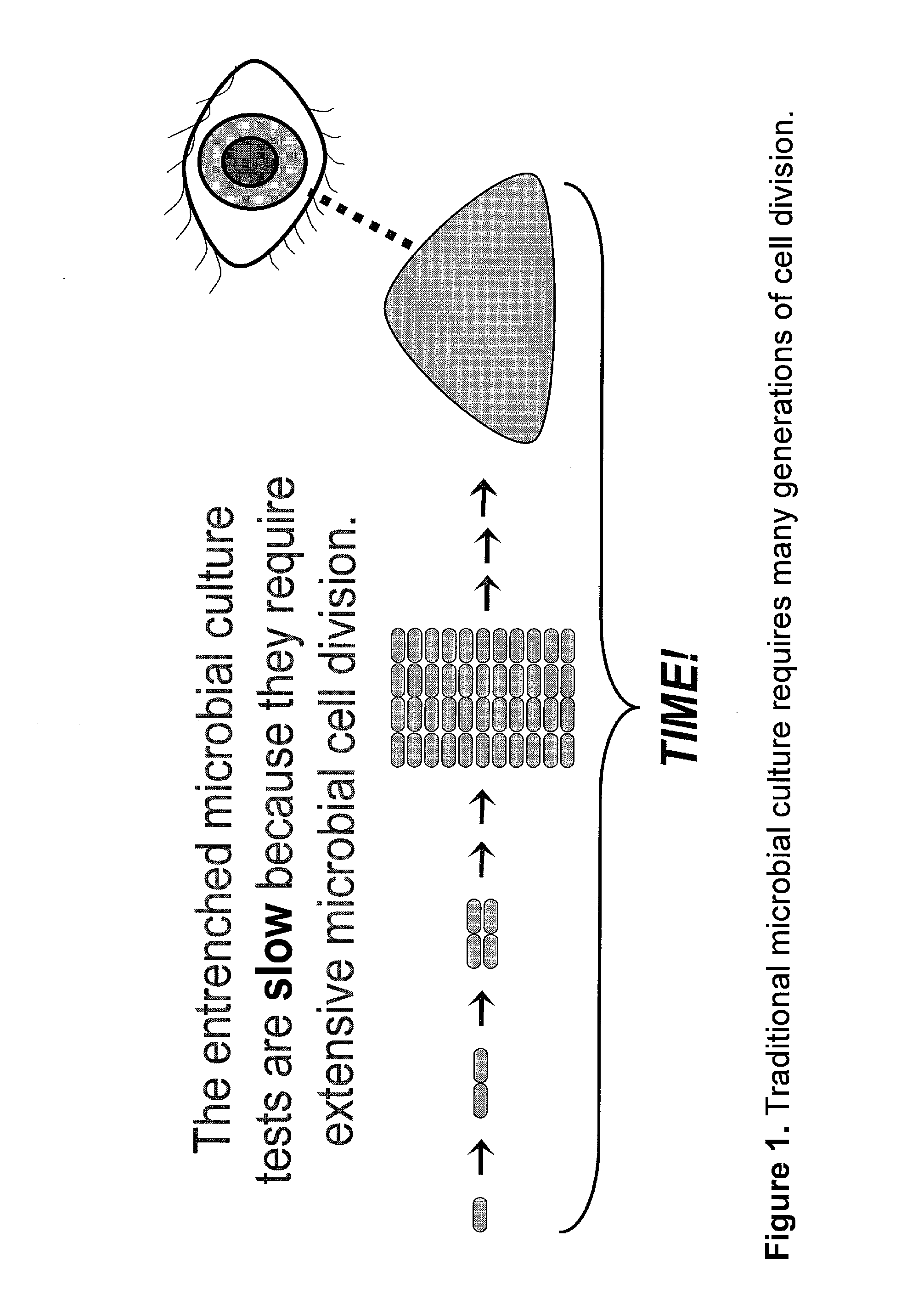
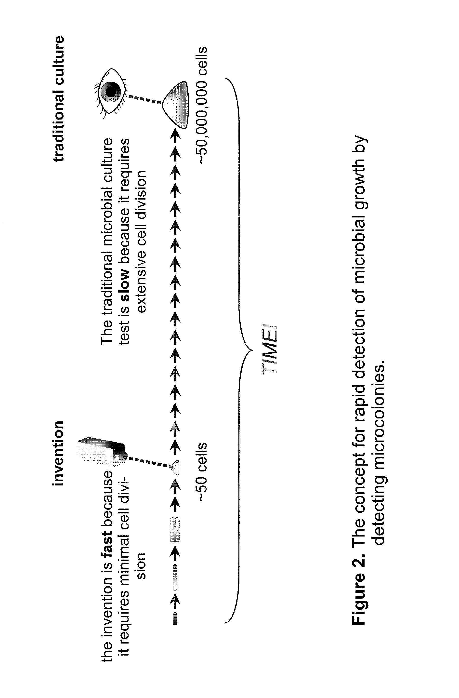
The most serious drawback of microbial culture is that it is slow—it takes time to generate the number of cells required for visual detection.
The long growth period required for microbial culture is a significant problem in both healthcare and industry.
The long time required for detecting M. tuberculosis can result in a patient with tuberculosis infecting many others with the highly contagious disease or the costly quarantine of patients who do not have tuberculosis.
In food manufacture, long testing cycles can increase food spoilage or result in moving inadequately tested material through subsequent processing steps.
Slow microbial culture also adversely impacts the production of biopharmaceuticals and vaccines.
Other disadvantages of microbial culture, such as tedious manual procedures and inability to culture some microbes, are considered less problematic than the long time required.
Most types of microbes found in the environment cannot be grown in the laboratory.
However, such microbes are often not harmful to humans or are destroyed in industrial manufacturing processes and are therefore ignored for most applications.
However, this method is not effective for testing large samples containing low numbers of microbial cells, because only a very small volume of sample can be observed in a microscopic field of view.
The low sensitivity of microscopic methods generally limits their usefulness to samples containing more than ten thousand bacterial cells per milliliter—these methods are much less sensitive than traditional microbial culture.
A drawback of the system is that the detection method kills the microbes, precluding isolation of pure cultures from the colonies.
The system also requires an expensive image intensifier module.
The method is limited to scanning for a particular microbe in one experiment.
Validating and instituting surrogate methods have been problematic since samples that are known to meet regulatory standards and that are scored as sterile using traditional plate culturing methods often have thousands of cells that score positive for the surrogate biochemical activity.
However, this approach often results in substantial “overcounting”—i.e., higher numbers of cells than are detected by traditional culture (Costanzo, S., et al.
Another disadvantage of the ScanRDI system is that it kills the microbes during the staining process precluding generation of pure cultures from the detected microbes.
Finally, laser scanning systems for cellular enumeration are complex and expensive (hundreds of thousands of dollars) making them difficult to justify for routine microbiological applications.
Flow cytometry is a more complex and expensive method than traditional culture.
Although faster than traditional culture, flow cytometry does not have a comparable limit of detection to the traditional method.
Furthermore, microbial targets are often killed by the staining methods used for detection, eliminating the ability to produce pure cultures.
The drawback of this technique is that it is many thousands of times less sensitive than microbial culture.
A disadvantage of this system is the great expense incurred because of the incorporation of the image intensifier and associated optics.
Furthermore, unlike microbial culture methods, the system can not detect any microbe, distinguish between living and dead microbes, or generate pure cultures.
Although some of these methods are substantially faster than microbial culture, none offers all of the features of culture that are critical to microbiologists.
For example, although numerous immunoassays for microbes have been commercialized, this technique is not inherently quantitative, is much less sensitive than microbial culture, and is not as powerful as culture for detecting many types of microbes in a single test.
Or, as another example, nucleic acid amplification methods can be as sensitive as microbial culture, but they do not distinguish between living and non-living cells and can not deliver pure cultures for antibiotic susceptibility testing.
Methods for biochemical analysis (e.g., of fatty acids, nucleic acids, or proteins) using electrophoresis, mass spectroscopy, and chromatography can be powerful for microbial identification, but such methods are usually inappropriate for microbial enumeration and are generally too expensive and complex for routine microbial diagnostics.
Microbial culture has the important advantages of being simple, ultra-sensitive, inexpensive, and quantitative but has the significant drawback of being slow.
The long time required for results has major costs in healthcare and in manufacturing.
More rapid methods have been developed, but while improving the time to results, they have sacrificed one or more of the critical advantages of microbial culture.

Method used


Image

  • Rapid detection of replicating cells
  • Rapid detection of replicating cells
  • Rapid detection of replicating cells

Examples


example 1

Detection and Identification of Bacterial Microcolonies Using Non-Magnified Large Area Imaging

[0211]Background and objectives: Detection of microbial growth is at the core of both clinical microbiology (e.g., bacterial identification and antimicrobial susceptibility testing) and industrial microbiology (e.g., mandated sterility testing), but the commonly used methods are slow. The consequent delays in analysis cause needless death and suffering in clinical situations and exact a large financial cost in industry.

[0212]Using non-magnified large area imaging to detect individual microcolonies exploits the advantages of microbial culture while avoiding the substantial disadvantages of traditional and emerging methods. Advantages of in situ replication analysis using the invention are: speed; ease of multiplexing (scanning for more than one microbe); and the ability to detect and identify without sacrificing microcolony viability (essential for efficient antimicrobial susceptibility test...

example 2

Autofluorescence-Based Detection of Bacterial Microcolonies Using Non-Magnified Large Area Imaging

[0216]Background and objectives: The importance of methods that detect microbial growth and the limitations of current methods are discussed in the Background section. This example demonstrates a very simple yet powerful method based on the present invention that rapidly detects the growth of bacterial microcolonies. The method relies on the intrinsic fluorescence (autofluorescence) of the target cells for generating detectable signal. Thus, this method does not use category-binding molecules or exogenous signaling moieties to achieve non-magnified large area imaging of microscopic target cells. The advantages of reagent-less non-destructive enumeration include generation of purified cultures (for microbial identification and antibiotic susceptibility testing, improved method validation, and the ability to follow microbial growth over time (for object discrimination and growth kinetics)...

example 3

A Simple Method for Validating a Rapid Reagent-Less Microbial Enumeration Test Using an Internal Comparison to the Traditional Culture Method

[0220]Background and objectives: Proving the equivalence of a new microbiological test to the “gold standard” method is an essential task for both the developers of new methods and their customers. Formalized validation requirements are generally codified in governmental regulations that guide the introduction of new microbiological methods in industry and healthcare. New methods for microbiological testing in the pharmaceutical industry have sometimes floundered because of the difficulty of proving equivalence to the accepted methods. The goal of this example is to demonstrate a simple method for proving the equivalence of a test based on the invention to the traditional microbial culture test.

[0221]Experimental methods. E. coli MG1655 cells were grown and analyzed as in Example 2. After imaging the microcolonies, the filter was re-incubated a...


PUM

PropertyMeasurementUnit
diameteraaaaaaaaaa
diameteraaaaaaaaaa
diameteraaaaaaaaaa

Abstract

The invention enables efficient, rapid, and sensitive enumeration of living cells by detecting microscopic colonies derived from in situ cell division using large area imaging. Microbial enumeration tests based on the invention address an important problem in clinical and industrial microbiology—the long time needed for detection in traditional tests—while retaining key advantages of the traditional methods based on microbial culture. Embodiments of the invention include non-destructive aseptic methods for detecting cellular microcolonies without labeling reagents. These methods allow for the generation of pure cultures which can be used for microbial identification and determination of antimicrobial resistance.

Description

CROSS-REFERENCE TO RELATED APPLICATIONS[0001]This application is a divisional of U.S. application Ser. No. 10 / 236,107, filed Sep. 6, 2002, now U.S. Pat. No. 7,582,415, which claims priority from U.S. Provisional Application No. 60 / 317,658, filed Sep. 6, 2001, both of which are hereby incorporated by reference.BACKGROUND OF THE INVENTION[0002]The invention relates to the detection, enumeration, and identification of replicating cells, especially microbial cells (e.g., bacteria, yeasts, and molds), in medical, industrial, and environmental samples. Microbial culture is the predominant methodology in these markets, because of its many attractive features. The invention addresses the chief drawback of microbial culture—the length of time needed to achieve results—while retaining the beneficial attributes of the method.Microbial Culture for Detecting and Enumerating Microbes[0003]During the 19th and 20th centuries an understanding emerged concerning the role of bacteria, yeast, and molds...

Claims


Application Information

Patent Timeline
Patent Type & Authority Applications(United States)
IPC IPC(8): H04N7/18C12M1/34C12Q1/00C12Q1/04C12Q1/06C12Q1/18C12Q1/68C12Q1/70G01N1/30G01N15/14G01N21/76G01N21/78G01N33/48G01N33/50G01N33/53G01N33/543G01N33/545G01N33/554G01N33/567G01N33/569G01N33/58
CPCB82Y5/00B82Y10/00B82Y20/00C12Q1/04C12Q1/18G01N15/1475G01N2021/6471G01N33/5008G01N33/56966G01N33/56983G01N33/58G01N33/582G01N2015/1486G01N33/5005G01N15/1433C12Q1/06G01N15/1429G01N21/6428G01N33/56916G01N33/56938G01N2021/6439G01N2333/195G01N2333/245G01N2333/31G01N2333/32G01N2333/33G01N2015/1488
Inventor STRAUS, DON
Owner RAPID MICRO BIOSYSTEMS INC
Learn More