
Flexible visually directed medical intubation instrument and method

a visual directed, medical intubation technology, applied in the field of medical instruments, can solve the problems of urethral obstruction, burden on the delivery of effective care through the healthcare system, and difficulty in more than forty percent of male urinary catheter placement, and achieve the effect of reducing the cost of the instrumen

Inactive Publication Date: 2009-12-24
PERCUVISION
View PDF72 Cites 90 Cited by
  • Summary
  • Abstract
  • Description
  • Claims
  • Application Information

AI Technical Summary

Benefits of technology

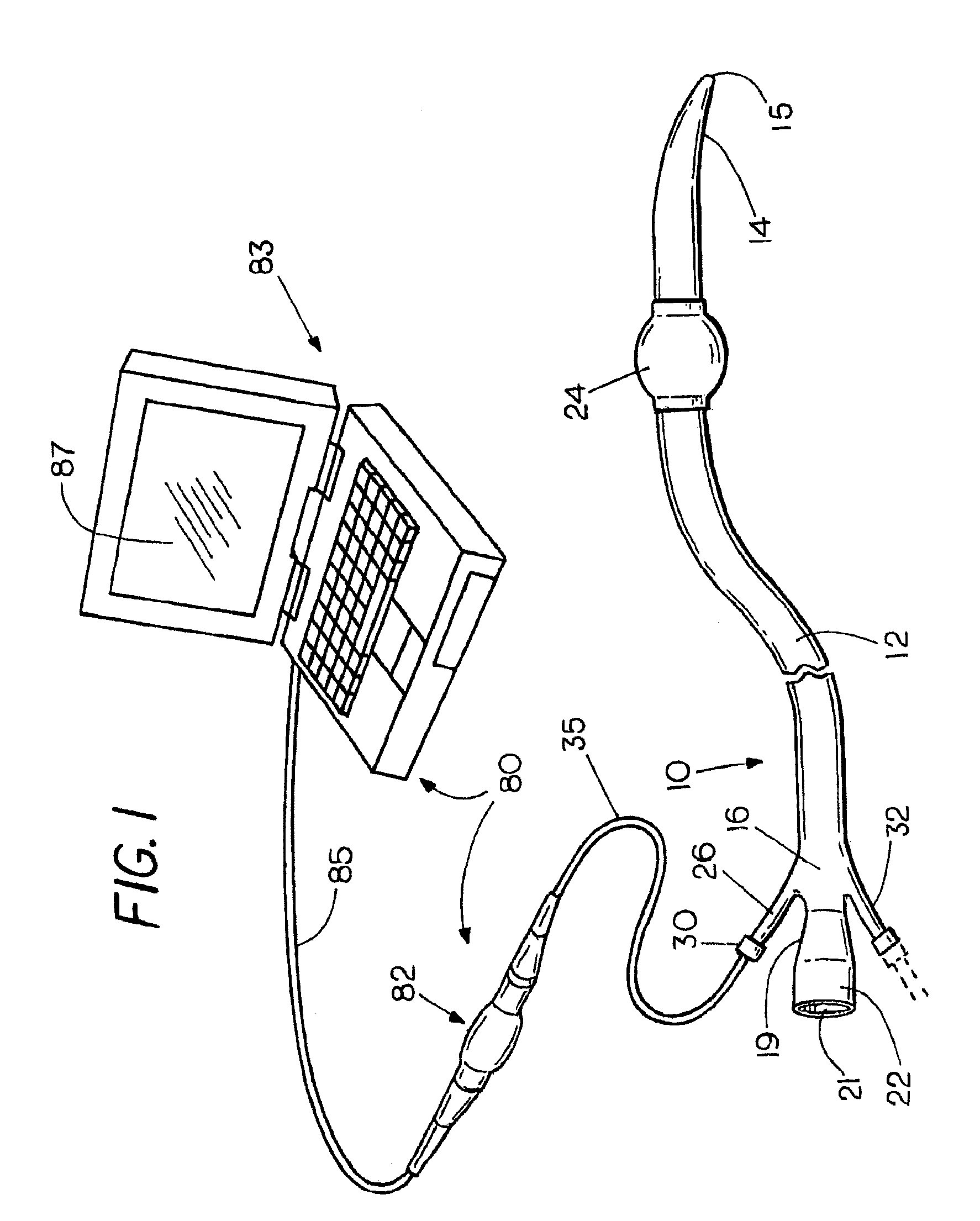
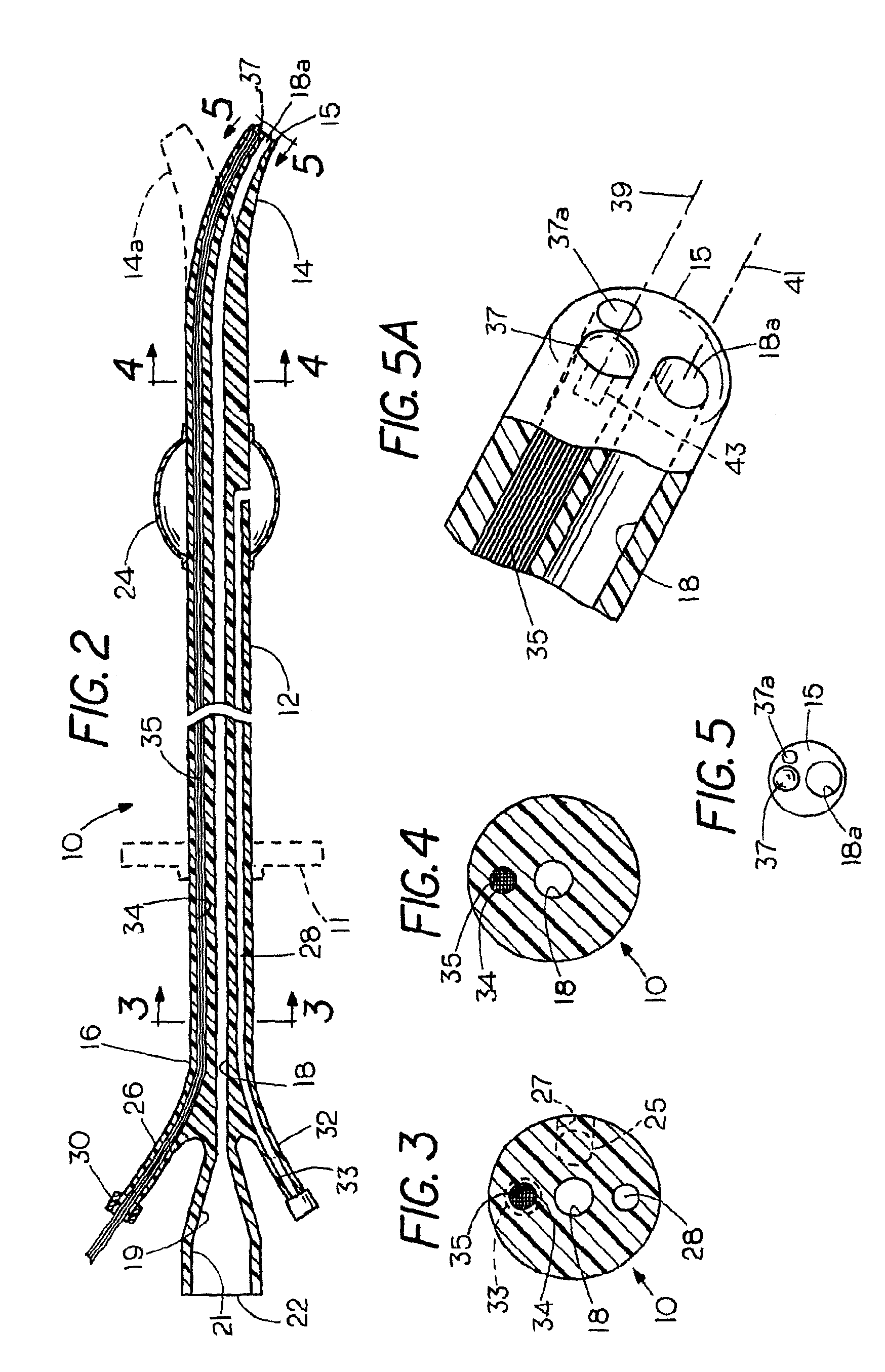
[0013]The present invention provides a method and apparatus for facilitating medical intubation procedures. In accordance with one aspect of the invention, there may be provided a flexible direct vision viewing instrument or viewer that includes a catheter or sheath formed from a highly flexible biocompatible polymer such as natural or synthetic rubber or plastic having a longitudinal working channel extending the length of the catheter with an outlet port that may be positioned in alignment with the channel at the distal end of the catheter. The catheter may have a second longitudinal channel or lumen that contains a flexible sensor cable such as viewing cable for optical sensing. In place of or in conjunction with an optical sensor, there can be provided any of various kinds of sensors such as a chemical sensor, a pH sensor, a temperature sensor, in vivo infection, or the like. In the case of a visual sensor, one of the channels contains an optical cable providing illumination in the proximity of the distal end of the catheter for enabling the body of the patient to be viewed during placement of the instrument through a body opening or percutaneously through a surgical opening. An objective optical sensor or other sensor at the distal end of the cable provides information, e.g. continuous viewing the body of the patient just ahead of the tip of the instrument during insertion of the instrument as well as after placement of the instrument within the body. The invention may be adapted to be produced in either a disposable version or a reusable version that can be sterilized after use.
[0014]The invention also provides a catheter that may be able to serve as a working sheath which can be thought of as a temporary and removable artificial tract or liner that is placed through an opening in the body of the patient at the beginning of a surgical procedure to facilitate endoscopic evaluation and treatment of the digestive tract, urinary tract, or other body cavity while minimizing trauma and patient pain. During use, it allows multiple insertions and removals, i.e., the interchange of endoscopic instruments, catheters, sensors, drains, etc. The viewing cable can act as a stiffener during insertion into the patient to provide a greater degree of firmness, especially when the sheath or catheter is relatively thin or tends to fold back upon itself during insertion. Once in place, the viewing cable can be removed and replaced by other sensors such as a temperature sensor, a pH sensor, or an infection sensor, or by other medical devices. At its proximal, i.e. exterior end, the lumen of the sheath may have an entry port for instruments with a removable cap that provides a nipple seal to preclude backflow of fluid from the body after the visual element or other sensor has been removed. The instrument can be placed into the stomach or other part of the digestive tract or the urethra under direct vision, i.e., with a flexible condition sensor extending through the sheath to act as a temperature, pH, or visual sensor. The sensor can include a sensor / actuator cable that provides an interoperable medium for transmitting optical or electrical signals, e.g. a fiber-optic bundle for illuminating and viewing a body cavity through the sheath, both during the insertion of the sheath and thereafter.
[0015]In some embodiments, the instrument may have catheter with a single longitudinal channel, and the sensor may occupy only a small portion of the longitudinal channel. In other embodiments, a catheter and sensor may have slightly differing working lengths, to help maintain a predetermined spatial arrangement between the catheter and sensor during stretching or compression of one of both of these elements. In yet another embodiment, a catheter wall thickness may vary in conjunction with a longitudinal channel diameter in order to maintain a constant over diameter of the catheter.

Problems solved by technology

While most placements proceed without problems, typically more than forty percent of male urinary catheter placements are difficult because of the problematic normal anatomy of the male lower urinary tract such as the external sphincter, the S-curve of the bulbous urethra and angulated prostatic urethra and / or pathologic conditions, such as urethral stricture disease, stones, trauma, tumors, enlarged prostate, iatrogenic false passages, and / or congenital disorders causing a substantial burden on the delivery of effective care through the healthcare system.
The most common problem is tetany, a spasm of the external urinary sphincter or stricture of the urethra.
Stones, and even clots descending from the bladder, also constitute urethral obstructions.
These costs to the healthcare system, hospitals, clinics and doctors' offices are substantial.
In addition, the delay in servicing urological catheter patients in a timely manner constitutes poor medical efficiency, delivery, and control.
When difficulty is encountered, the resulting frustration among healthcare professionals, especially nurses, physician extenders and physician assistants, creates a very real feeling of ineffectiveness on the part of these healthcare workers, to say nothing of the dissatisfaction on the part of the patients caused by the delay and added discomfort.
Difficult catheterizations can also be a source of urinary tract infection subsequent to damage of the urinary epithelium.
While the dollar cost to the healthcare system is not the only concern, such elements as added labor and material costs, time delays for patient rectification, excess space and equipment required, catheter kit value, nurse technician and physician costs constitute an expense to the healthcare system of surprising proportions.
Of these, conservatively about 40% are difficult which means that they require multiple advances and pull-backs of the urinary catheter to negotiate the urethra, multiple catheters on the same patient, several staff workers attending to the same patient, or special instrumentation such as filoforms / followers, cystoscope or radiologic services.
Other instruments may be suited for urological use but not be suited for use in gastroenterology.
Certain intubation devices such as the Councill catheter are only capable of a blind insertion and must rely on a guide wire to navigate to the bladder.
Consequently, if the Councill catheter encounters resistance during insertion, there is no way to know its cause.
Any obstructions, anomalous conditions, or anatomical idiosyncrasies can interfere with successful insertion of the tube.
Moreover, no provision is made for sensing conditions at or near the distal tip of the intubation instrument with traditional analog sensors and / or actuators or smart digital sensors or actuators.

Method used


Image

  • Flexible visually directed medical intubation instrument and method
  • Flexible visually directed medical intubation instrument and method
  • Flexible visually directed medical intubation instrument and method

Examples


Embodiment Construction

[0047]Refer now to the FIGS. 1-29 wherein the same numerals refer to corresponding parts in the several views. The invention will be described by way of examples, all of which are intended for illustration only and not for limitation, which illustrate a visually directed intubation instrument in accordance with the invention that can be placed into the body of the patient under direct and continuous visual control in any of a variety of different surgical specialties. The invention is especially versatile and can be dimensioned and configured for use in urology, in gastroenterology, and in other surgical fields. The various embodiments illustrate the versatility of the invention since it can be employed as a drain or for exploratory purposes as well as a working channel to be used during a surgical operation or even in the field of gastroenterology as a feeding tube.

[0048]In certain embodiments, the instrument 10 comprises a flexible catheter 12 formed from natural or synthetic rubb...


PUM


Abstract

A flexible medical intubation instrument provided for placement into an animal or human patient comprises a catheter with at least one longitudinally extending lumen or channel. A fixed or slideably removable sensor cable is at least partially contained within the channel, having a sensor for sensing a characteristic or condition. While enabling observations through the sensor channel, the working channel may simultaneously function as a drain or an irrigation duct, a feeding tube, or to provide a passage for the insertion of one or a succession of surgical devices such that the catheter serves as a protective artificial tract or liner as surgical devices are inserted and removed through it in succession so as to minimize tissue trauma.

Description

CROSS-REFERENCE TO RELATED APPLICATIONS[0001]This application is a continuation-in-part application of, and claims priority to, U.S. patent application Ser. No. 12 / 214,944, filed Jun. 23, 2008.STATEMENT REGARDING FEDERALLY SPONSORED RESEARCH OR DEVELOPMENT[0002]Not Applicable.FIELD OF THE INVENTION[0003]This invention relates to medical instrumentation and more particularly to a method and apparatus for facilitating intubation of an animal or human patient.BACKGROUND OF THE INVENTION[0004]In many medical procedures it is often necessary to place an instrument into the body of the patient for drainage, for viewing a part of the body, or for performing a surgical operation such as the endoscopic removal of a tumor, to take a biopsy, or for feeding the patient. The invention may have general application in medicine including the field of urology as well as in the field of gastroenterology and in other medical and surgical specialties. The placement of a catheter in the urethra for the ...

Claims


Application Information

Patent Timeline
IPC IPC(8): A61B6/12A61M25/00
CPCA61B1/00082A61B1/00165A61B1/012A61B1/05A61B1/01A61B5/0084A61B5/036A61M25/04A61M25/10A61B1/07
Inventor SINGH, ERROLSTOCK, ALLENRUSSELL, WALTER
Owner PERCUVISION
Learn More