Method and reagent for protein analysis

a protein and reagent technology, applied in the field of protein analysis, can solve the problems of inability to quantitatively, low detection sensitivity, and significant variation in detection sensitivity, and achieve the effect of convenient and rapid performance of highly sensitive qualitative or quantitative protein analysis

Inactive Publication Date: 2010-01-07
NAT INST OF ADVANCED IND SCI & TECH
View PDF6 Cites 1 Cited by
  • Summary
  • Abstract
  • Description
  • Claims
  • Application Information

AI Technical Summary

Benefits of technology

[0013]The present invention makes it possible to conveniently and rapidly perform highly sensitive qualitative or quantitative protein analysis.

Problems solved by technology

Among these conventional techniques, the CBB method can be carried out conveniently and rapidly, but it has problems that its detection sensitivity is low (detection limit: 50 ng to 100 ng) and its detection sensitivity varies significantly depending on protein type.
The silver staining method is a highly sensitive method such that the detection limit ranges from 1 ng to 10 ng, but it has drawbacks in that it cannot be quantitative and requires special processing of a waste solution containing silver.
A method using a fluorescent dye such as Sypro Ruby (Invitrogen Corporation) may exhibit higher detection sensitivity than that of the silver staining method; however, when the method is applied to SDS-PAGE, such fluorescent dye tends to be affected by SDS remaining on the carriers, so that a target protein cannot be detected in some cases.
Accordingly, the method has problems that it cannot be performed rapidly and leads to a significant burden on operators.
However, all of the fluorescent compounds specifically described in the Examples of the above documents are hardly soluble in water, so that insoluble matter is frequently formed during staining and storage.
Therefore, insoluble matter is deposited on electrophoresis carriers and then false positive spots appear during the detection step, which may disturb the detection.

Method used


Image

  • Method and reagent for protein analysis
  • Method and reagent for protein analysis
  • Method and reagent for protein analysis

Examples


example 1

(Example 1) Synthesis of compound 1 (2,2′-(1E, 1′E)-2,2′-(4-(dicyanomethylene)-4H-pyrane-2,6-diyl)bis(ethene-2,1-diyl)bis(sodium benzenesulfonate)salt)

[0047]

[0048]Sodium 2-sulfo benzaldehyde (2.42 g (11.6 mmol)) was added to a 50-mL three-neck flask and then suspended in 30 mL of ethanol:methanol=1:1 (v / v). 4-(dicyanomethylene)-2,6-dimethyl-4H-pyrane (1.0 g (5.81 mmol)) and piperidine (495 mg (5.81 mmol)) were added and then the resultant was refluxed under an argon atmosphere for 12 hours. The precipitate was collected by filtration. The thus obtained yellow powder was washed with water and then subjected to drying under reduced pressure, so that 1.43 g of a desired compound was obtained (yield: 44.5%).

[0049]ESI-MS (+): [M+Na—H]+=574.6

[0050]1H-NMR (400 MHz, DMSO-d6) σ 6.92 (2H, s), 7.20 (2H, d, J=16 Hz), 7.37-7.46 (4H, m), 7.77-7.88 (4H, m), 8.62 (2H, d, J=16 Hz).

[0051]13C-NMR (100.53 MHz, DMSO-d6) σ 56.6, 106.5, 115.5, 120.3, 127.2, 127.4, 129.0, 129.3, 132.5, 138.1, 146.4, 156.1,...

example 2

Measurement of Maximum Absorption Wavelength and Maximum Fluorescence Wavelength

[0061]Compound 1 and Compound 2 were separately dissolved in purified water at a concentration of 25 μg / mL. The absorption spectra at a wavelength ranging from 200 nm to 800 nm and the fluorescence spectra at a wavelength ranging from 450 nm to 600 nm were measured. The absorption spectra were measured using U3310 (Hitachi High-Technologies Corporation) and the fluorescence spectra were measured using RF-1500 (Shimadzu Corporation). The results are shown in Table 1.

TABLE 1Comparison of optical characteristics of compoundsMaximum absorptionMaximum fluorescencewavelengthwavelengthCompound 1293 nm516 nm (excitation: 444 nm)380 nmCompound 2388 nm511 nm (excitation: 440 nm)

example 3

Protein Detection

(1) Preparation of Samples

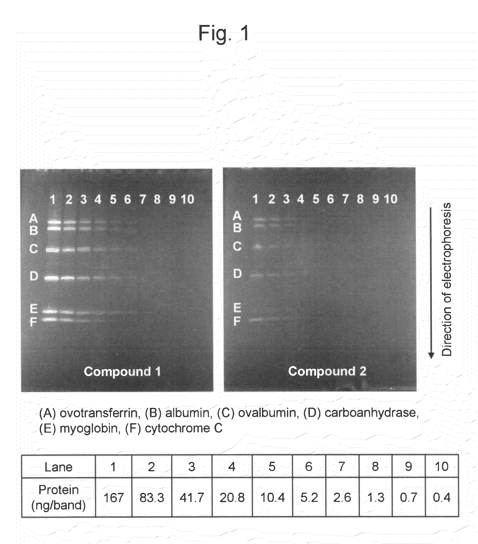
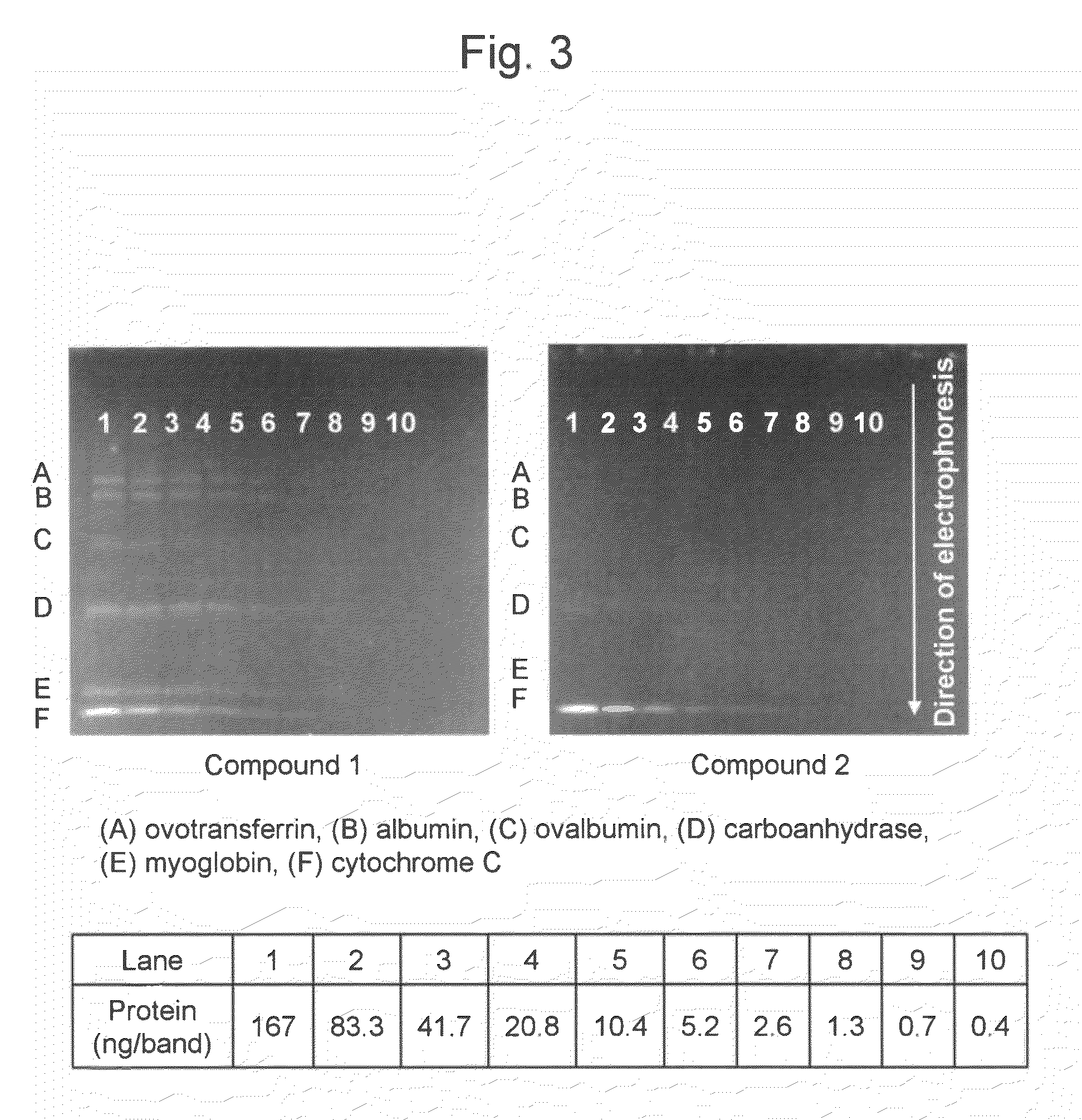
[0062]Two (2) mL of a stock buffer (a mixture of purified water (4.2 mL), 0.5 M Tris-hydrochloric acid buffer (pH 8.6) (1.0 mL), glycerol (0.8 mL), 100 g / L sodium dodecyl sulfate (1.6 mL), and 2-mercaptoethanol (0.4 mL)) was added to 2 mg of a commercially available molecular weight marker (Protein Standard Mixture IV; Merck & Co., Inc.). The mixture was heated in boiling water for 2 minutes and then cooled. Thirty (30) μL each of the resultant was dispensed in a 0.5-mL microtube and then cryopreserved at −80° C. until the use thereof. A sample diluent (a mixture of purified water (3.8 mL), 0.5 M Tris-hydrochloric acid buffer (pH 8.6) (1.0 mL), glycerol (0.8 mL), 100 g / L sodium dodecyl sulfate (1.6 mL), and 5 g / L bromophenol blue (0.8 mL)) was added to the thawed molecular weight marker, so that a dilution series with protein concentrations ranging from 0.4 ng / μL to 200 ng / μL was prepared.

[0063]The marker contained, in terms of mass, equal ...


PUM

PropertyMeasurementUnit
concentrationaaaaaaaaaa
fluorescence wavelengthaaaaaaaaaa
pHaaaaaaaaaa

Abstract

A convenient, rapid, and highly sensitive protein detection method is provided. According to the present invention, an easily water-soluble compound of formula I is provided:
[wherein, R1 and R2 are same or different, each of which is an aryl or heteroaryl group substituted with one or more anionic substituents].

Description

BACKGROUND OF THE INVENTION[0001]1. Field of the Invention[0002]The present invention relates to a method for protein analysis using a water soluble fluorescent compound and the use of the compound as a protein analysis reagent.[0003]2. Background Art[0004]As a method for protein analysis, an electrophoretic methods are widely employed. The electrophoretic methods are advantageous in that these allow proteins to be resolved based on their molecular weights so that the molecular weights can be estimated, these enable semi-quantitative measurement of amounts of proteins, and the methods can be carried out using a relatively simple apparatus, for example.[0005]Examples of a technique for protein analysis using electrophoresis include a technique that involves staining proteins existing in a sample before electrophoresis or directly staining proteins existing on electrophoresis carriers such as gel and then visually detecting the proteins, a technique that involves transferring proteins...

Claims


Application Information

Patent Timeline
Patent Type & Authority Applications(United States)
IPC IPC(8): G01N27/447C07D315/00G01N33/00
CPCC07D309/34
Inventor SUZUKI, YOSHIOYOKOYAMA, KENJITAKAGI, NOBUYUKICHIMURO, TOMOYUKISHINOHARA, ATSUSHI
Owner NAT INST OF ADVANCED IND SCI & TECH
Why Eureka
Try Eureka