[0005]Prior to this invention, gastric anchoring methods have required stapling, suturing, or other modification of the
anatomy to hold a device in place. In contrast, the invention described herein exploits asymmetric geometry of gastric
anatomy to maintain a gastric
implant in a relatively
fixed position within the
gastrointestinal tract without attachment to the
gastric wall. Specifically, the present invention is directed to a gastric
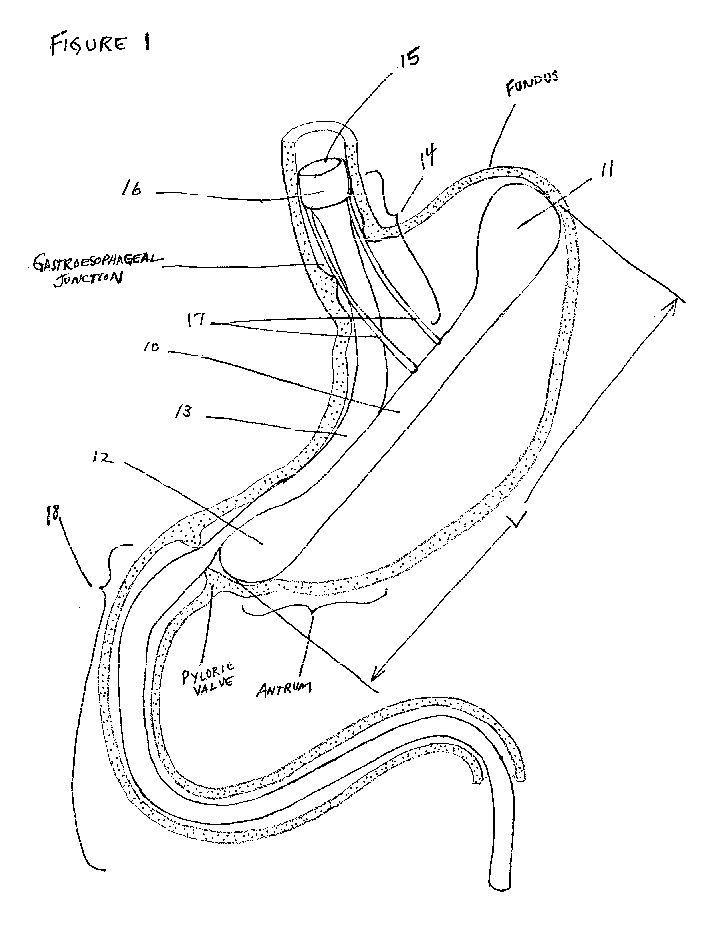
implant that comprises an intragastric anchor and a therapeutic or diagnostic device coupled to the anchor. The intragastric anchor of the invention limits the movements of any device attached to it to the displacement available to the anchor within the stomach. In one embodiment, the intragastric anchor has an elongate shape extending along the
long axis of the stomach substantially from the
antrum to the fundus such that displacement of the anchor along the
long axis due to gastric
contractility or other causes is limited by the gastric
anatomy of the fundus and
pylorus. Similarly, the elongate anchor is configured to be longer than the transverse width of the stomach and thus too long to be flipped end over end within the stomach by gastric
contractility. Thus, the intragastric anchor provides a relatively stable platform on which to anchor bariatric or other therapeutic or diagnostic devices. Once deployed within the stomach, the anchor is configured to be larger than the pyloric valve to prevent its passing out of the stomach and into the small bowel. This configuration enables the intragastric anchor to maintain a relatively
fixed position and orientation within the
gastrointestinal tract. Additionally, an extension from the anchor into the
esophagus may be present, which defines a plane such that the anchor may also prevent rotation within the stomach. The present invention is further directed to a method for anchoring a therapeutic device or a diagnostic device within the stomach in a relatively
fixed position, that is with a relatively stable position and orientation, while being free from attachment to the stomach wall, the method comprising positioning an anchor according to the invention in the stomach of a patient between the fundus and the pyloric valve; and
coupling a therapeutic device or a diagnostic device to the anchor.
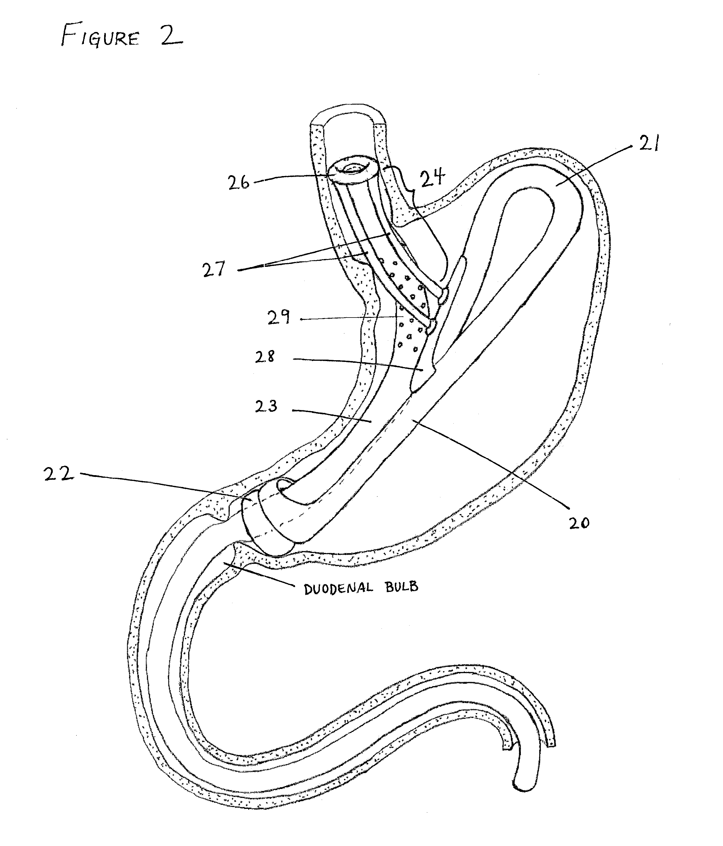
[0006]Having secured the anchoring implant within the stomach, any number of devices may be attached to it. In one embodiment, the device is a therapeutic device. For example, a bariatric sleeve may be secured to extend from the esophagus to the
jejunum, supported by a geometrically-fixed intragastric anchor. Similarly, a jejunal bypass sleeve extending from the
pylorus into the
jejunum may be supported by such an anchor. Also similarly, devices restricting gastric inflow and / or outflow may be supported by an intragastric anchor. Alternatively, a removable
esophageal stent deliberately designed for
sliding contact within the esophagus, so as to avoid hyperplastic
tissue ingrowth, may be supported by the intragastric anchor. The combination of elongate intragastric anchor and sliding
esophageal stent improves upon conventional stenting by avoiding the tendency of esophageal stents to migrate or to become unremovable through hyperplastic
tissue ingrowth and scarring.
[0007]Similarly, diagnostic devices, such as for example a pH sensor, may be supported within the
gastrointestinal tract by an intragastric anchor. By way of example, when configured to maintain a pH sensor within the esophagus, an intragastric anchor may include an esophageal extension, extending from the body of the anchor through the lower
esophageal sphincter and into the esophagus, to support the sensor in a relatively fixed position within the esophagus. Since the esophagus serves to introduce oral intake into the stomach, the pH sensor is preferably held relatively apposed to the
esophageal wall to avoid luminal blockage. Furthermore, since the intragastric anchor will move in a restricted fashion during normal gastric
contractility and likewise move the pH sensor affixed to it, the pH sensor is preferably slidably coupled to the esophagus by an atraumatic sliding
apposition structure. Regular sliding along the
esophageal wall reduces the possibility of hyperplastic
tissue ingrowth, minimizes pressure on healthy mucosa, and enhances the removability of the sensor. The combination of elongate intragastric anchor and sliding
apposition structure improves upon stenting, stapling, suturing, and other fixation methods for implanted diagnostic devices by avoiding the tendency of such implants to migrate or to become unremovable through tissue ingrowth and scarring.
[0008]One application of the intragastric anchor revolves around bariatric therapy. A specific implementation described in this disclosure mimics the mechanisms by which Roux-en-Y
gastric bypass surgery is thought to operate while avoiding surgical changes to the patient's anatomy. An anchored bariatric sleeve reduces the
effective volume of the stomach and provides a mechanism for early satiety through stimulation of stretch receptors in the fundus, reduces
exposure to
gastric juices by bypassing the stomach, delays
exposure to digestive enzymes by bypassing the ampulla of Veter, and reduces
nutrient absorption by bypassing a section of the small bowel.
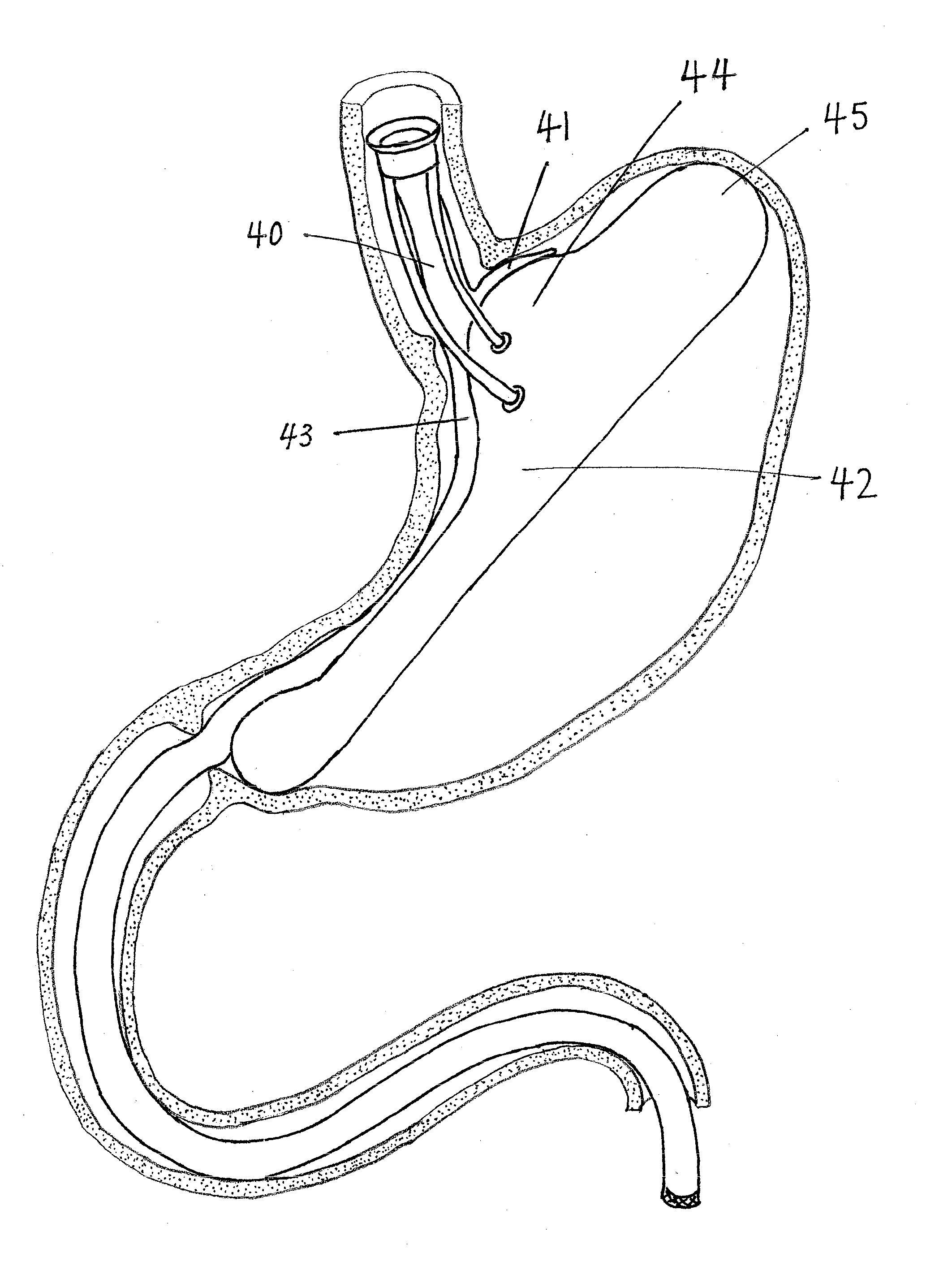
[0009]Configured to secure an anchored bariatric sleeve within the gastrointestinal tract, the intragastric anchor supports the proximal opening of the sleeve in the esophagus proximal to the
gastroesophageal junction such that it does not migrate into the stomach. Since the intragastric anchor will move in a restricted fashion during normal gastric contractility and will likewise move the bariatric sleeve affixed to it, the proximal opening of the anchored bariatric sleeve is preferably slidably coupled to the esophagus by an atraumatic sliding esophageal seal. The combination of elongate intragastric anchor and sliding esophageal seal improves upon stenting, stapling, suturing, and other fixation methods by avoiding the tendency of gastrointestinal tract implants to migrate or to become unremovable through tissue ingrowth and scarring.
[0012]In a first specific aspect of the present invention, an intragastric implant comprises an elongate anchor adapted to extend substantially from the fundus to the pyloric valve in a patient, and a therapeutic or diagnostic device, such as a bariatric sleeve, coupled to the anchor. By “extends substantially from the fundus to the pyloric valve” is meant that the anchor may reach from the proximal stomach in the fundal area to the distal stomach in the antral area. The anchor may be shorter than the stomach but generally reaches from one end to the other, so that the ends sit in the
antrum and fundus but do not necessarily contact the
pylorus or the end of the fundus. The elongate anchor will usually be adapted to remain positioned in the stomach without the need for suturing, stapling, or other forms of attachment. The length and geometry of the anchor will typically be selected to assure that the anchor remains within the stomach without being ejected through the pyloric valve or otherwise adversely affecting the patient. The therapeutic or diagnostic device will be either fixedly or movably coupled to the anchor, and may be coupled at one or more points. In the case of a bariatric sleeve, the bariatric sleeve will be configured to act in a manner similar or analogous to the Roux-en-Y gastric bypass, typically having a central passage with an upper or proximal opening positionable in the esophagus and a lower or distal outlet positionable in the intestines, or in some instances within the stomach. The bariatric sleeve may have a variety of particular configurations: it may be either rigid, flexible, or have portions of each; it may be either straight, curved, or have other combination geometries; and / or it may comprise nestable,
interlocking or hinged links in order to have a shape-lock configuration which facilitates introduction and subsequent reconfiguration within the stomach. The elongate anchor will usually have upper and lower atraumatic ends, where the atraumatic end may be a bulbous geometry, a looped structure, or the like.

