

Drug delivery systems and methods for treating neovascularization
a delivery system and neovascularization technology, applied in the direction of antibody medical ingredients, drug compositions, biocide, etc., can solve the problems of excessive growth of blood vessels and corneal neovascularization, and achieve the effect of effective treatment of corneal neovascularization, reducing abnormal vessels, and reducing pannus formation
- Summary
- Abstract
- Description
- Claims
- Application Information
AI Technical Summary
Benefits of technology
Problems solved by technology
Method used
Image

Examples
example 1
Rapid Drug Clearance from Sub-Tenon Space
[0128]We hypothesized that the lymphatic system and blood vessels present within the conjunctiva and sclera is able to eliminate small and large molecular weight drugs (such as anti-VEGF monoclonal antibodies) from an intraocular (i.e. intra-scleral, such as sub-tenon) site to which a low viscosity, aqueous drug solution is administered. Elimination being to the regional lymph nodes and thence out of the eye.
[0129]We had previously determined that PLGA microspheres (not in a high viscosity vehicle) injected in the sub-Tenon's space cleared rapidly (within six hours) out of the sub-tenon's space, thereby limiting the value of such microspheres to treat an ocular disease.
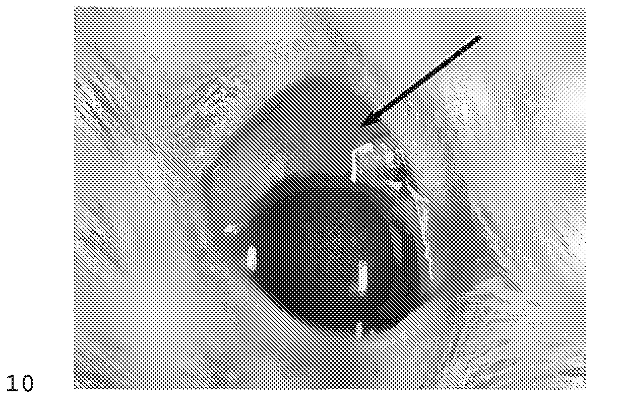
[0130]Thus, to evaluate the clearance mechanisms of an aqueous solution a tracer dye was injected in the sub-Tenon's space in a rabbit eye and the time to disappearance of the dye was determined as follows. A 2-3 kg New Zealand Rabbit was given general anesthesia. The right eye...
example 2
Intraocular Durability of Cross Linked Hvaluronic Acid in Sub-Tenon Space
[0131]An experiment was carried out demonstrating that a cross-linked, polymeric hyaluronic acid has long-term durability and tolerability in the sub-Tenon's space and therefore suitability to act as a drug carrier for sustained drug delivery.
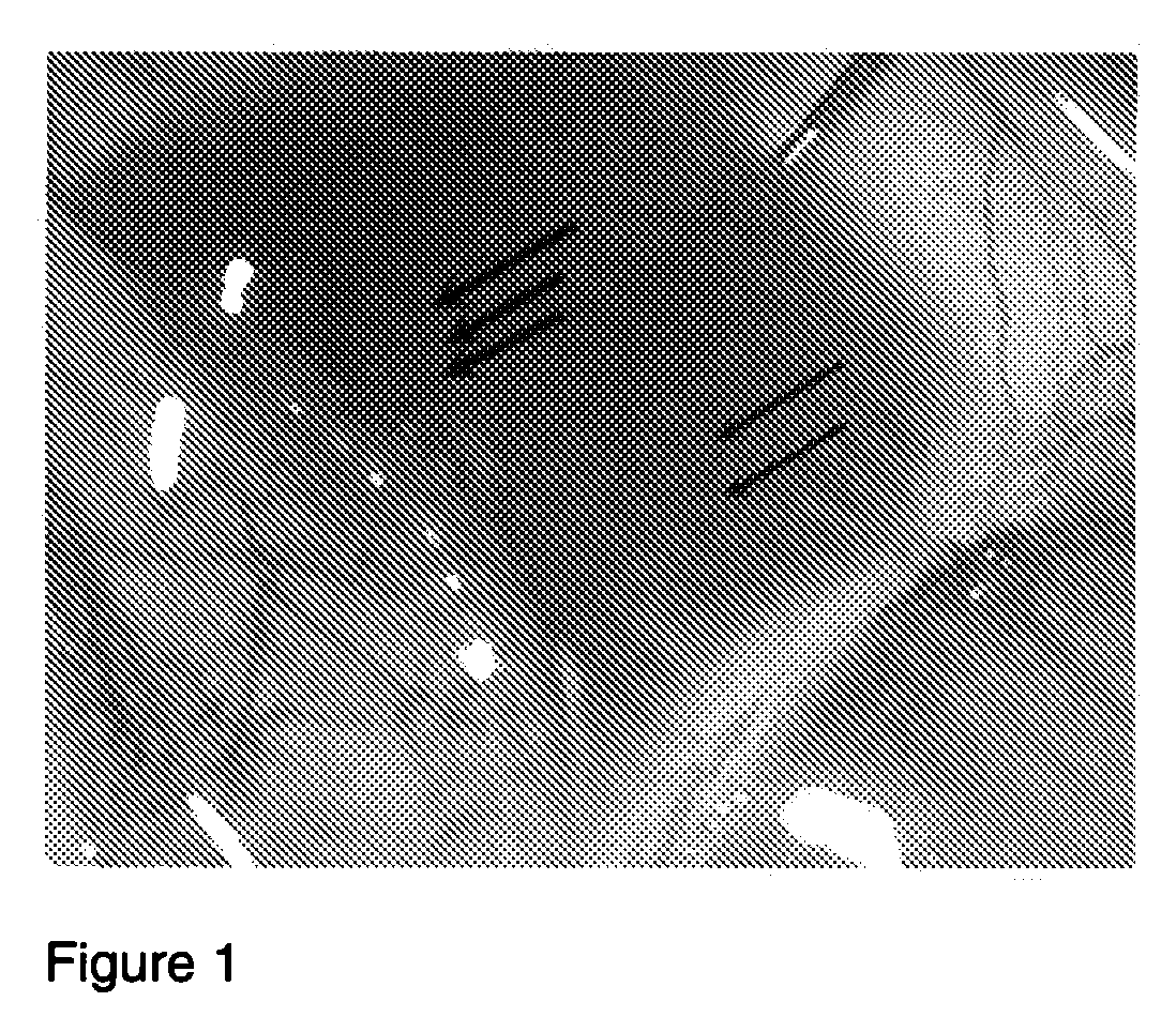
[0132]A 300 gram Sprague-Dawley Rat was placed under general anesthesia and one eye was injected in the sub-Tenon's space with 10 μl of the polymeric hyaluronic acid formulation Juvederm Ultra Plus (Allergan, Irvine, Calif.). The polymeric hyaluronic acid can be used at a concentration of about 20 mg / ml. An alternate polymeric hyaluronic acid which can be used comprises about 95% crosslinked hyaluronic acid and 5% uncrosslinked (free-flowing) hyaluronic acid. The uncrosslinked hyaluronic acid can have an average molecular weight between 600-1,500 kDa, and the cross linked HA component can have an average molecular weight of greater than about 400 kDa. Magnetic resonance im...
example 3
Intraocular Durability of Microspheres in Hyaluronic Acid in Sub-Tenon Space
[0137]An experiment was carried out demonstrating that a polymeric hyaluronic acid (HA) can be used as a vehicle for and can retain microspheres in an intraocular depot formulation over a period of at least a 1 month period.
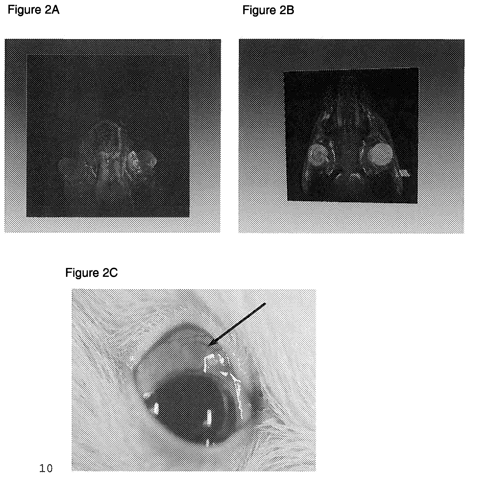
[0138]Thus, a cross linked, polymeric HA (the same HA used in Example 2, that is Juvederm Ultra Plus) was used to investigate its ability to retain surrogate drug microspheres in following injection into the sub-Tenon's space. The experiment was carried out as follows. A 2-3 kg New Zealand Rabbit was given general anesthesia. Colored microspheres were used as surrogate for similar sized microspheres that are used clinically for drug delivery. The microspheres used were “Dye-trak microspheres” with an average diameter of 15 microns, obtained from Triton Technology Inc. as part number 145-0672. The right eye of the rabbit was injected with 15 μm diameter Dye-Trak Microspheres (Triton Tech, ...
PUM
| Property | Measurement | Unit |
|---|---|---|
| Temperature | aaaaa | aaaaa |
| Percent by mass | aaaaa | aaaaa |
| Percent by mass | aaaaa | aaaaa |
Abstract
Description
Claims
Application Information