Separation device for use in the separation, characterization and/or identification of microorganisms

a technology for separation devices and microorganisms, which is applied in the direction of fluorescence/phosphorescence, instruments, and analysis material containers, etc., can solve the problems of bloodstream infections, high morbidity and mortality, and take several days to perform

Inactive Publication Date: 2010-05-13
BIOMERIEUX INC
View PDF27 Cites 53 Cited by
  • Summary
  • Abstract
  • Description
  • Claims
  • Application Information

AI Technical Summary

Benefits of technology

[0010]In another embodiment, the separation device can be sealed, for example, the device can be hermetically sealed. Such devices can provide safety advantages when handling potentially infectious agents. In other possible embodiments, the separation device can provide a means to access the separated, isolated, or pelleted microorganism sample, thereby allowing the sample to be removed from the separation device prior to interrogation, or for additional testing.

Problems solved by technology

Bloodstream infections are associated with high morbidity and mortality, yet current diagnostic methods, of culture followed by biochemical identification and antibiotic susceptibility testing, can take several days to perform.
Typically, empiric therapy is initiated based on clinical symptoms, and test results only impact clinical decisions when the initial therapy fails.
Molecular amplification methods have been proposed to fill this need, but serious challenges to this approach remain.
These direct-from-the-bottle tests are not appropriate for all microorganisms (e.g., Gram-positive cocci), are not validated by the test manufacturers, and generally take 3-8 hours to provide results.
Optical spectroscopy methods, such as intrinsic fluorescence (IF), infrared spectroscopy (FTIR), or Raman spectroscopy, and mass spectrometry methods such as MALDI-TOF, have the potential to allow for identification of microorganisms very quickly, but may encounter interference from the many highly fluorescent and absorptive compounds present in liquid microbiological culture media and in clinical samples such as blood or combinations thereof.
However, these methods have several drawbacks.
The resultant microbial preparation often contains contaminating red blood cells, platelets, lipid particles, plasma enzymes and cellular debris, which can cause poor results in traditional phenotypic ID tests.
These methods are also very labor-intensive and unsafe due to steps which can result in aerosol exposure of potentially dangerous pathogens to the user.

Method used


Image

  • Separation device for use in the separation, characterization and/or identification of microorganisms
  • Separation device for use in the separation, characterization and/or identification of microorganisms
  • Separation device for use in the separation, characterization and/or identification of microorganisms

Examples


example 1

Devices and Methods for the In Situ Identification of Purified Microbial Pellet

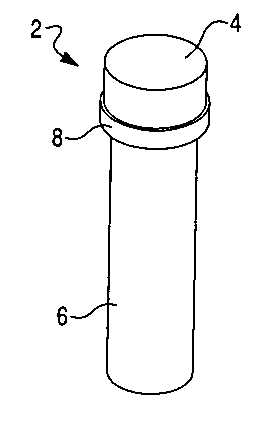
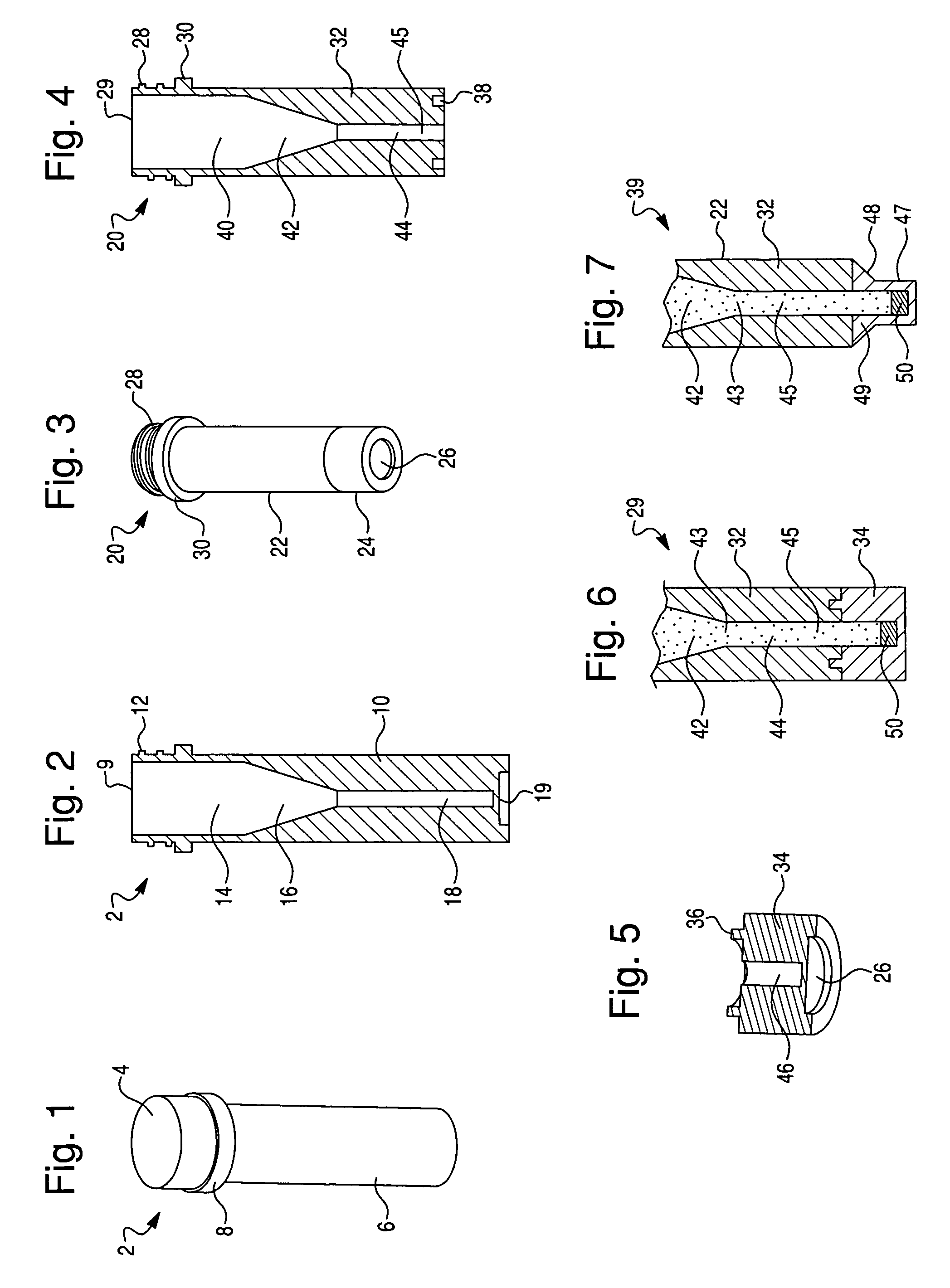
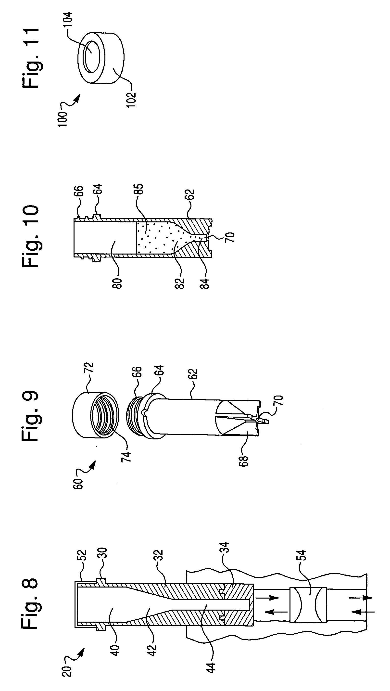
[0050]To explore the potential of the rapid in situ separation and identification of microorganisms in a separation device, several devices were designed and molded from UV-transparent plastic, in accordance with this invention. These devices contained several common features, including a closure, sample reservoir and a tapered optical quality lower region to enable spectroscopic interrogation of the sedimented microbial pellet from below and / or the side, and features that facilitated the coupling of the device to a spectrofluorimeter. The devices must also be capable of withstanding relatively high g-forces during the separation step. Several iterations of this tube were designed to improve microbial recovery, fluorescence reproducibility and reduce contamination by stray scattered light. The tube was also designed to be hermetically sealed.

[0051]Optical interrogation of the sedimented microbial pellet w...


PUM

PropertyMeasurementUnit
volumeaaaaaaaaaa
volumeaaaaaaaaaa
volumeaaaaaaaaaa

Abstract

The present invention is directed to a separation device or container that can be used in the separation, isolation or pelleting of microorganisms from a test samples known to contain or suspected of containing said microorganisms. Subsequently, the separated, isolated or pelleted microorganism sample can undergo one or more interrogation steps to provide measurements useful for the characterization and/or identification of microorganism. In one aspect of the present invention, the interrogation steps can occur in situ in the separation device or container described herein.

Description

CROSS REFERENCE TO RELATED APPLICATION[0001]This application claims the benefit of U.S. Provisional Patent Application No. 61 / 110,187, entitled, “Method and System for Detection and / or Characterization of a Biological Particle in a Sample”, filed Oct. 31, 2008, which is incorporated herein.FIELD OF THE INVENTION[0002]The present invention is directed to a separation device for the separation of microorganisms. In particular, the device of the present invention can be used to separate microorganisms for characterization and / or identification.BACKGROUND OF THE INVENTION[0003]The detection of pathogenic microorganisms in biological fluids should be performed in the shortest possible time, in particular in the case of septicemia for which the mortality remains high in spite of the broad range of antibiotics which are available to doctors. The presence of biologically active agents such as a microorganism in a patient's body fluid, especially blood, is generally determined using blood cu...

Claims


Application Information

Patent Timeline
Patent Type & Authority Applications(United States)
IPC IPC(8): C12M1/34
CPCC12Q1/04G01N21/35G01N21/3581G01N21/47G01N21/359G01N21/65G01N33/6848G01N21/03G01N21/64B01L3/50C12M1/00C12M1/24C12M1/34B01L3/5021B01L2300/042B01L2300/0838B01L2300/0861C12Q1/24C12Q1/37C12Q1/6816
Inventor WALSH, JOHNHYMAN, JONES M.RONSICK, CHRISTOPHERLINK, JOHNROBINSON, RONWILSON, MARK
Owner BIOMERIEUX INC
Why Eureka
Try Eureka