
Gold nanoparticle based protease imaging probes and use thereof

Inactive Publication Date: 2010-05-20
KOREA INST OF SCI & TECH
View PDF1 Cites 18 Cited by
  • Summary
  • Abstract
  • Description
  • Claims
  • Application Information

AI Technical Summary

Benefits of technology

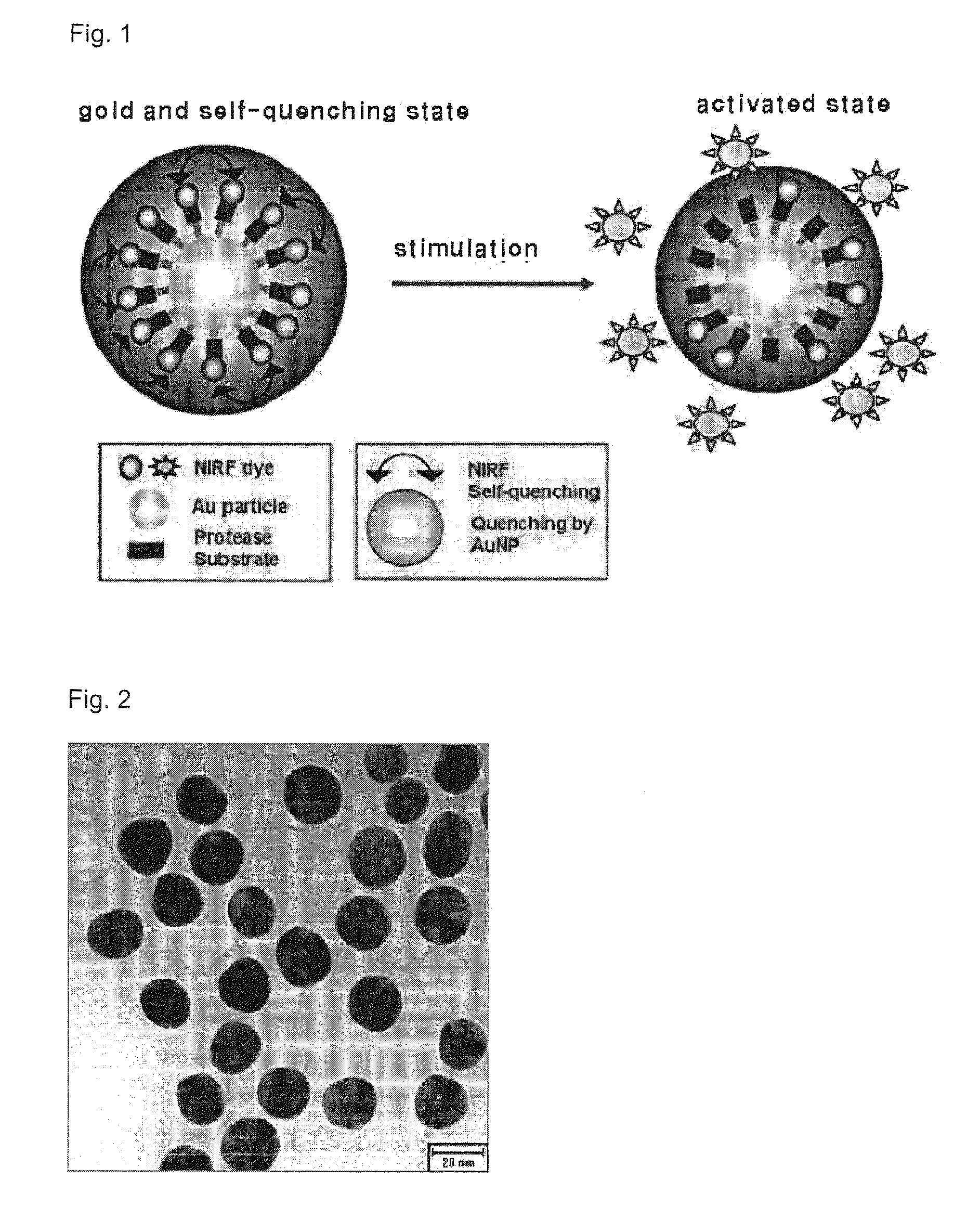
[0016]As described above, the present invention relates to a metal nanoparticle onto which peptide and fluorophore are chemically modified, in order to image the protease expressed in a cell and in a tissue. The metal nanoparticle has excellent bio-compatibility. The peptide is degraded by a specific protease, whereby the fluorophore exhibits fluorescence when the peptide is degraded. Hence, it is allowed for the metal nanoparticle of the present invention to rapidly screen activation and inhibition of the protease by imaging the protease. Also, the metal nanoparticle can selectively be absorbed in a cell and in a tissue, so as to be effectively used in the real-time cell images, the early diagnosis of non-invasive diseases and screening of drugs which inhibit the overexpression of the protease.

Problems solved by technology

For example, deficiency or overexpression of a spatiotemporal protease causes fatal results, such as cancers, arthritides, neurodegenerative diseases, cardiovascular diseases and the like.
However, no technique has not yet given to analyze the amount of specific protease activated and expressed in a quantitative imaging manner or to non-invasively image the degree of protease expressed in vivo, so associated technique development is in urgent need.
Such methods, however, require multi-leveled measurement protocols such that it cannot be efficient to be used in screening many kinds of drugs, like developing new drugs.
Moreover, such methods are not possible to be used in the early diagnosis of diseases because they detect the expression of unique protease in humans or quantitatively analyze the amount of expression thereof.

Method used


Image

  • Gold nanoparticle based protease imaging probes and use thereof
  • Gold nanoparticle based protease imaging probes and use thereof
  • Gold nanoparticle based protease imaging probes and use thereof

Examples


example 1

Preparation of Gold Nanoparticle Exhibiting Specific Fluorescence with Respect to Protease

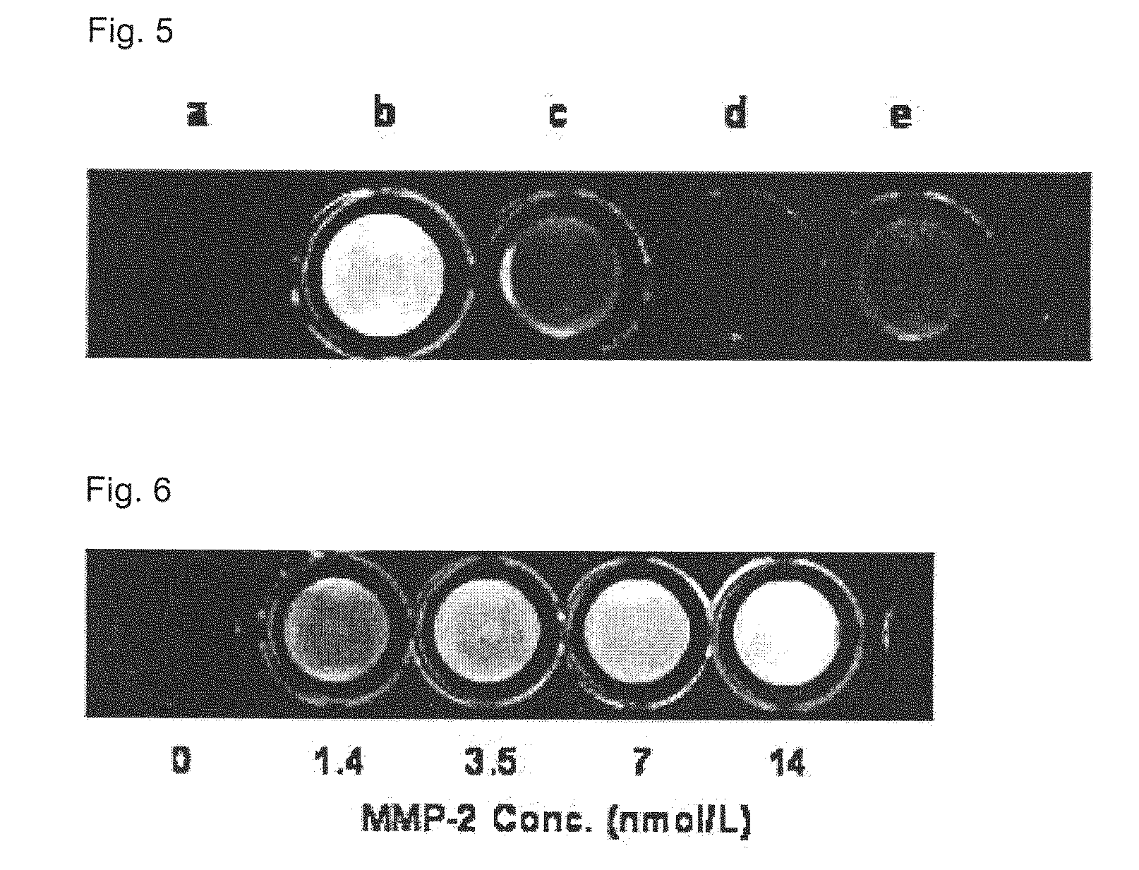
[0039]A gold nanoparticle was prepared in a manner of reducing gold salt (HAuCl4) to sodium borohydride (NaBH4) in an aqueous solution. Peptide-fluorophore derivatives Cy5.5-Gly-Pro-Leu-Gly-Leu-Phe-Ala-Arg-Cys which are specifically degraded by MMP-2 were chemically bonded to the surface of gold, so as to prepare a nanoparticle exhibiting specifically near-infrared fluorescence onto MMP-2.

[0040]1-1. Preparation of Gold Nanoparticle

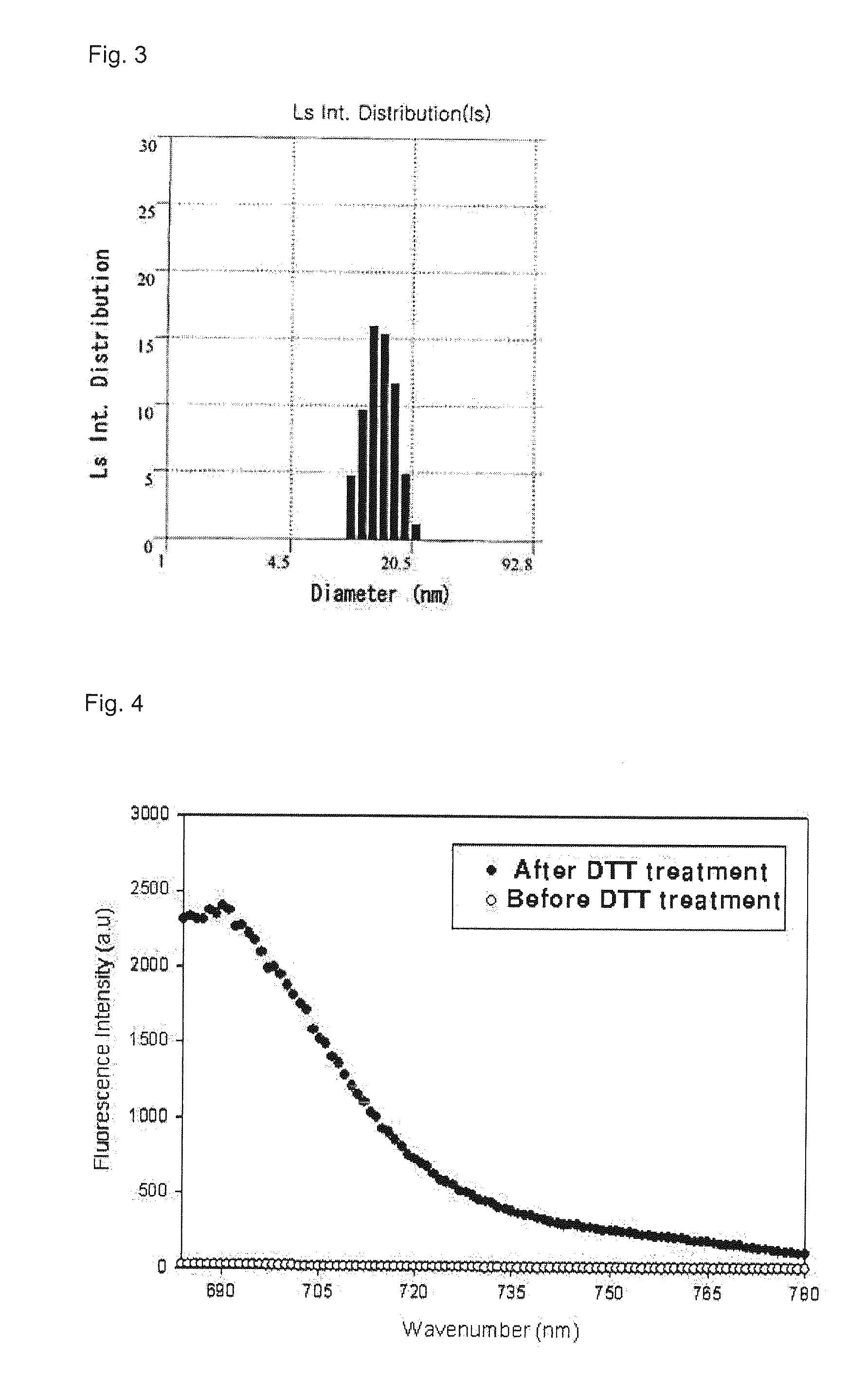
[0041]0.01 g of gold salt (HAuCl4, 3H2O) was slowly mixed into 100 mL of boiling distilled water, and 2 mL of sodium citrate (C6H5Na3O7-2H2O, 1 wt %) was added into the gold salt solution, to prepared gold nanoparticle. The shape and size of the gold nanoparticle were controlled by adjusting the concentration of the gold salt solution and the amount of the sodium citrate. The gold nanoparticle was prepared to have a diameter of 20 μm in the present invention.

[0042]1-2...


PUM


Abstract

Disclosed are a metal nanoparticle onto which a peptide substrate specifically degraded by protease and fluorophore are chemically modified for selectively imaging protease expressed in cell and in tissue in a human body, and the use thereof. Also, a quantitative analysis method of protease using the metal nanoparticle, a cell imaging method and a drug screening method of inhibiting a protease overexpression are provided. In detail, the present invention is directed to a metal nanoparticle having a peptide substrate and fluorophore coupled thereto, the peptide substrate and the fluorophore being specifically degraded by due to a protease activated in various ways in cell and in a human body to exhibit fluorescence. Hence, the metal nanoparticle can be used to rapidly screen activation and inhibition of the protease in the imaging manner. Also, the metal nanoparticle is capable of being selectively absorbed into a cell and a tissue so as to be possibly used as a sensor for real-time cell imaging and early diagnosis of non-invasive diseases.

Description

TECHNICAL FIELD[0001]The present invention relates to a metal nanoparticle having a peptide substrate specifically degraded by a protease and fluorophore coupled to the peptide substrate, capable of selectively imaging protease expressed in cell and in tissue in a human body, and the use thereof.[0002]Also, the present invention relates to a quantitative analysis method of protease using the metal nanoparticle, a cell imaging method, and a drug screening method of inhibiting protease overexpression.BACKGROUND ART[0003]Proteolytic Enzymes (proteases) inhibit proteins activation or their fate based on hydrolysis of irreversible peptide bonds. Examples of such inhibition include the designation of positions of proteins inside or outside a cell, separation of proteins from a cellular surface, activation or deactivation of various types of growth factor hormone, cytokine and protease, conversion of agonist of receptor into antagonist, and the like. Thus, the protease serves to control va...

Claims


Application Information

Patent Timeline
IPC IPC(8): C12Q1/37C07K2/00
CPCA61K49/0056A61K49/0065A61K49/0093A61K49/0032C12Q1/37C01G23/04C04B35/465B82B3/00
Inventor KWON, ICK-CHANCHOI, KUI-WONKIM, KWANG-MEYUNGAHN, CHEOL-HEEYOUN, IN-CHANLEE, SEUL-KIPARK, KYEONG-SOON
Owner KOREA INST OF SCI & TECH
Learn More