Real-time monitoring of age pigments and factors relating to transmissible spongiform encephalopathies and apparatus
- Summary
- Abstract
- Description
- Claims
- Application Information
AI Technical Summary
Benefits of technology
Problems solved by technology
Method used
Image

Examples
examples 1 (
EXAMPLES 1(A) AND 1(B)
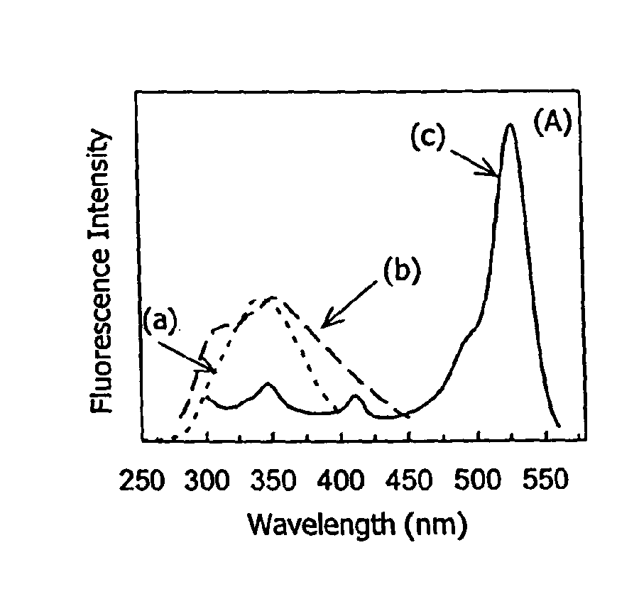
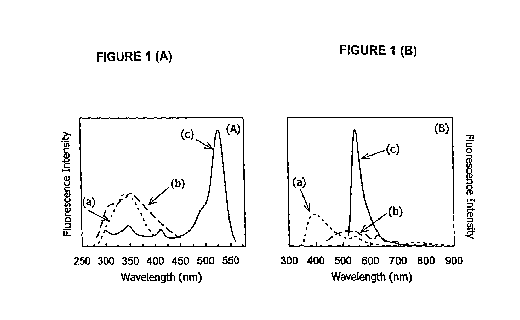
[0039]The present invention can be understood with reference to the spectra of emission and excitation light that are detected when a sample of spinal cord tissue is tested under conditions of varying excitation light or varying emission light. In Examples 1(A) and 1(B), spinal cord tissue is placed in a mixture of chloroform to methanol at a 2:1 volumetric ratio for testing.
[0040]In Example 1(A), the sample of spinal cord tissue extract is exposed to a broad range of excitation wavelengths, and the result emissions are viewed through a monochromator set at a series of fixed emission wavelengths, including (a) 420 nm, (b) 480 nm, and (c) 580 nm. FIG. 1(A) depicts the resulting excitation spectra. For the sake of presenting the graphical data in a viewable form, the excitation spectra corresponding to the 420 nm emission has been scaled by a factor of twenty.
[0041]In Example 1(B), the same sample was exposed to a series of three discrete excitation wavelengths, ...
example 2
[0044]The present invention can also be appreciated from the visual results of the application of one embodiment when it is used to excite a meat substrate (such as a piece of steak) upon which are placed samples of both spinal cord and feces. The steak and feces serve as controls in Example 2.
[0045]In FIG. 2(A), there is shown the visible, non-fluorescence-based image of the meat substrate. The spatial arrangement of the spinal cord and feces on the meat substrate are shown. The arrow at the left points to the spinal cord tissue; the one to the right, the feces.
[0046]Using a CHEM IMAGER 4000, Alphalnnotech Corp., San Leandro, Calif., in conjunction with an actinic blue aquarium light from Energy Savers Unlimited Inc., Harbor City, Calif. fitted with a 430-nm, 10-nm bandpass, interference filter from CVI Laser Corp., Albuquerque, N.M., the sample is exposed and the visual results are presented in FIGS. 2(B)-(C).
[0047]FIG. 2(B) depicts the meat sample when imaged by means of lipofusc...
example 3
[0052]The literature indicates that abnormal TSE prions also display characteristic optical spectra. Our own preliminary data (FIG. 3) indicate that the fluorescent spectra of scrapic-infected sheep brain is substantially different from that of non-infected sheep brain. We hypothesize that this spectral difference is the result of altered lipofuscin and / or prion spectral properties.
[0053]In Example 3, sheep brain tissue, both healthy and infected with scrapie, a form of TSE to which sheep are susceptible, is excited at 280 nm using a front-faced excitation geometry. The resulting fluorescence emission spectra are shown in FIGS. 3(A)-(B), with all spectra normalized to unity at approximately 340 nm.
[0054]FIG. 3(A) depicts the spectra of untreated brain tissue not treated with formalin (a formaldehyde containing solution) and fixed on a glass slide, whereas FIG. 3(B) depicts the spectra of brain tissue treated with formalin and fixed on a glass slide. The region from 520 to 580 nm is ...
PUM
 Abstract
Description
Claims
Application Information
 - R&D
- Intellectual Property
- Life Sciences
- Materials
- Tech Scout
- Unparalleled Data Quality
- Higher Quality Content
- 60% Fewer Hallucinations
Browse by: Latest US Patents, China's latest patents, Technical Efficacy Thesaurus, Application Domain, Technology Topic, Popular Technical Reports.
© 2025 PatSnap. All rights reserved.Legal|Privacy policy|Modern Slavery Act Transparency Statement|Sitemap|About US| Contact US: help@patsnap.com