Nanoparticles for Cytoplasmic Drug Delivery to Cancer Cells

a technology of nanoparticles and cancer cells, applied in the field of nanoparticles, can solve the problems of insufficient selectivity of anticancer drugs for cancer, insufficient cytoplasmic drug delivery to cancer cells, and insufficient cytoplasmic drug delivery, so as to enhance the absorption of nanoparticles, prolong the circulation time, and increase the effect of anticancer drug levels

Active Publication Date: 2010-08-12
UNIVERSITY OF WYOMING
View PDF1 Cites 16 Cited by
  • Summary
  • Abstract
  • Description
  • Claims
  • Application Information

AI Technical Summary

Benefits of technology

[0017]The invention consists of layered nanoparticles for delivering high levels of anticancer drugs to cancer cells. The nanoparticles include an outer shell, an inner core, and an intermediate layer. The core is comprised of the anticancer drug or drugs and polymer chains that are soluble at the lysosomal pH of the cancer cell. The intermediate layer is comprised of polymer chains that are insoluble at the pH of healthy tissue, but soluble at the pH of the cancer interstitium. The outer shell is comprised of water-soluble polymer chains to shield the nanoparticle from recognition by the reticuloendothelial systems so as to provide the nanoparticle with a long circulation time in the bloodstream of a subject. In an alternative embodiment, the outer shell also includes folic acid moieties that enhance the absorption of the nanoparticles by cancer cells that are over-expressing folate receptors.
[0021]Yet another purpose of the invention is to provide nanoparticles with more than one active moiety on the surface to improve uptake of the nanoparticles by cancer cells of different characteristics.
[0022]Still another purpose of the invention is to provide nanoparticles for the rapid release of anticancer drugs to cancer cells that, upon entry into the cancer cell, quickly release the full load of anticancer drugs so as to raise the level of anticancer drug in the cell above the toxic level.

Problems solved by technology

Unfortunately, these anticancer drugs have limited selectivity for cancer and are inherently toxic to both cancer and normal tissues.
As a result, anticancer drugs can cause severe side effects and damage to healthy tissues.
However, it has significant toxicity, in particular, acute as well as chronic nephrotoxicity [von Hoff et al., 1979; Pinzani et al., 1994].
Other common side effects of anticancer drugs include decrease in the number of white blood cells (increasing risk of infection), red blood cells (losing energy) and platelets (risk for bruising and bleeding) as well as nausea, vomiting, hair loss, etc.
Furthermore, the high glomerular clearance of the anticancer drugs leads to an extremely short circulation period in the blood compartment [Siddik et al., 1987].
Most importantly, treatments in conventional dosage form of these drugs may lead to initial cancer regression, but soon the cancer becomes insensitive to the drugs, causing cancer progression and death.
As a consequence of the slowed drug entry but efficient drug removal by the P-gp pumps and the drug consumption by other forms of drug resistance, the effective drug concentration in cytoplasm is well below the cell-killing threshold, resulting in a limited therapeutic efficacy.
Thus, a continuing challenge in cancer treatment is to develop new methodologies that have great drug selectivity for cancer and overcome the cancer drug resistance to simultaneously enhance the therapeutic efficacy and reduce toxicity to healthy tissues.
In addition, cancer also has much fewer lymphatic capillaries than healthy tissues, such that the lymphatic drainage of macromolecules from cancer tissues is inadequate.
First, the premature burst release of drugs in bloodstream is a general problem of existing nanoparticle drug carriers.
A typical drug-release profile of nanoparticles suggests that the nanoparticles would immediately release drug into the bloodstream upon intravenous administration and thus only a portion of drugs reach the tumors, causing non-targeted drug release, low drug efficiency, toxicity to healthy tissues and less drug being available to cancer [Liu et al., 2001].
Such nanoparticles, however, showed low or completely no anticancer activity [Yokoyama et al., 1998], because chemically bound DOX is not released due to the absence of hydrolysable link between the drug and the polymer chains of the core.
A second issue is their slow drug release.
The cores of existing nanoparticles are made of solid polymers and the drugs have to diffuse from the core to the outside and thus the drug release is inevitably slow.
A third issue of the nanoparticles is their slow cellular uptake by cancer cells.
Because cancer cells have the P-gp pumps located in the membrane that can effectively transport the drugs out of the cell while they are in the membrane, drugs released in cell interstitium cannot effectively enter the cell plasma through the membrane.
Nevertheless, nanoparticles functionalized with folic acid only may not be sufficiently effective to overcome the cancer drug resistance.
Thus, the internalization of nanoparticles only via FR-mediated endocytosis (FR-Endoc) may not be fast enough to build up a cytoplasmic drug concentration exceeding the capacity of the cell's drug resistance.
Furthermore, not all cancers express folate receptors.
These cells cannot effectively take up the folic acid-functionalized nanoparticles and thus survive the treatment, causing relapse.
The drug concentration in cancer cells is thus still lower than the cell-king threshold concentration and cannot effectively induce cell death.

Method used


Image

  • Nanoparticles for Cytoplasmic Drug Delivery to Cancer Cells
  • Nanoparticles for Cytoplasmic Drug Delivery to Cancer Cells
  • Nanoparticles for Cytoplasmic Drug Delivery to Cancer Cells

Examples


example 1

Slow- and Fast-Release Nanoparticles with a Core and a Single-Layer Corona

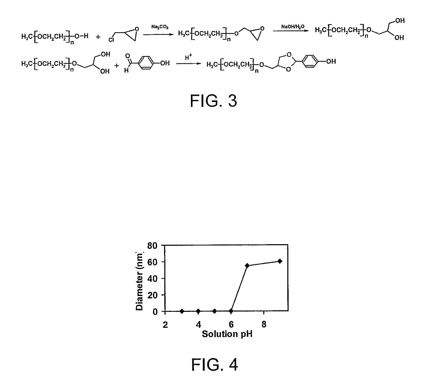
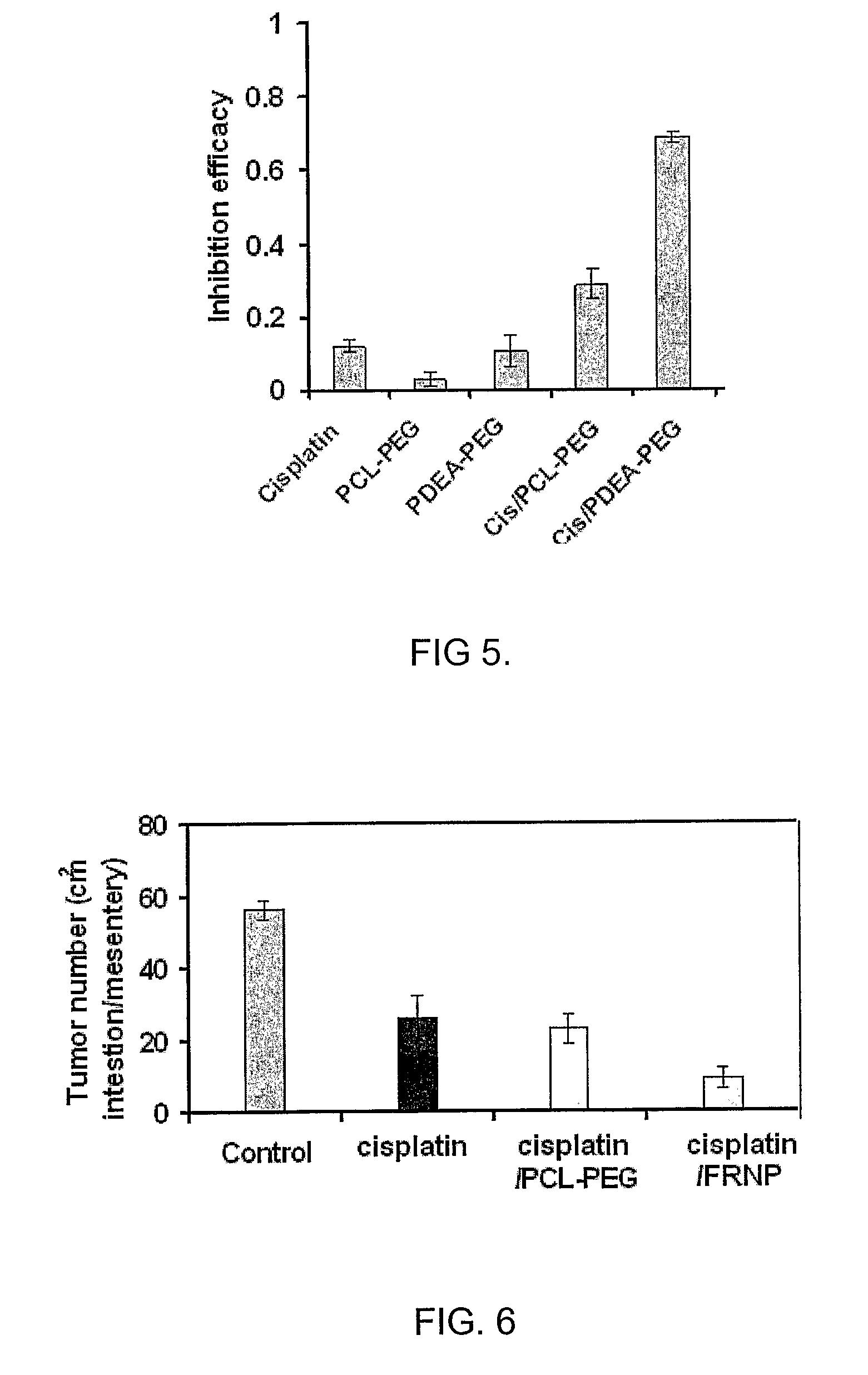
[0041]An example of a fast-release single-layer nanoparticles are nanoparticles with pH-responsive poly[2-(N,N-diethylamino)ethyl methacrylate) (PDEA), synthesized from PDEA-block-poly(ethylene glycol) (PDEA-PEG) copolymer using a solvent-displacement (acetone-water) method. On the other hand, nanoparticles with pH-resistant poly(ε-caprolactone) (PCL) cores, synthesized from PCL-block-PEG (PCL-PEG), is an example of slow-release single-layer nanoparticles. We provide the latter as a point of reference for evaluating the rate of release.

example 2

Cationic-Charged Functionalized Nanoparticles, for Example, Tumor-Cell-Triggered Instant-Intracellular-Drug-Releasing (IIDR) Nanoparticles

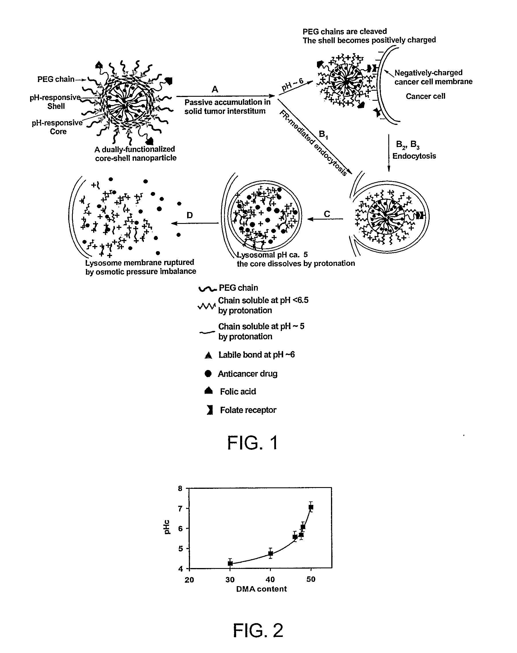
[0042]The tumor-cell-triggered instant-intracellular-drug-releasing (IIDR) nanoparticles are characterized in vitro and in vivo and evaluated for enhanced cancer chemotherapy by overcoming cancer drug resistance. An example of a nanoparticle according to the present invention and its action are shown in FIG. 1.

[0043]The IIDR nanoparticle has a three-layer-onion structure with a diameter around between about 10 and 500 nm, preferably between about 50 and 300 nm and most preferably between about 75 and 150 nm. Its core-surface or shell-surface is crosslinked for improved stability. The outer layer consists of water-soluble polymer chains. These chains shield the nanoparticle from RES recognition and give the nanoparticle a long circulation time in bloodstream [Kwon, 1998; Brigger et al., 2002]. The water-soluble chains are linked to the shell with a...

example 3

Folic Acid, Cationic-Charged Dually-Functionalized Fast-Release Nanoparticles

[0046]An alternative embodiment of the invention makes use of the folic acid moieties to enhance the uptake of the dually functionalized nanoparticles by the cancerous cells. The dually functionalized nanoparticle has a three-layer-onion structure with folic acid groups tethered on the outer layer of the nanoparticles shown in Example 2.

[0047]After intravenous administration, the nanoparticles extravasate and are trapped in the tumor interstitium (FIG. 1A). Subsequently, three endocytosis processes may occur: 1) The folic acid moieties on the nanoparticle surface may bind folate-receptors on cell membranes and trigger FR-Endo (B1); 2) In the acidic tumor interstitium, the acid-labile bonds (▴) begin to break and the PEG chains are shed off from the nanoparticle surface to expose the shell layer, the shell layer is positively charged at this pH and thus adsorbed on the negatively-charged cell membrane, leadi...


PUM

PropertyMeasurementUnit
sizeaaaaaaaaaa
sizeaaaaaaaaaa
sizeaaaaaaaaaa

Abstract

The invention is a nanoparticle that contains an anticancer drug that is released in cancer cells when administered to a subject. The nanoparticles have a core including the anticancer drug and polymer chains that are soluble at the pH of the cancer cell. The core is surrounded by a layer of polymer chains that are insoluble at the pH of healthy tissue but soluble at the pH of the cancer interstitium. An outside layer is made of water-soluble polymer chains to shield the nanoparticle from RES recognition and give the nanoparticle a long circulation time in the bloodstream of the subject. The outside layer may also include folic acid moieties that bind folic acid receptors on the surface of the cancer cell.

Description

[0001]This application claims priority to U.S. Patent Application Ser. No. 60 / 643,623 filed Jan. 13, 2005, and U.S. Patent Application Ser. No. 60 / 608,599 filed Sep. 10, 2005.BACKGROUND OF THE INVENTION[0002]1. Field of the Invention[0003]The invention relates generally to nanoparticles that are used to deliver drugs into the cytoplasm of cancer cells and, more specifically, to the rapid delivery of the drugs by nanoparticles that have an inner core containing an anticancer drug and a polymer that is soluble in cancer cells.[0004]2. Background of the Art[0005]Cancer is the second leading cause of death in the United States. Each year more than 1.2 million Americans are diagnosed with cancer, and less than half can survive five years. Annual medical costs for cancer treatment account for billions of dollars in the US alone. Chemotherapy, which uses chemical agents (anticancer drugs) to kill cancer cells, is one of the primary methods of cancer treatment. Unfortunately, these anticanc...

Claims


Application Information

Patent Timeline
Patent Type & Authority Applications(United States)
IPC IPC(8): A61K9/14A61K33/24A61P35/00
CPCA61K9/5146A61K9/1075A61K9/51A61K9/5021A61K9/5073A61K9/5138A61P35/00
Inventor RADOSZ, MARCIEJXU, PEISHENGSHEN, YOUQING
Owner UNIVERSITY OF WYOMING
Why Eureka
Try Eureka