
Method and apparatus for ocular surface imaging

a technology of ocular surface and ocular surface, applied in the field of methods, can solve the problems of invariability and reliability of these assessments when performed by individual physicians, the invariance of purkinje image with image analysis and feature detection, and the difficulty of quantitative image analysis of ocular surface images

Active Publication Date: 2010-08-12
ALCON INC
View PDF15 Cites 45 Cited by
  • Summary
  • Abstract
  • Description
  • Claims
  • Application Information

AI Technical Summary

Benefits of technology

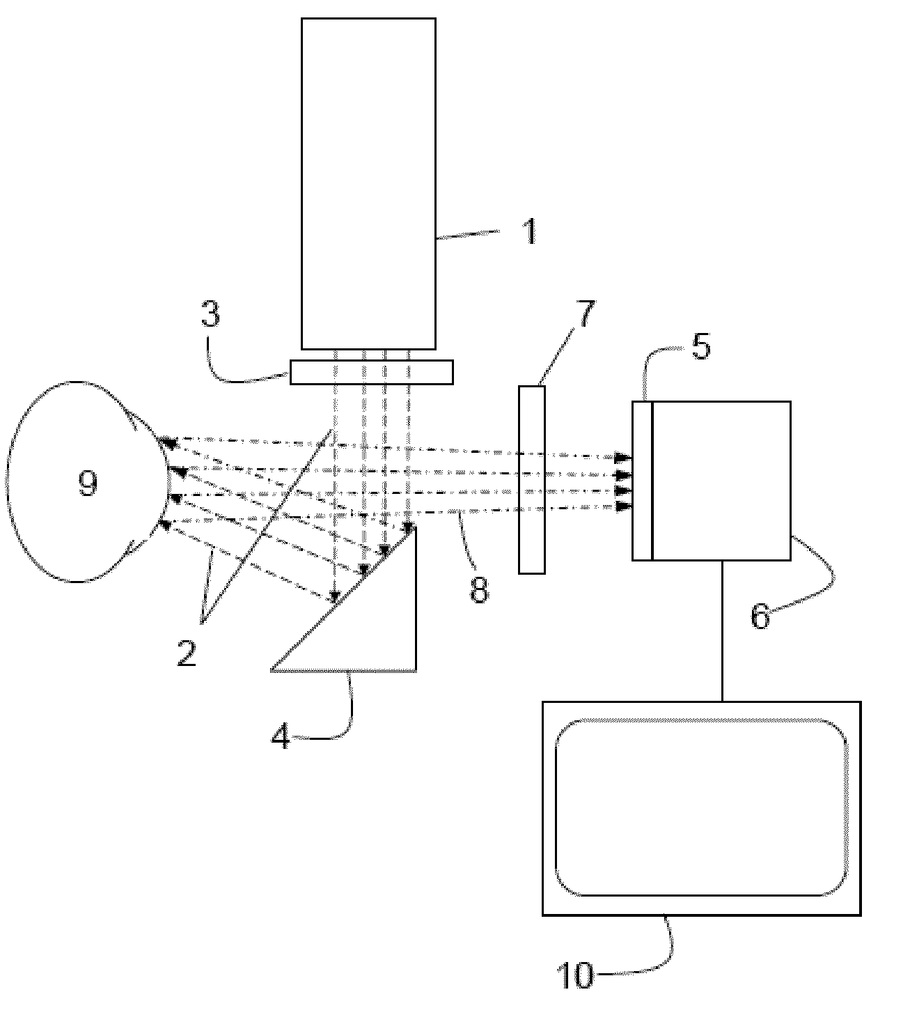
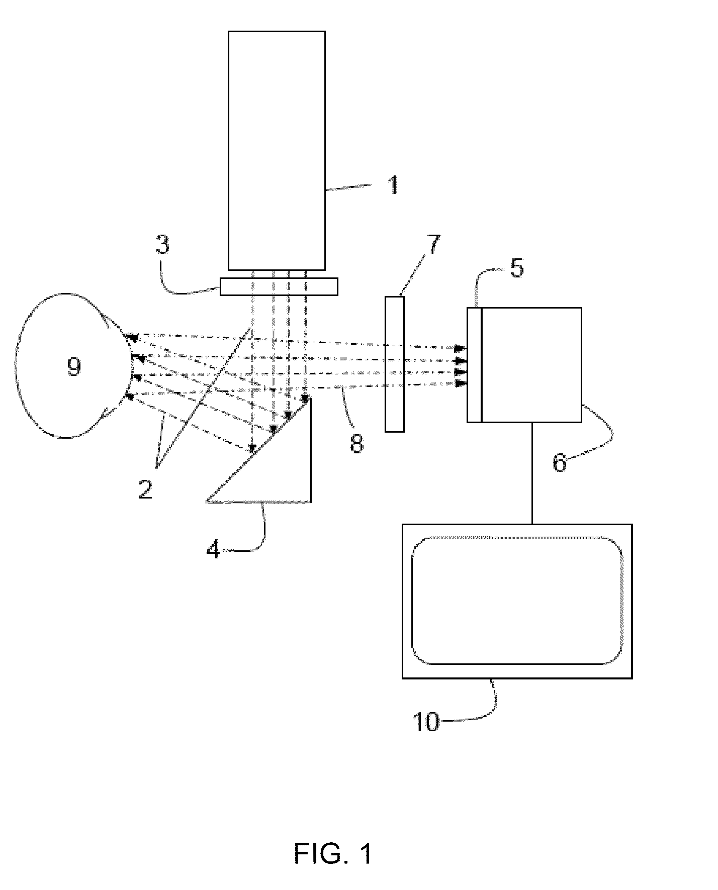
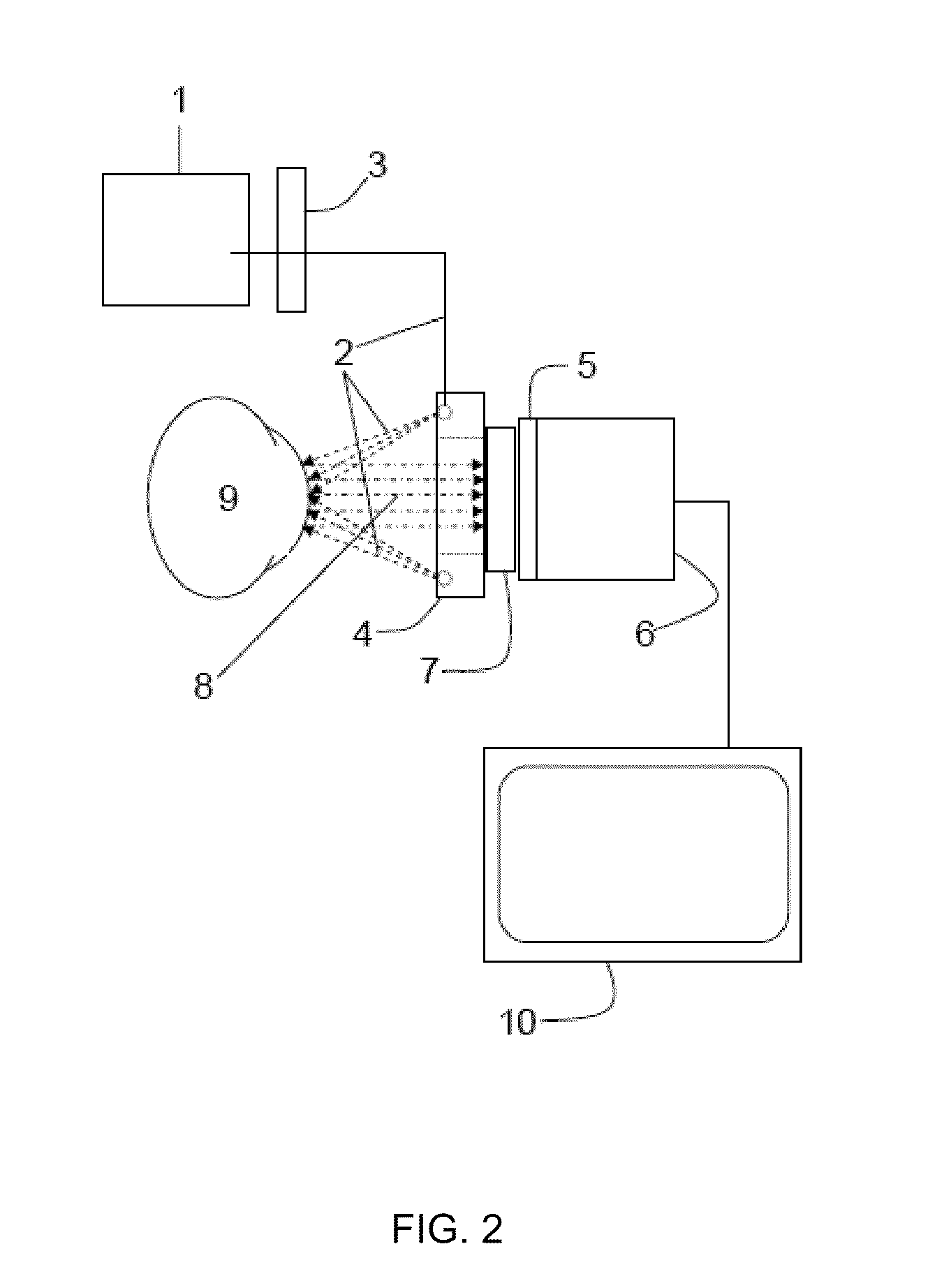
The invention provides methods and apparatus for detecting ocular surface diseases using a light source, illumination filter, and imaging filter. The light source illuminates the ocular surface and the illumination filter transmits light in a specific wavelength range to excite a contrast agent bound to defects on the ocular surface. The imaging filter then detects a light signal emitted from the contrast agent and transmits it to an image capture device. The image capture device analyzes the intensity and distribution of the contrast agent to provide quantitative data on the disease. The invention can be used to detect various ocular surface diseases such as glaucoma, corneal abrasions, and dry eye.

Problems solved by technology

However, the variability and reliability of these assessments has been poor when performed by individual physicians.
Heretofore, quantitative image analysis of ocular surface images has proven difficult for at least three reasons.
The Purkinje image invariably interferes with image analysis and feature detection.
Since it is not constant from one image to the next, such specular images are impossible to control for and must be removed prior to image analysis.
A second difficulty in accurately detecting and measuring corneal stain relates to the methods used to capture corneal images.
Hence any given stained corneal image from such a system can not possibly have all stained objects in sharp focus for subsequent analysis.
A third difficulty in accurately detecting and measuring corneal stain relates to the methods used to separate surface stained objects from surrounding image elements for subsequent measurement.

Method used


Image

  • Method and apparatus for ocular surface imaging
  • Method and apparatus for ocular surface imaging
  • Method and apparatus for ocular surface imaging

Examples


example 1

Spectral Filtering Removes Purkinje Images of the Illumination Source from Images of Specular Surfaces

[0053]The ability to spectrally remove the specular or Purkinje image from ocular surface fluorescence images was demonstrated with an artificial cornea prepared from a hard contact lens. For this a hard contact lens made of polymethylmethacrylate was coated with green fluorescent beads, (Bangs Laboratories, Inc, Fishers, Ind.) to simulate stained spots on a curve corneal surface. The spectral properties of these beads were similar to fluorescein. The artificial cornea was position in front of a Nikon digital single lens reflex camera and illuminated with a fiber optic ring light mounted to the lens of the camera. The fiber optic ring illuminator was configured such that band pass filters could be used to spectrally define the light emanating from the ring. Images were collected through either a green bandpass filter optimized for fluorescein emission wavelengths, (532 + / −45 nm) or ...

example 2

Spectral Filtering Removes Purkinje Images of the Illumination Source from Images of the Ocular Surface

[0055]A severe dry eye patient and a non-dry eye patient were subjected to corneal staining with 5 μL of 2% sodium fluorescein for 3 minutes, followed by a saline wash (Unisol 4 Saline Solution, Alcon Laboratories, Inc., Fort Worth, Tex.). The stained eyes were imaged with a Haag-Streit BX-900 slit lamp. The Haag-Streit slit lamp consisted of a stereo biomicroscope, a combination flash lamp and modeling lamp for illuminating the eye and a Canon 40D digital camera and computer for image capture. The slit lamp was modified by the addition of a fluorescein excitation bandpass filter to the flash lamp output and the insertion of a fluorescein emission filter in the light path to the between the eye and the camera. The fluorescein excitation filter had a bandpass (transmission>90%) of 483 + / −32 nm and the emission filter had a bandpass of 536 + / −40 nm. The out-of-band optical densities ...

example 3

Fourier Analysis of Ocular Surface Defects of a Dry Eye Patient

[0057]The effect of the Fourier bandpass filtering process was illustrated in FIG. 5 and FIG. 6. The fluorescein corneal image in FIG. 5A was obtained with a camera based imaging system equipped with fluorescein spectral band pass filters as described above. The two dimensional Fourier transform of the image in FIG. 5A was then calculated using an unpadded fast Fourier transform algorithm found in most image analysis software. The transform was then multiplied by the Fourier transform of a low pass Gaussian filter and the inverse transform of the product was calculated to produce the image in FIG. 5B. As expected, the Fourier processing removed the low background brightness of the conjunctiva and background stain leading to an enhancement of the small speckled stained structures in the cornea (compare image in FIG. 5A and FIG. 5B).

[0058]The utility of the imaging method and the Fourier bandpass filtering process in measu...


PUM


Abstract

The invention provides apparatuses and methods for detecting corneal surface defects. The methods and / or an apparatus of the invention can be used to detect corneal surface diseases, such as dry eye.

Description

CROSS-REFERENCE TO RELATED APPLICATION[0001]This application claims priority under 35 U.S.C. §119 to U.S. Provisional Patent Application No. 61 / 151,880 filed Feb. 12, 2009, the entire contents of which are incorporated herein by reference.FIELD OF THE INVENTION[0002]The invention relates to apparatuses and methods for detecting corneal surface defects. An apparatus and method of the invention can be used to detect corneal surface diseases, such as dry eye.BACKGROUND OF THE INVENTION[0003]The diagnosis and prognosis of ocular surface diseases, such as keratoconjunctivitis sicca (dry eye), involves several tests. One of the tests includes staining the ocular surface with a dye, followed by visual examination for stained surface defects. Lesions in the epithelial cell layers of the cornea adsorb stain and allow for the penetration of stain into interstitial spaces. The resultant bright speckled appearance of the otherwise black cornea is a hallmark pathology of cornea disease. Visual e...

Claims


Application Information

Patent Timeline
Patent Type & Authority Applications(United States)
IPC IPC(8): A61B6/00
CPCA61B3/101A61B5/0059G06T7/0012G06T2207/30041G06T2207/10056G06T2207/10064G06T2207/10012A61B3/107A61B3/14
Inventor ORNBERG, RICHARD L.WILLIAMS, GARY W.
Owner ALCON INC
Learn More