
Compositions and methods for joining non-conjoined lumens

Active Publication Date: 2010-08-12
TAUTONA GROUP
View PDF74 Cites 5 Cited by
  • Summary
  • Abstract
  • Description
  • Claims
  • Application Information

AI Technical Summary

Benefits of technology

[0027]This invention is directed to the discovery of novel thermoreversible sol-gel compositions for use to stabilize the geometry of the distal portions of tubular tissues or lumens to facilitate joining such non-conjoined lumens. The novel compositions of this invention are the result of an unexpected discovery that

Problems solved by technology

Because of the complexity and judgment required in suturing, automated techniques are not well accepted.
Calcified and diseased vessels provide mechanical challenges.
Sutures may, in some instances, cause a reaction resulting in long term stenosis or fibrosis.
Frequently, this is troublesome to the practitioner because the end of an tubular tissue—such as a clamped blood vessel devoid of blood—is not a perfectly round circle; rather it is in its unpressurized, deflated-looking state where a cross-sectional view of the circumference may be a circle, an oval or irregular, and, of course having no structural support from within, is unstable in any shape (unless the surrounding tissue possesses structural strength).
Microvascular anastomosis is often performed by hand under a microscope, and is tedious and painstaking work.
As a result, it can take many hours to complete just the microvascular anastomosis required to reconnect a severed body part or transplant an organ.
This system, however, is limited to use with two blood vessels having substantially the same diameter.
Further, manual eversion of a blood vessel having a diameter on the order of one millimeter is difficult and painstaking, particularly when the eversion is to be substantially even around the circumference of the ring.
This can slow or stop blood flow through the heart's blood vessels, leading to chest pain or a heart attack.
Multiple practitioners, additional complexity, and, as a practical matter, additional health care cost is involved over surgical procedures involving fewer practitioners and procedures.
Moreover, blood quality may be degraded as the heart-lung machine repetitively pumps the patient's blood through the systemic circulation.
The blood may embolize or clot in the distal circulation, or form clots which migrate to the distal vasculature, and cause a stroke.
Frequently, because the graft must be performed on arteries in locations directly affected by the beating heart, stabilizing mechanisms are not thoroughly effective, and the practitioner must suture the graft while the graft is moving in conjunction with the heart beat, at least to some extent.
Thus, the graft quality may be compromised.
After an hour, the risk of patient morbidity is thought to increase perhaps due to the heart-lung machine degrading the quality of the blood as it is circulated through the systemic circulation.
Bypass surgeries, however, often last three hours or longer.
Moreover, where the aorta is clamped and blood therefore cannot pass through, the blocked blood is thought to cause additional issues.
Anastomosis is time-consuming.
In “off pump” procedures where the heart remains beating, the difficulty of suturing an anastomosis graft on a moving surface of the heart may degrade the quality of such grafts completed on patients.
It can be appreciated that some of the sutures are easily made from on top of the conduit or blood vessel graft, while others are more difficult to complete as they are beneath the conduit.
It can be further appreciated that performing such complex suturing procedures on a moving platform, such as the beating heart, further increases the difficulty associated with such suturing procedures.
Improperly connecting blood vessel grafts to the patient may present substantial post-operative complications and / or increase operating room time spent correcting the improperly connected graft.

Method used


Image

  • Compositions and methods for joining non-conjoined lumens
  • Compositions and methods for joining non-conjoined lumens
  • Compositions and methods for joining non-conjoined lumens

Examples


example 1

Preparation and Property Determination of a Sol-Gel Composition of this Invention

[0282]Presented here is a protocol for preparing 100 mL (100 g) of a thermoreversible sol-gel composition of this invention.

[0283]Materials:[0284]Phosphate buffered saline (“PBS”): 64 g, pH 7.4, with Ca2+ and Mg2+;[0285]Poloxamer 407 (BASF Pluronic® F 127): 14.4 g;[0286]Poloxamer 188 (BASF Pluronic® F 68): 21.6 g.

[0287]Procedure:

[0288]The above materials were mixed at 4° C. under high shear to facilitate dispersing the solid poloxamer in the solvent. The heterogenous mixture is then rocked at 0.5 Hz and 4° C. for at least 12 hours to allow for complete dissolution of the poloxamer in the solvent. The mixture is then further stirred to ensure homogeneity.

[0289]Water or other aqueous solutions may replace PBS as the solvent. It is preferred that the solvent has an ionic concentration and pH that are close to that of the body fluid so that the resulting thermoreversible sol-gel compositions do not cause su...

example 2

Anastomosis Procedure Using a Sol-gel and Cyanoacrylate Glue

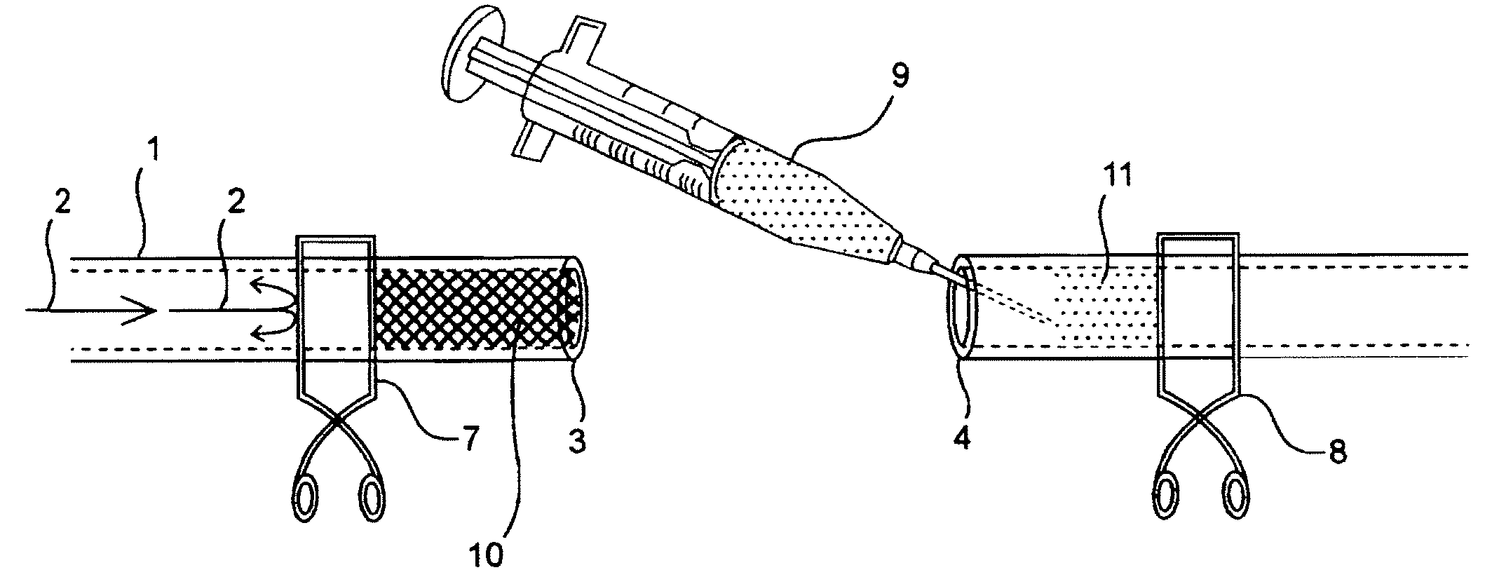
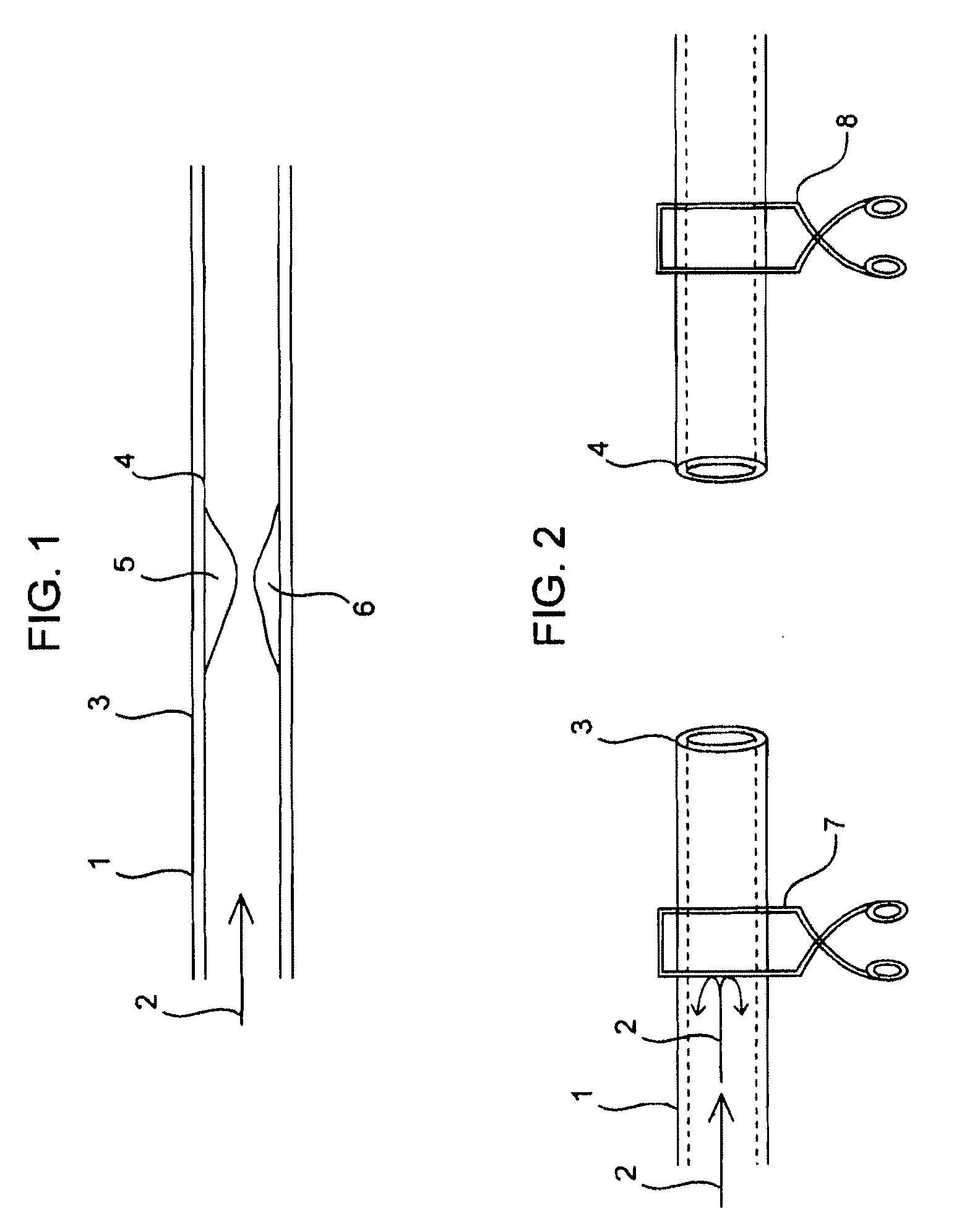
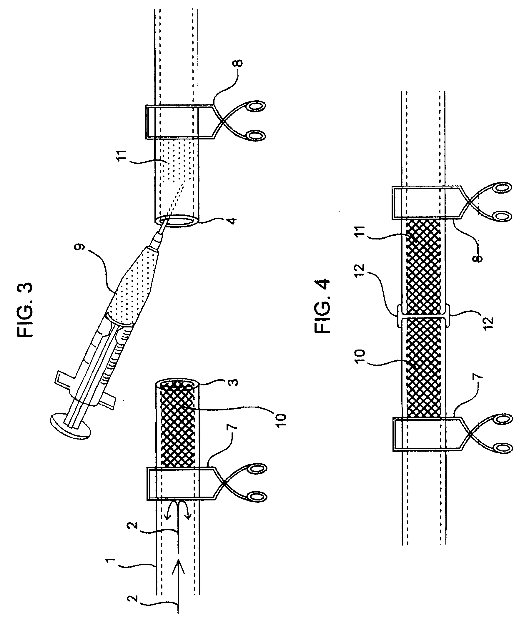
[0295]Dogs, which were treated and cared for in accordance with all applicable laws and regulations, and in accordance with good laboratory practices, were anesthetized and placed in dorsal recumbency. The femoral artery and vein were exposed bilaterally, and the peri-adventitial tissues on both artery and vein were cleaned off at the site of anastomosis. Two clamps were placed on the artery, distal and proximal to the target vessel site, and one clamp was placed on the vein, proximal to the target vessel site. The vein was ligated and incised distally and an arteriotomy was made with an arteriotomy punch. The target vessels were flushed with heparinized saline to remove blood and the vessel ends were approximated with four stay sutures. The surgical area was heated to about 35-45° C. and a sol-gel composition of this invention was warmed to about 35-45° C. A sol-gel composition, Composition 2, was injected into the lumens ...

example 3

Anastomosis Procedure Using a Sol-Gel and Biological Glue

[0296]Dogs, which were treated and cared for in accordance with all applicable laws and regulations, and in accordance with good laboratory practices, were anesthetized and placed in dorsal recumbency. The femoral artery and vein were exposed bilaterally, and the peri-adventitial tissues on both artery and vein were cleaned off at the site of anastomosis. Two clamps were placed on the artery, distal and proximal to the target vessel site, and one clamp was placed on the vein, proximal to the target vessel site. The vein was ligated and incised distally and an arteriotomy was made with an arteriotomy punch. The target vessels were flushed with heparinized saline to remove blood and the vessel ends were approximated with four stay sutures. The surgical area was heated to about 35-45° C. and a sol-gel composition of this invention was warmed to about 35-45° C. A sol-gel composition, Composition 2, was injected into the lumens of ...


PUM

PropertyMeasurementUnit
Fractionaaaaaaaaaa
Fractionaaaaaaaaaa
Fractionaaaaaaaaaa

Abstract

Disclosed are compositions, methods, and kits for joining together non-conjoined lumens in a patient's body including vascular lumens. More particularly, in various aspects, this invention provides compositions, methods, and kits for joining such non-conjoined lumens, including small lumens typically requiring microsurgical technique.

Description

FIELD OF THE INVENTION[0001]This invention relates generally to compositions, methods, and kits for joining together non-conjoined lumens in a patient's body including vascular lumens. More particularly, in various aspects, this invention provides compositions, methods, and kits for joining such non-conjoined lumens, including small lumens typically requiring microsurgical techniques.BACKGROUND OF THE INVENTION[0002]Non-conjoined lumens arise in a variety of settings including surgical settings where the non-conjoined lumens are intentionally created or arise from lacerations or puncture wounds. Intentionally created non-conjoined lumens include those arising during surgical repair of e.g., treatment of a blockage in a lumen by a bypass procedure, attaching a synthetic graft or during free tissue transfer in cosmetic surgical settings. Anastomosis is conducted to surgically reconnect the open ends of the lumen. Examples of anastomosis procedures include anastomotic procedures on the...

Claims


Application Information

Patent Timeline
IPC IPC(8): A61B17/11A61K47/10C07C43/13A61K47/04
CPCA61L24/001A61L24/046C08L71/02
Inventor RAPPLEYE, C. TRAVISRIMSA, JOSEPHHOFFMANN, DREW A.
Owner TAUTONA GROUP
Learn More