Endothelial scaffolds

a technology of endothelial cells and scaffolds, applied in the field of endothelial cells, can solve the problems of corneal opacity improvement, corneal thickening, and patient wearing thick glasses for correction, and achieve the effect of reducing the risk of stroma formation

Active Publication Date: 2010-08-26
WAKE FOREST UNIV HEALTH SCI INC
View PDF13 Cites 51 Cited by
  • Summary
  • Abstract
  • Description
  • Claims
  • Application Information

AI Technical Summary

Problems solved by technology

When this spreading is inadequate, vision-impairing corneal opacity results.
Problems encountered from the stromal thickness include unpredictable refractive error, which may require the patient to wear thick glasses for correction.
Presence of the stroma can also create interface haze or scarring.

Method used


Image

  • Endothelial scaffolds
  • Endothelial scaffolds
  • Endothelial scaffolds

Examples


example 1

Cornea Decellularization

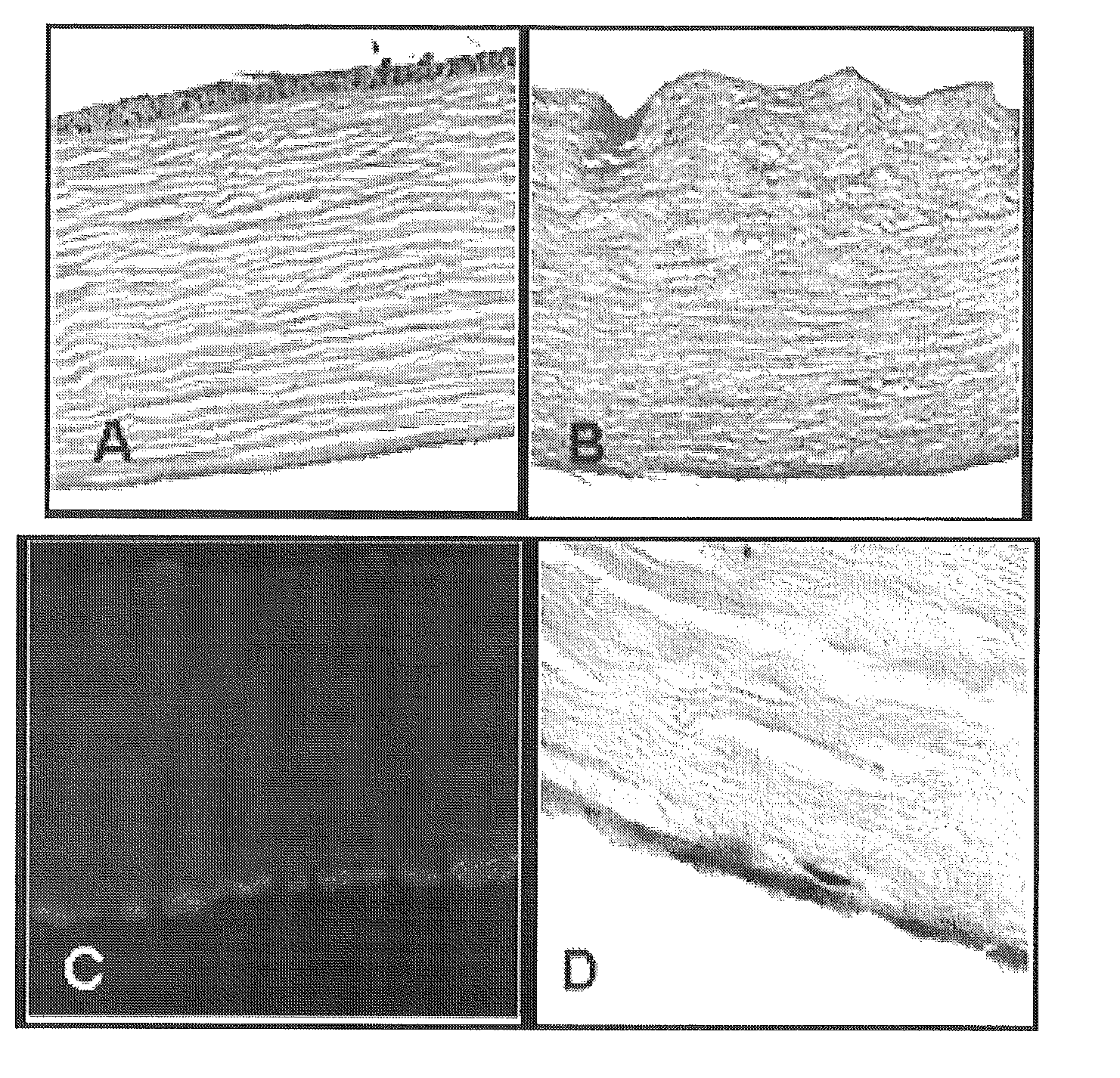
[0059]Corneal scaffolds were made using a microtome to cut whole corneas (obtained from the eye bank) into 125 micron-thick slices cut parallel to the endothelial surface. These corneal slices were placed in decellularization solution (2% Triton X100, 0.1% NH4OH) for 3 days at 4° C. A native cornea (A) and a decellularized cornea (B) are shown in FIG. 1. The decellularization process removed the epithelial, endothelial, and 95% of the stromal cells. These decellularized sections were stored in PBS and kept at a 4° C. until they were used for cell seeding experiments.

example 2

Corneal Endothelial Cells (CECs)

[0060]Human CECs were collected from the basal surface of the limbus of cadaver corneas of donors 14-75 years old. The corneas were placed in a petri dish containing 0.02 g collagenase II in 10 mL PBS for each rim and incubated at 37° C. for 90 minutes. Sterile forceps and a spatula were then used to gently scrape the scleral rim of Descemet's membrane with intact endothelium. Cells removed by this process were centrifuged for 5 minutes at 1500 rpm and then resuspended in 2.5 mL EGM-2 complete culture medium containing 10% FBS and plated in the wells of a 6-well tissue culture dish.

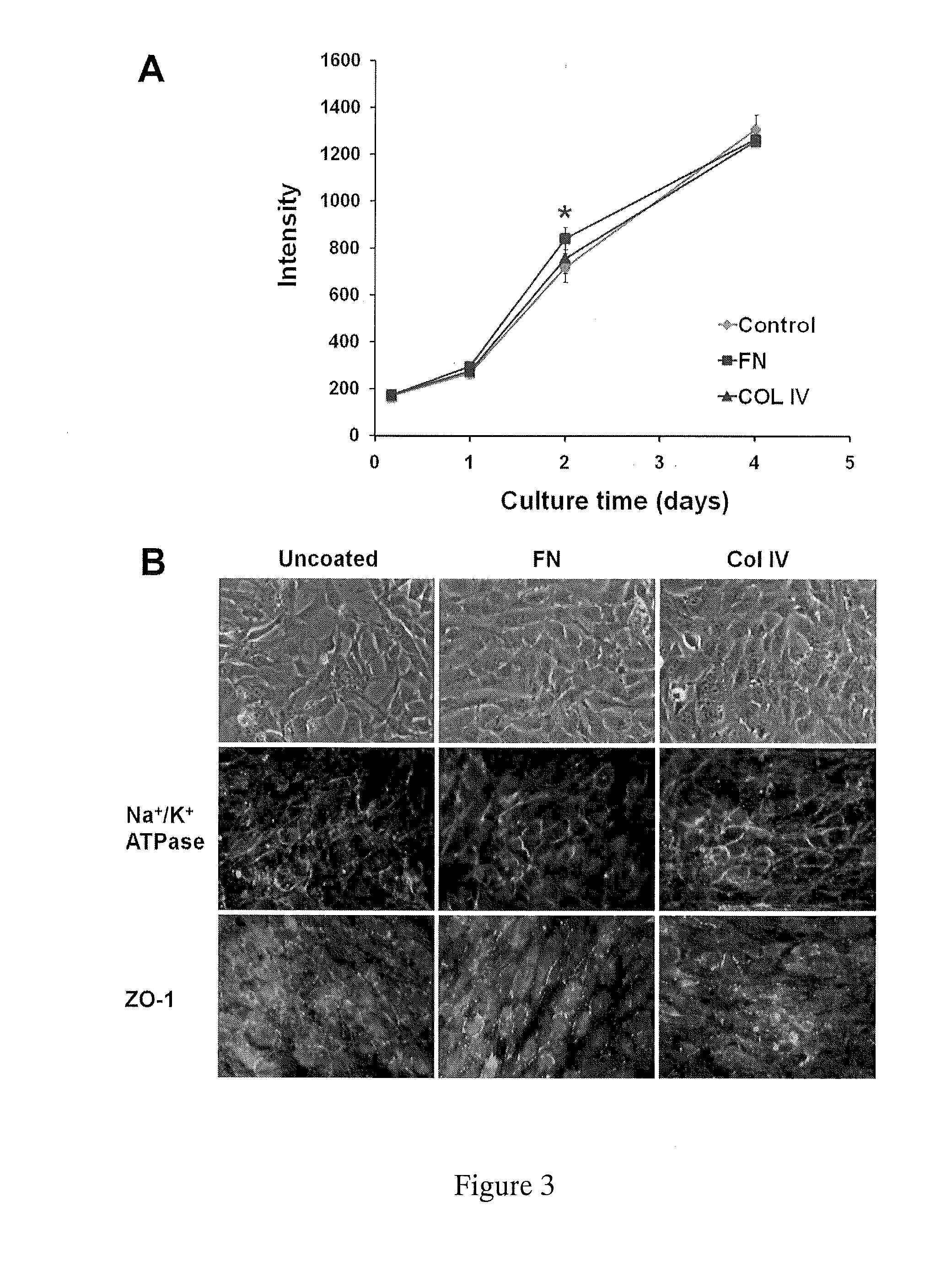
[0061]Type IV collagen is a major component of the corneal stroma and fibronectin is an adhesive protein that is often used to coat endothelial cell-seeded culture plates. To test if the presence of extracellular matrix components affected growth rates, we plated cells on petri plates with no coating, fibronectin coating, or collagen type IV coating. There were no differenc...

example 3

Seeding the Decellularized Corneal Scaffolds

[0067]Approximately 30,000 CECs were seeded on the decellularized scaffold (8 mm diameter). Acellular corneas were placed in the bottom of a well of a 48-well dish, and the cell suspension was layered on top, allowing the seeding to take place by simple gravitational settling.

[0068]The construct was placed in EGM-2 media (Lonza, Walkersville, Md.) supplemented with fetal bovine serum (to a final concentration of 10%) for seven days, and then analyzed.

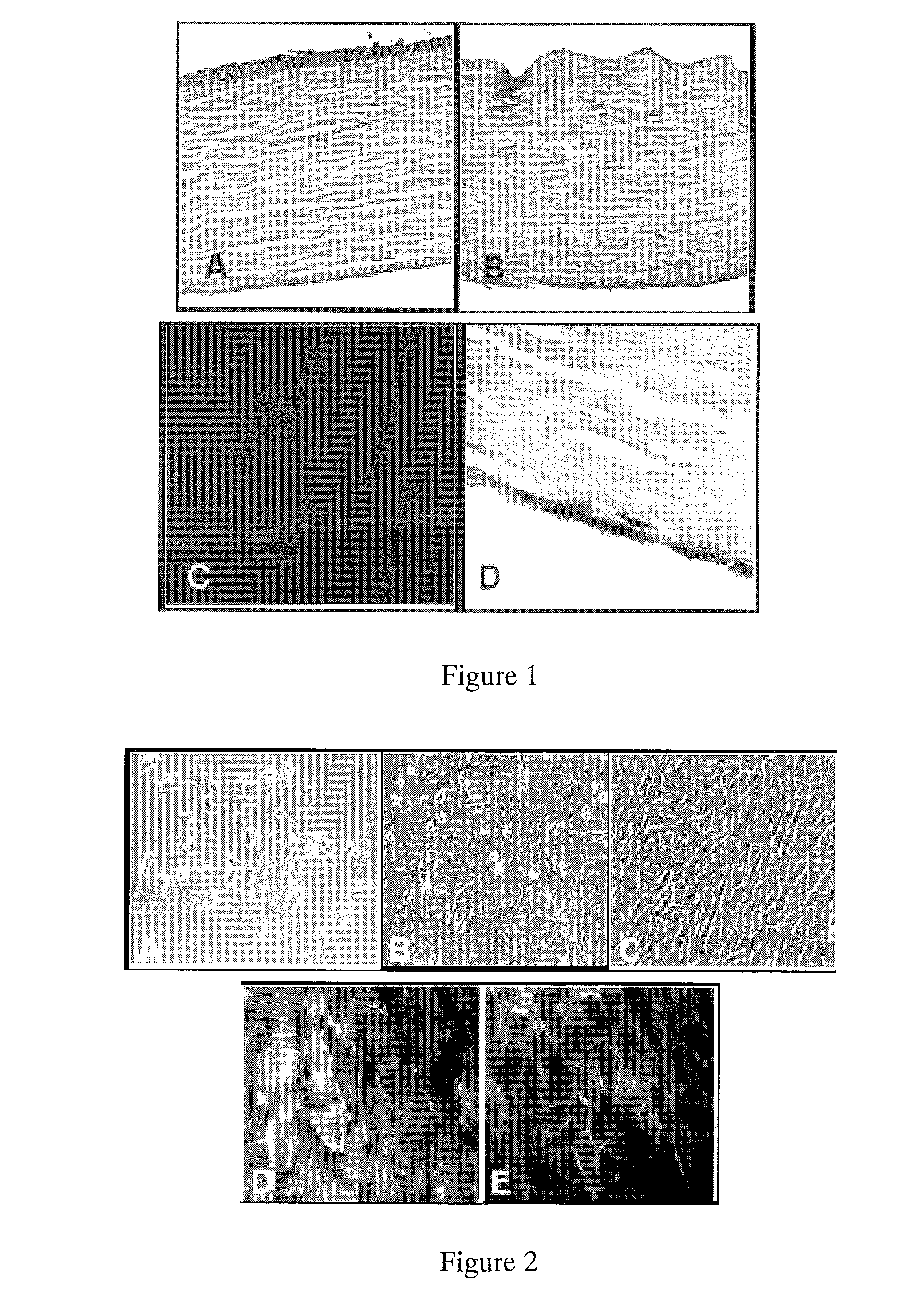
[0069]Scanning electron microscopy (SEM) was performed on fresh (frozen) samples to determine CEC coverage of the stroma. Isolated hCEC formed confluent monolayers with a phenotype typical of endothelial / epithelial cells (FIGS. 2,A&B). The decellularization process efficiently removed each of the stromal, epical and basal cell layers (FIG. 1). When seeded on the surface of decellularized corneas, hCEC formed a single cell monolayer, similar to native cornea (FIG. 1, right upper and lower panel...


PUM


Abstract

Provided herein is an endothelial scaffold comprising, consisting of, or consisting essentially of decellularized corneal stroma. In some embodiments, the scaffold has cultured endothelial cells seeded thereon. Methods of treating a patient in need of corneal endothelial transplant are also provided, including implanting the scaffold as described herein onto a cornea of the patient (e.g., by deep keratectomy).

Description

RELATED APPLICATIONS[0001]This application claims the benefit under 35 U.S.C. §119(e) of U.S. Provisional Patent Application Ser. No. 61 / 155,760, filed Feb. 26, 2009, the disclosure of which is incorporated herein by reference in its entirety.FIELD OF THE INVENTION[0002]The present invention involves scaffolds derived from endothelial tissue and processes for making and using the same.BACKGROUND[0003]The inner layer of the cornea is a single layer of neural crest-derived endothelial cells (CECs), which form a barrier between the cornea and the aqueous humor and transport water from the corneal stroma. CEC loss can result from eye injuries, complications from cataract surgery (pseudophakic bullous keratopathy (PBK) or aphakic bullous keratopathy (ABK)), and in an inherited condition known as Fuchs dystrophy.[0004]These cells do not divide during adult life. Rather, existing CECs simply spread to compensate for loss or damage. When this spreading is inadequate, vision-impairing cornea...

Claims


Application Information

Patent Timeline
Patent Type & Authority Applications(United States)
IPC IPC(8): A61K9/00A61F2/14A61K35/12A61P27/02
CPCA61F2/142A61K9/0051A61K35/44A61L27/3604A61L27/3808A61K35/12A61L2430/16A61P27/02
Inventor SOKER, SHAYWILLIAMS, J. KOUDYLABER, PATRICKGREVEN, MARGARETWALTER, KEITH A.
Owner WAKE FOREST UNIV HEALTH SCI INC
Why Eureka
Try Eureka