Methods and system for analyzing cells

a cell and method technology, applied in the field of methods and systems for analyzing cells, can solve the problems of limiting the identification of nucleus, the prior method is limited to identification, and the planer solid tissue cells pose a unique problem, so as to control the correctness of separation and high the risk of metastasis

Inactive Publication Date: 2010-11-04
TISSUE GNOSTICS GMBH
View PDF4 Cites 22 Cited by
  • Summary
  • Abstract
  • Description
  • Claims
  • Application Information

AI Technical Summary

Benefits of technology

[0006]The objective of the invention is the automated recognition of irregular shaped planer solid tissue single cells and their components such as nucleus, cytoplasm, and cell membrane in dense, continual tissue structures and the exact measurement of such components without loss of spatial relations to the cells or the ability to identify their localization within the tissue. The present invention is a method that provides for and describes the forward and backward connection implemented into the software utilized in analysis according to the present invetnion. The present invention provides methods that allows the user to control cellular analysis of irregular shaped cells of a specific feature that is located in a tissue and conversely, the present invention allows a user to see and analyze cells at which position in a dataset of a population a given cell is located. In addition, the present invention allows for analysis without loss of spatial relations to the cells or the ability to identify their location within the tissue. The present invention also provides that the tissue sample is not only divided into single cells, but is also divided into different regions such as epithelial structures, muscle structures, interstitial areas and the like. This division by structures is used on the one hand to classify for example a normal glandular pattern by analyzing size, orientation and frequency to divid

Problems solved by technology

Planer solid tissue cells pose a unique problem in analysis due to the high level of irregularity of these cells.
However these prior methods are limited only to identification of a nucleus or a membrane.
Methods of nucleic staining are limited to identification of nucleus only.
This method does not even provide identification of nuclei as such.
This is not tragic in the case of negative cells but when it comes to partial loss of Her-2neu membrane reactivity, which is important for treatment and therefore clinically relevant this methods is not able to provide information how much parts of the membrane lack the staining.
Although other methods known

Method used


Image

  • Methods and system for analyzing cells
  • Methods and system for analyzing cells
  • Methods and system for analyzing cells

Examples


Embodiment Construction

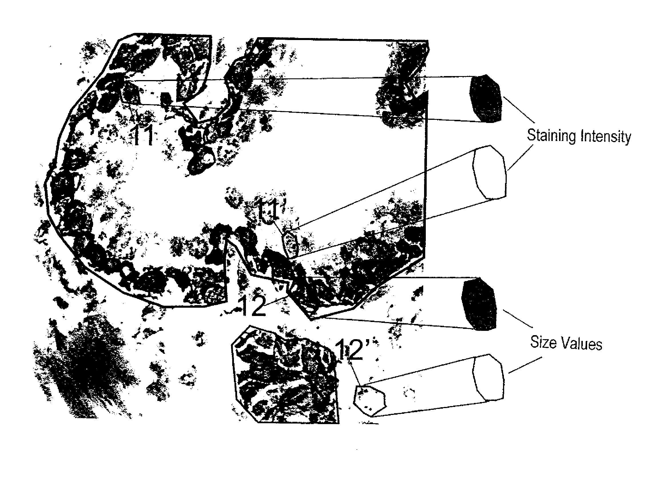
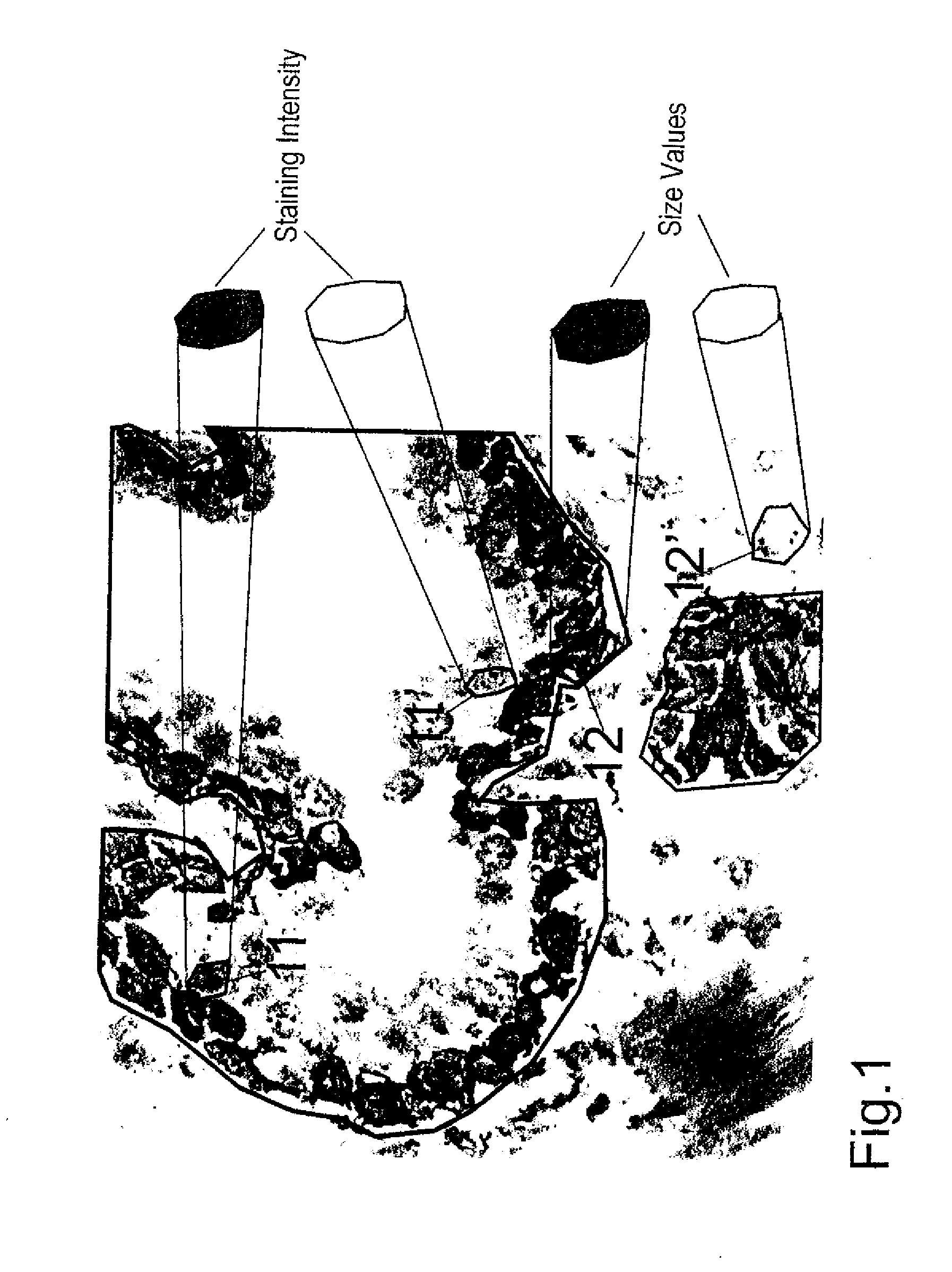
[0029]FIG. 1 shows a kidney section as an example of a tissue specimen. Cell nuclei (identity stain, in blue) and cytoplasm (target structure stain) were stained. After the staining of tissue specimens parts are defined and scanned, for example, with the help of an Eppendorf micromanipulator. Using a laser-scanning microscope, for example, two scans are performed in the z-plain. One of these is a rough scan, the other a fine scan both of which are guided along a horizontal line in the middle of the image. The focus of the scan can be adjusted to the brightest region. The scan takes, for example, 4 seconds per microscopic field of view, where each section is scanned only once using an argon laser (488 nm). It is possible to undertake multiple scans in sequence. In order to obtain distinctive stains it would be possible, for example, to perform a first scan using 543 nm He / Ne laser so as to measure the fluorochromes cyanine 5 (Cy5) or Cy3. This may be followed by a scan using a 488 nm...


PUM


Abstract

The invention relates to a method for analyzing cells that are present as closed clusters. According to said method, a planar tissue preparation is subjected to an identification staining of the cell nuclei and a target structure staining of cell objects that is different from the identification staining. Digital images are recorded of the stained tissue preparation by means of an electronic image recording device and at least one image of a subsection of the tissue cut is displayed in at least one coloration. According to the inventive method, at least one parameter of the cell nuclei and at least one parameter of the cell objects labeled by target structure staining is restricted to a predetermined range of values. Cell nuclei and cell objects whose parameters correspond to the respective parameter range(s) are detected and optionally displayed using image processing algorithms in the image of said subsection. The image content of at least one image detected for the cell nuclei is correlated with the image content of at least one image detected for the target-structure stained cell objects to detect the individual cells. On the basis of the cell nuclei identified a cell growth or a cell enlargement is induced using a predetermined arithmetic algorithm to reconstruct the individual cells. In doing so it is made sure that neighboring cells do not fuse. The number of reconstructed individual cells is determined and/or the individual cells are divided into populations according to certain parameters.

Description

CROSS-REFERENCE TO RELATED APPLICATION[0001]This is a continuation-in-part application of application Ser. No. 10 / 415,179, filed Apr. 24, 2003; which was a continuation application, under 35 U.S.C. §120, of International application PCT / AT2001 / 00343, filed Oct. 23, 2001; the application also claims the priority, under 35 U.S.C. §119, of Austrian patent application No. A 1821 / 2000, filed Oct. 24, 2000; the prior applications are herewith incorporated by reference in their entirety.[0002]The invention concerns a procedure in accordance with the following: A process for the examination, in particular the identification of cells in preferably dense, cohesive irregular shaped cell complexes and solid tissues, in which a solid planar tissue sample, especially frozen or paraffin sections, cell smears, cytospin preparations or similar are processed with one or a number of different, especially complete, preferably plane identity stain / s of the cell nucleus, predominantly all nuclei, in whic...

Claims


Application Information

Patent Timeline
IPC IPC(8): G01N1/30
CPCG01N15/1475G01N2015/1006G01N2015/1497G01N2015/1486G01N2015/1472
Inventor STEINER, GEORG E.ECKER, RUPERT CH.
Owner TISSUE GNOSTICS GMBH
Why Eureka
Try Eureka