Self-assembling micelle-like nanoparticles for systemic gene delivery

a technology of micelle-like nanoparticles and gene delivery, which is applied in the direction of powder delivery, microcapsules, drug compositions, etc., can solve the problems of limited “critical micelle concentration” and the difference between nanoparticles, and achieves simple and reproducible one-step procedure, high loading capacity, and facilitates the self-assembly process.

Inactive Publication Date: 2010-11-11
NORTHEASTERN UNIV
View PDF9 Cites 13 Cited by
  • Summary
  • Abstract
  • Description
  • Claims
  • Application Information

AI Technical Summary

Benefits of technology

[0011]Nanoparticles according to the invention, a novel construct for gene delivery, are non-toxic, long-circulating, and effective for the in vivo transfection of therapeutic nucleic acids to both RES sites and other organs. This invention combines polymer-based gene delivery systems with lipid-based gene delivery systems, resulting in a new approach for using a chemical conjugate of phospholipids and polymer. The conjugation of polyethylenimine (PEI) at the distal end of phospholipid alkyl chain leads to a new chemical entity, a phospholipid-polyethylenimine (PLPEI) conjugate. The PLPEI possesses two functional domains for i) DNA binding and ii) membrane-formation, attributed to PEI and PL moieties, respectively. The PLPEI self-assembles, in the presence of DNA, into nanoparticles via electrostatic interaction of polycationic PEI with poly-anionic DNA. The self-assembly process is also facilitated by hydrophobic interaction between lipids moieties. The self-assembled nanoparticles possess a unique supramolecular structure in which the PEI / NA polyplex core and lipid monolayer envelope are connected by chemical bonds. The nanoparticle is different from, e.g., liposomal nanoparticles, where lipids form a bilayer instead of monolayer. The nanoparticle is also different from micelles, which assemble solely by hydrophobic interaction and are subject to “critical micelle concentration” limitation.
[0012]This invention provides advantages of a simple and reproducible one-step procedure in combination with a high loading capacity compared to other liposomal nanoparticle entrapping PEI / DNA polyplexes such as bioPSL and pSPLP. Nanoparticles according to the invention also provide for a high DNA loading capacity of around 25% (w / w), which is about 10-fold higher than values reported in the literature for other systems. As used herein, the term “DNA loading capacity” or “nucleic acid loading capacity” refers to the amount of DNA or other nucleic acid that can be incorporated into nanoparticles according to the invention.

Problems solved by technology

The nanoparticle is also different from micelles, which assemble solely by hydrophobic interaction and are subject to “critical micelle concentration” limitation.

Method used


Image

  • Self-assembling micelle-like nanoparticles for systemic gene delivery
  • Self-assembling micelle-like nanoparticles for systemic gene delivery
  • Self-assembling micelle-like nanoparticles for systemic gene delivery

Examples


example i

Preparation of Micelle-Like Nanoparticles (MNP)

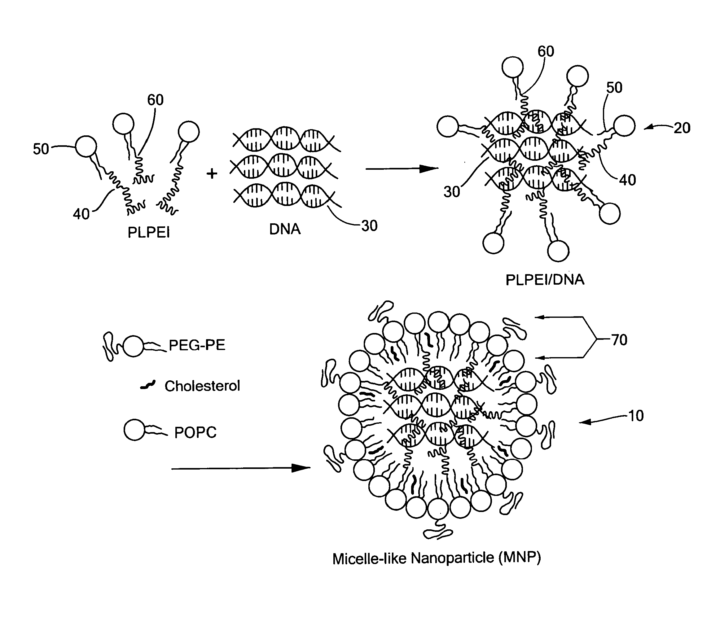
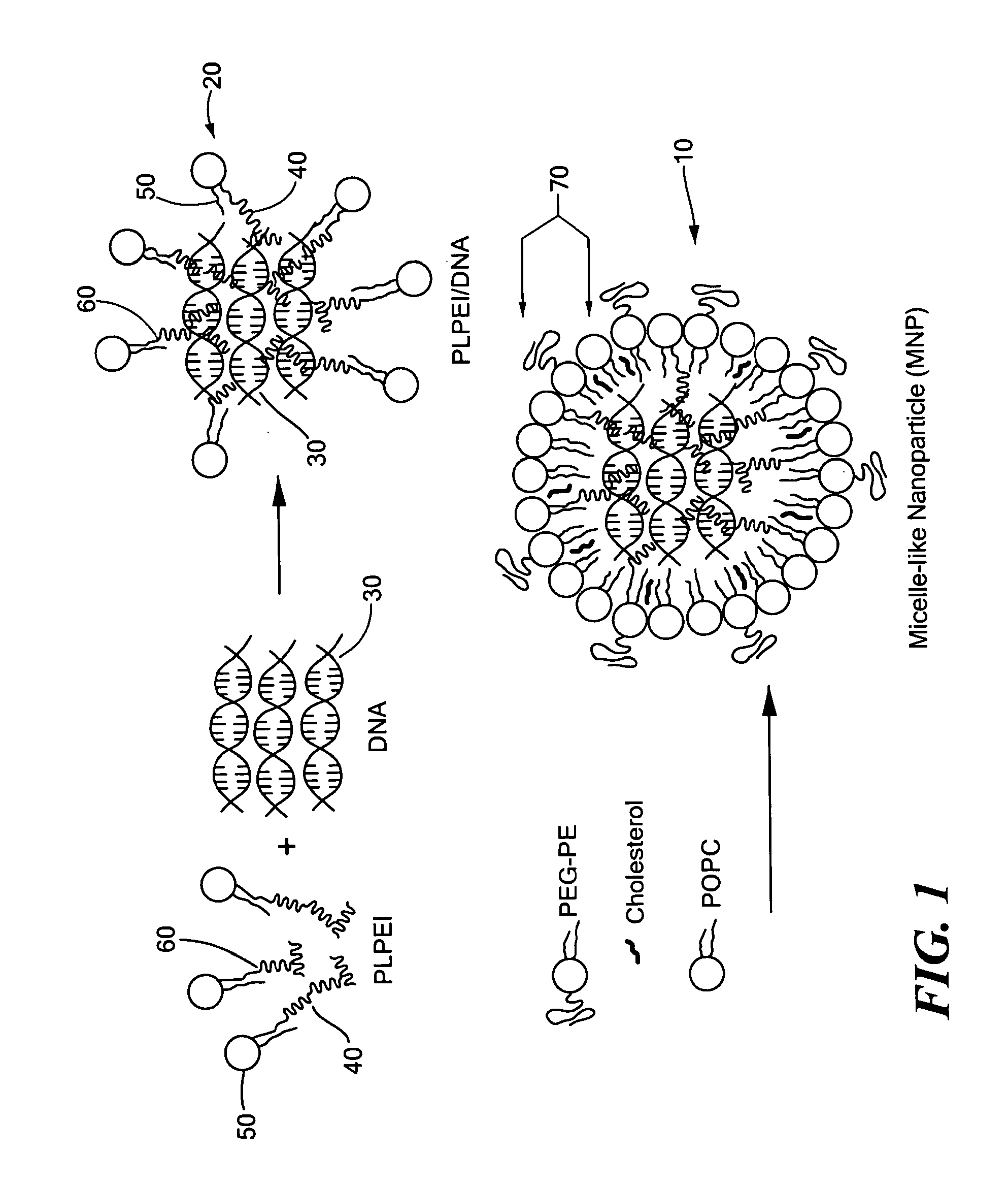
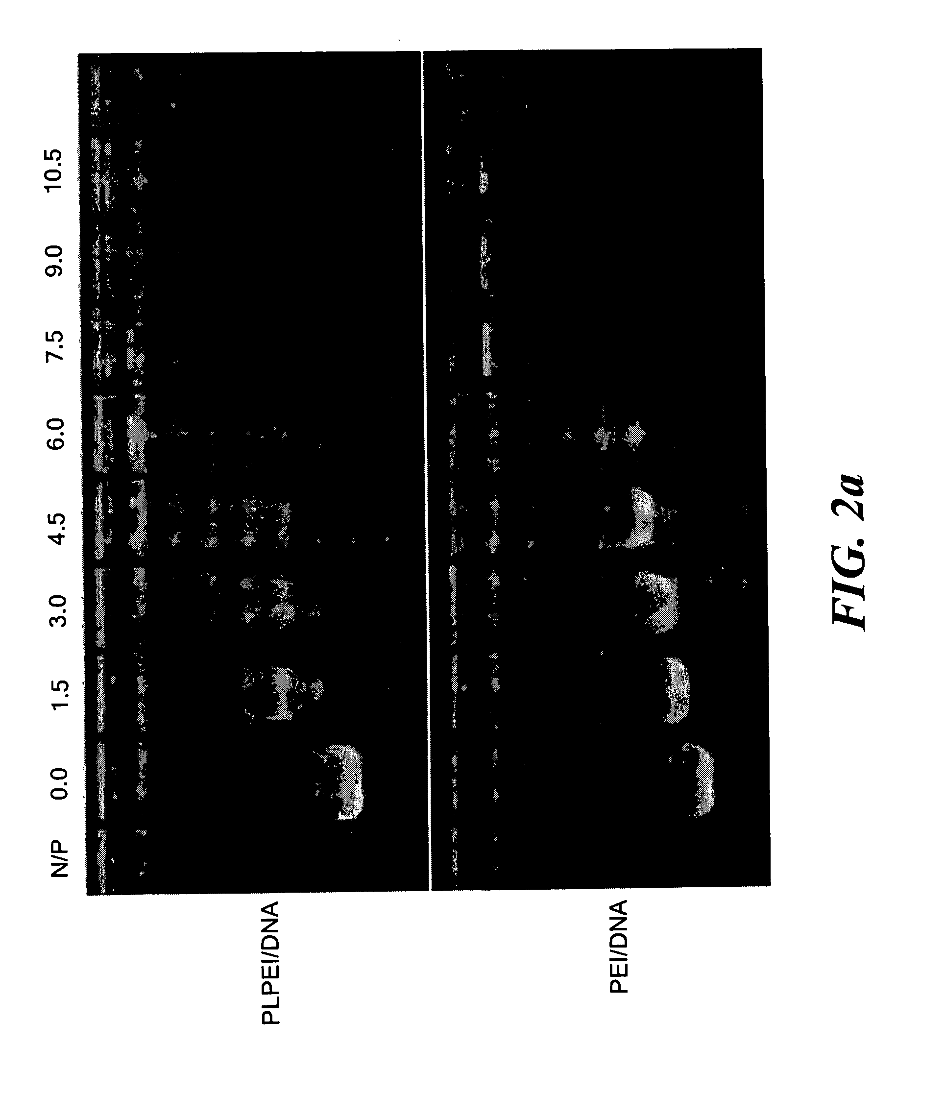
[0063]The micelle-like nanoparticles (MNP) were prepared by complexing plasmid DNA with PLPEI and then enveloping the preformed complexes with a lipid layer containing also PEG-phosphatidylethanolamine conjugate (PEG-PE) (FIG. 1). As for the complexation, the optimal ratio of PLPEI to DNA was determined based on the amounts of amine required to completely inhibit DNA migration on an agarose gel, since the complex formation hinders the migration of DNA, retaining the DNA in the wells. Constant amounts of plasmid DNA were mixed with PLPEI at varying amine / phosphate (N / P) ratios and analyzed by agarose gel electrophoresis. The bound fraction of DNA was increased as the N / P ratio increased and the most DNA was bound at an N / P ratio higher than 6. The complexation profile of PLPEI was comparable to that of the unmodified PEI, indicating that the PEI capacity for DNA complexation was not diminished by lipid conjugation (FIG. 2a). An N / P ratio...

example ii

Physicochemical Properties of MNP

[0066]Traditional PEI / DNA polyplexes tend to aggregate rapidly under physiological high salt conditions [8]. To demonstrate the stabilizing effect of the lipid envelope against the salt-induced aggregation, NaCl was added to complex formulations to a final concentration of 0.15M while monitoring the hydrodynamic diameter. As expected, PEI / DNA polyplexes aggregated immediately after adding NaCl with continuous increases in hydrodynamic diameter up to almost 20-folds over a 24 hour period. The intermediate PLPEI / DNA complexes without free lipids and PEG-PE showed a two-fold increase immediately after adding NaCl and then remained relatively constant over the 24 hours. At the same time, MNP remained stable with no significant aggregation upon salt addition for 24 hours (FIG. 3a).

[0067]Zeta potential measurement revealed that MNP have a favorable neutral surface charge of −2.1±0.86 mV (mean±s.e.m., n=5), while PEI / DNA polyplexes have a more toxic positiv...

example iii

In Vivo Biodistribution and Gene Expression

[0070]To demonstrate the prolonged circulation time of MNP in the blood and thus the feasibility of their enhanced delivery to target tissues such as tumors, pharmacokinetic and biodistribution studies were performed with MNP loaded with 111In-DNA in mice. The radioactivity in major organs after i.v. bolus administration of MNP loaded with 111In-DNA was measured and compared to that of control PEI / 111In-DNA complexes. After 10 min, as much as 30% ID / ml of MNP remained in the blood compared to about 10% ID / ml for PEI / DNA polyplexes. At 1 hour post-injection, about 20% ID / ml of MNP was still present in the blood, while only about 5% ID / ml of PEI / DNA polyplexes was detected in the circulation (FIG. 5a).

[0071]The slower clearance and thus more prolonged circulation of DNA in MNP compared to PEI / DNA were also confirmed by pharmacokinetic parameters. The half-life (t1 / 2 beta) was estimated by fitting the blood concentration data colleted to 60 mi...


PUM

PropertyMeasurementUnit
diameteraaaaaaaaaa
molecular weightaaaaaaaaaa
diameteraaaaaaaaaa

Abstract

Nanoparticles containing nucleic acid and suitable for use as in vivo delivery agents for nucleic acids are provided. The nanoparticles use a covalent conjugate of a polycation such as polyethylenimine and phospholipids. The final DNA-containing nanoparticle has a vesicular structure with a polyplex core surrounded by a mixed lipid / PEG-lipid monolayer envelope and offers simple preparation, high loading capacity, and in vivo stability. The nanoparticles have good in vivo stability and a prolonged blood circulation time and can effectively deliver a gene to a biological target such as a tumor.

Description

CROSS REFERENCE TO RELATED APPLICATIONS[0001]This application claims the priority of U.S. Provisional Application No. 61 / 002,626 filed Nov. 9, 2007 entitled, NANOPARTICLES FOR GENE DELIVERY, the whole of which is hereby incorporated by reference herein.STATEMENT REGARDING FEDERALLY SPONSORED RESEARCH OR DEVELOPMENT[0002]The research leading to this invention was carried out with United States Government support provided under a grant from the National Institute of Health, Grant No. RO1 HL55519. Therefore, the U.S. Government has certain rights in this invention.BACKGROUND OF THE INVENTION[0003]In vivo gene therapy depends on the delivery of DNA-based drugs, either in the form of oligonucleotides (antisense oligodeoxyribonucleotides (ODN), siRNA) or entire genes (plasmid DNA) to their cellular site of action. With few exceptions, where local administration may be feasible, progress towards broad clinical application of gene therapies requires the development of effective non-invasive...

Claims


Application Information

Patent Timeline
Patent Type & Authority Applications(United States)
IPC IPC(8): A61K31/7088A61K9/14C12N15/63C12N15/64C08G73/00A61K9/127A61P35/00
CPCA61K9/1075A61K9/5146C12N15/88A61K47/488A61K47/48192A61K47/59A61K47/6907A61P35/00A61P43/00
Inventor KO, YOUNG TAGKALE, AMITTORCHILIN, VLADIMIR
Owner NORTHEASTERN UNIV
Why Eureka
Try Eureka