Automatic geometrical and mechanical analyzing method and system for tubular structures

a geometrical and mechanical analysis and tubular structure technology, applied in the direction of image analysis, instruments, details involving processing steps, etc., can solve the problems inability to automatically and comprehensively solve the problem of inability to analyze vascular bifurcations, and model applicability that is very limited, so as to improve the quality of the fe mesh

Inactive Publication Date: 2010-11-18
VASCOPS
View PDF2 Cites 33 Cited by
  • Summary
  • Abstract
  • Description
  • Claims
  • Application Information

AI Technical Summary

Benefits of technology

[0040]Some embodiments of the invention also provide automatic hexahedral-dominated meshing of the volume of the vascular body, and hence, it allows the application of efficient mixed finite elements, e.g., the so called Q1P0 formulation, see Simo and Taylor, 1991, Quasi-incompressible finite elasticity in principal stretches. Continuum basis and numerical algorithms. Comp Meth Appl Mech Engrg. 85. 273-310. This is essential to represent the incompressibility properties of vascular tissue in a numerically efficient and proper way.
[0041]Some embodiments of the invention also provide for automatic 2D and 3D mesh smoothing and optimization to improve the quality of the FE mesh, and hence, the quality of the predicted results.

Problems solved by technology

However, this model's initialization requires the presence of an ILT and it needs furthermore the presence of a high threshold between the lumen and the ILT, and it does not allow for the analysis of vascular bifurcations, as it is frequently required.
Hence, this model has very limited practical applicability.
However, presently there is no automatic and comprehensive system available, which integrates all structural relevant anatomical objects and provides information regarding the mechanical loading conditions of e.g. vascular tissue.
However, the method and system disclosed in US2006 / 0100502-A1 are limited to provide a single surface mesh of a vascular lesion, and hence, only shell-like structural effects of the vessel wall can be considered, e.g., using Shell Finite Elements.
Consequently, in case of AAAs, the structural impact of the ILT is neglected, which causes unrealistic and unreliable mechanical predictions of the AAAs.
Hence, an unreliable diagnosis and prediction of risk of rupture may be made, which is unsatisfactory, at least from a point of patient safety.
This is both inconvenient for a user technically challenging as an interface between the different software products has to be guaranteed in a reliable and safe manner, which in practice is difficult to ensure.
Again, it is limited to model the AAA's outer surface by means of shell-like structural effects, and the thick-walled structure (or volume effect) of the vascular wall is neglected.
However, the methods and systems disclosed in US2006 / 0100502-A1 or Raghavan et al., 2005, apply mesh-smoothing strategies, which change the geometry of the objects, such that the vascular body's (outer) geometry cannot be captured accurately, i.e. a mismatch between the model geometry and the underlying image data exits.
The proposed concept therein requires expert knowledge in structural modeling and again several steps are involved, which is again inconvenient for technically challenging applications.
1) In-plane segmentation using, e.g., NURBS representation of the edges, where deformable models are used on a single image slice, such that out-of-plane information of the set of image data is neglected. This concept can only be applied to a sub-class of geometries, and excludes, for example, saccular aneurysm, see FIG. 0, which are of important clinical relevance. Also, this concept cannot be applied to vascular bifurcations.
2) Generation of a solid model based on the edge information defined by the segmentation. Here always smoothing of the segmented curves is required, in particular to avoid scatter along the out-of-plane direction. This naturally alters the geometry, and hence, the geometry of the vascular body, as defined by the set of image data, cannot be maintained.
3) Meshing the solid model, which for realistic (clinically relevant) geometries of vascular bodies need to be subdivided into smaller bodies, which are simple enough to allow automatic meshing. This is usually a time-consuming task requiring engineering expert knowledge in mesh generation and structural analysis. Most important, in case the geometry is too complicated, even modifications of the solid model might be required to facilitate a meshing of the structure. Hence, the geometry of the vascular body, as defined by the set of image data, cannot be maintained.
In summary, currently known approaches are characterized by severe manual interactions and necessary engineering expert knowledge of the user, which does not allow their clinical application.

Method used


Image

  • Automatic geometrical and mechanical analyzing method and system for tubular structures
  • Automatic geometrical and mechanical analyzing method and system for tubular structures
  • Automatic geometrical and mechanical analyzing method and system for tubular structures

Examples


Embodiment Construction

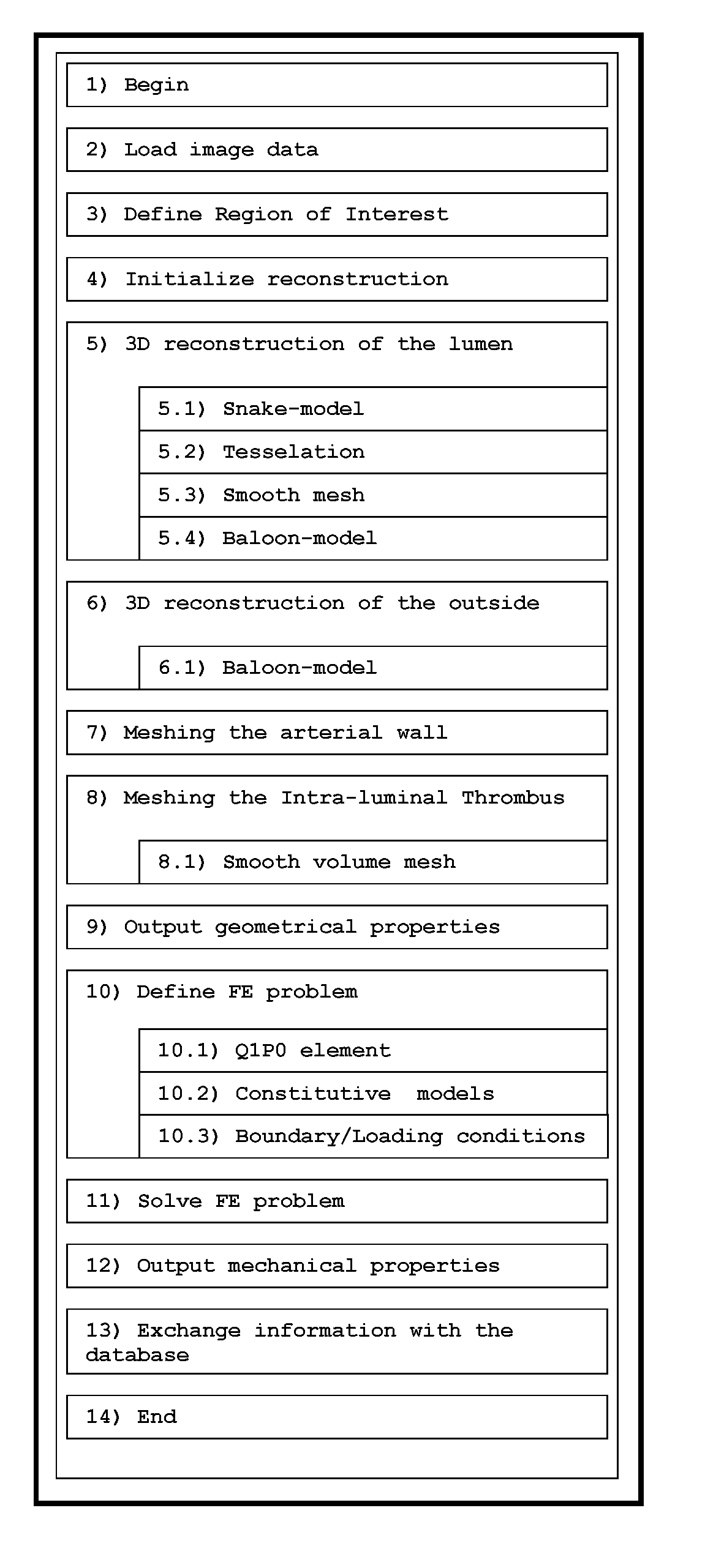
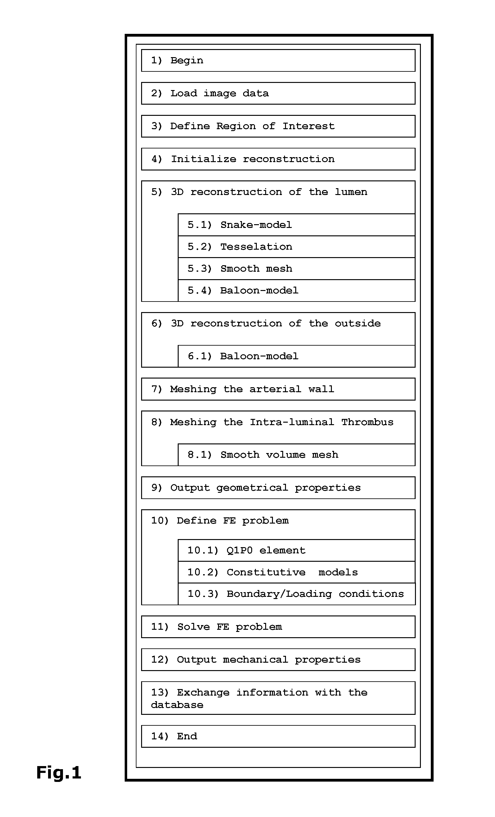
[0060]Specific embodiments of the invention will now be described with reference to the accompanying drawings. This invention may, however, be embodied in many different forms and should not be construed as limited to the embodiments set forth herein; rather, these embodiments are provided so that this disclosure will be thorough and complete, and will fully convey the scope of the invention to those skilled in the art. The terminology used in the detailed description of the embodiments illustrated in the accompanying drawings is not intended to be limiting of the invention. In the drawings, like numbers refer to like elements.

[0061]The following description focuses on embodiments applicable to analyze vascular lesions and in particular to AAAs or carotid stenoses. However, it will be appreciated that the invention is not limited to this specific application but may in some embodiments be applied to many other tube-like internal organs including for example other blood vessels, the ...


PUM


Abstract

A method and system for analyzing tubular structures, such as vascular bodies, with respect to their geometrical properties and mechanical loading conditions is disclosed. To this end, geometrical and structural models of vascular bodies are generated from standard sets of image data. The method or system works automatically and the tubular structure is analyzed within clinical relevant times by users without engineering background. Most critical in that sense is the integration of novel volume meshing and 3D segmentation techniques. The derived geometrical and structural models distinguish between structural relevant types of tissue, e.g., for abdominal aortic aneurysms the vessel wall and the intra-luminal thrombus are considered separately. The structural investigation of the vascular body is based on a detailed nonlinear Finite Element analysis. Here, the derived geometrical model, in-vivo boundary/loading conditions and finite deformation constitutive descriptions of the vascular tissues render the structural biomechanical problem. Different visualization concepts are provided and allow an efficient and detailed investigation of the derived geometrical and mechanical data. In addition, this information is pooled and statistical properties, derived from it, can be used to analyze vascular bodies of interest.

Description

[0001]This invention relates to the field of diagnostic systems, and more specifically to computer-based diagnostic systems for hollow structures, such as elongated hollow structures, such as tubular structures, such as for instance vascular structures comprising vascular tissue. The diagnostic systems provide analysis and information data for instance related to the geometry and mechanics of the elongated hollow structures.BACKGROUND OF THE INVENTION[0002]Many procedures, e.g. interventions and diagnostics concerning vascular tissue must be carried out at an internal anatomical site. The physician's information for these medical procedures is enriched by image data acquired by image modalities, for instance a scanning device, e.g. based on Computer Tomography (CT) or Magnetic Resonance (MR). In general this provides a plurality of two-dimensional (2D) images, also called slices, of the patient's anatomical structure. Some scanning devices include computer hard and software for buil...

Claims


Application Information

Patent Timeline
Patent Type & Authority Applications(United States)
IPC IPC(8): G06K9/00G06T15/00
CPCG06T7/00G06T7/0012G06T2210/41G06T2200/08G06T2207/30101G06T17/20
Inventor GASSER, CHRISTIAN T.AUER, MARTIN
Owner VASCOPS
Why Eureka
Try Eureka