Prevention of staphylococcus biofilm formation

a technology of staphylococcus and biofilm, applied in the field of prevention of staphylococcus biofilm, can solve the problems of failure of conventional antibiotic therapy, limited understanding of the mechanism of detachment process, etc., and achieve the effects of preventing biofilm formation, preventing biofilm formation, and preventing biofilm formation

Inactive Publication Date: 2010-11-18
HERMANS PETER WILHELMUS MARIA +1
View PDF9 Cites 17 Cited by
  • Summary
  • Abstract
  • Description
  • Claims
  • Application Information

AI Technical Summary

Benefits of technology

[0012]The invention further comprises a method for preventing biofilm formation by Staphylococcus on a medical device comprising coating said medical device with an antibody according to the invention. Said medical device is preferably an intravascular device such as a stent or a cannula, a prosthesis, such as an orthopaedic endoprothesis or a prosthetic valve, a pacemaker, a catheter, a drain, an endovascular graft or a cerebrovascular shunt. This

Problems solved by technology

However, our understanding of the mechanism of the detachment process is very limited.
This antibiotic resistance of biofilms often leads to the failure of conventional antibiotic t

Method used


Image

  • Prevention of staphylococcus biofilm formation
  • Prevention of staphylococcus biofilm formation
  • Prevention of staphylococcus biofilm formation

Examples


example 1

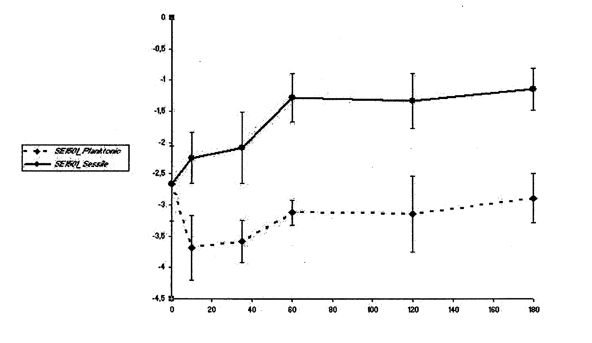
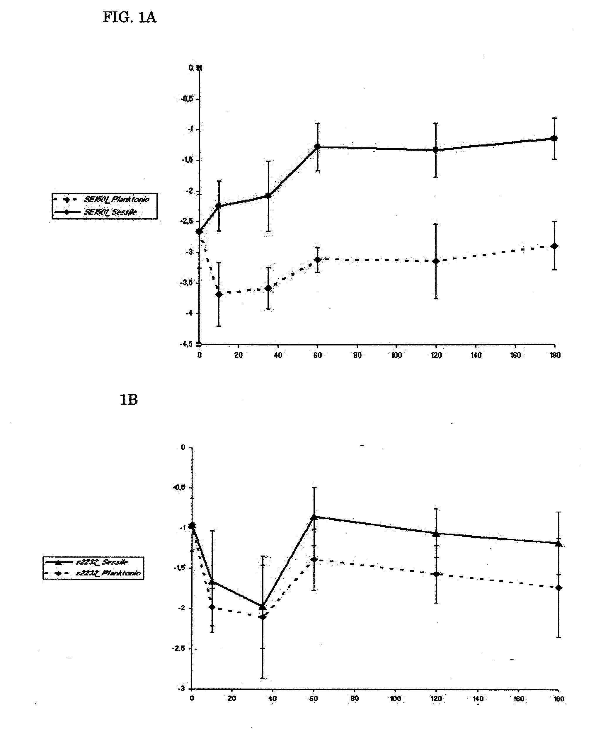
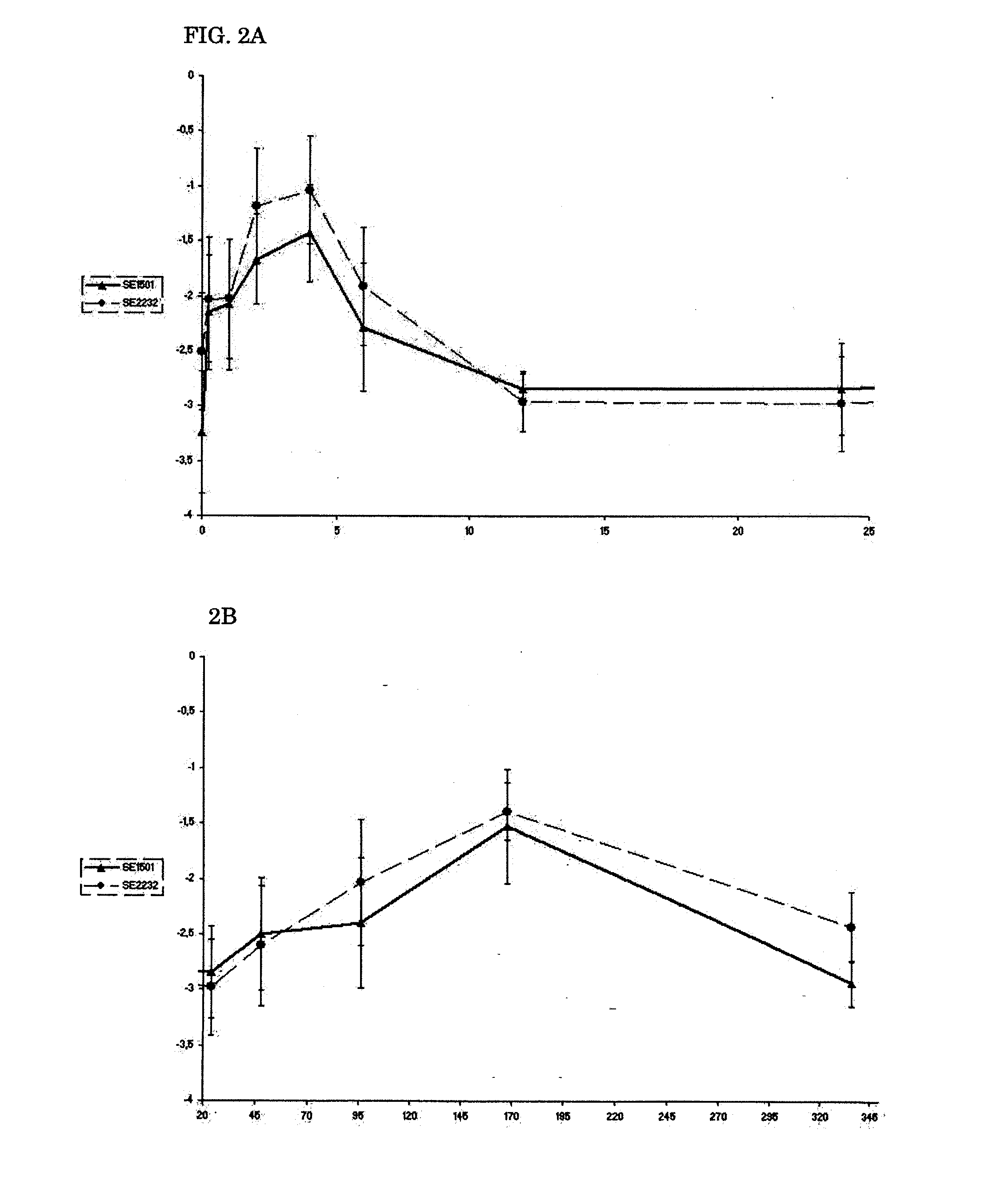
Expression of SE2232 and SE1501 In Vitro and In Vivo

Bacteria Used.

[0088]For all experiments, a previously well-characterized biofilm-producing S. epidermidis strain, strain 10b, was used. S. epidermidis 10b was isolated from a patient with a proven catheter-related bloodstream infection (31, 32, 33, 34). It is homogeneously oxacillin resistant and has a 100% infection rate in the rat model used (34).

Gene Identification.

[0089]The sequences of the genes studied were retrieved from the National Center for Biotechnology Information (NCBI) GenBank (Table 1). On the basis of the sequences, primers were designed with Primer Express 2.0 software (Applied Biosystems Division of Perkin-Elmer) and used to amplify part of the corresponding genes in S. epidermidis 10b. The following thermal cycling conditions were used: 5 min at 95° C., followed by 25 repeats of 30 s at 95° C., 30 s at related annealing temperatures (Table 1) and 1 min at 72° C. followed by 7 min at 72° C. and holding at 4° C. P...

example 2

Production of Antibodies

Cloning, Expression and Purification of Recombinant Proteins.

[0105]We amplified the genes for production of the recombinant proteins by PCR with primers of SE1501F (5′ CACGTGCTAGCGCTGAATCAAACACTTCAGTTTCTTCT 3′), SE1501R (5′ATGCGGATCCTAGTGATGGTGATGGTGATGCATACCTGTATTTGGTAAT AG 3′), SE2232F (5′ ACGTGCTAGCGCAGATTCAGAAAGTACATC 3′) and SE2232R (5′ATGCGGATCCTAGTGATGGTGATGGTGATGATCAGCTGTAGCTGTTCC 3′), which incorporate flanking NheI and BamHI restriction sites and sequence coding for a C-terminal His6 tag. The genes were cloned into a pET11c expression vector (Stratagene, LaJolla, Calif.) and we electrotransformed the recombinant plasmids into E. coli BL21 (DE3). The expression of recombinant protein was induced by the addition of IPTG, and the recombinant proteins were purified by Ni+ affinity chromatography with the HisTrap™ Kit (Amersham Pharmacia, Uppsala, Sweden) according to the manufactures recommendations. We determined the purity of the recombinant proteins ...

example 3

Biofilm Inhibition

Biofilm Inhibition Assay.

[0114]The amount of biofilm formed was determined by a semiquantitive microtiter plate method as described (18, 38). A frozen culture of S. epidermidis 10b was grown to the end-exponential growth phase, pelleted, and resuspended in 0.9% NaCl, diluted to an OD600 of 0.05 (˜5×10⁷ cells) and mixed with polyclonal antibodies. Sixplicate samples (each containing 0.1 ml) were added to individual wells of a 96-well flat-bottom microwell plate (comp). Microwell plates were incubated 2 h at 4° C. and then overnight at 37° C. After three washes with PBS, any remaining biofilm was stained with safranin O dye for 1 min and washed with PBS again. Optical density at 492 nm (OD492) was determined with a 96-well plate spectrometer reader. Percent inhibition of biofilm formation was calculated by using the following formula: (A492, positive−A492, antibody) / (A492, positive−A492, negative)×100%. (18). 0.9% NaCl medium without bacteria was used as negative con...


PUM

PropertyMeasurementUnit
Fractionaaaaaaaaaa
Compositionaaaaaaaaaa
Immunogenicityaaaaaaaaaa

Abstract

The present application discloses antibodies directed to protein SE2232 (SesC) of Staphylococcus epidermis, and homologus proteins which are useful in preventing biofilm formation by said micro-organism. The antibodies can be used to coat medical devices which are inserted or implanted into the mammalian, preferably human body. Further these antibodies or vaccines comprising said proteins can be used to vaccinate subjects to prevent or treat natural occurring biofilm formation during infection. Also covered are medical devices coated with the antibody.

Description

[0001]The invention relates to the field of medical microbiology, more specifically the prevention of biofilm formation by bacteria, more specifically wherein said bacteria are Staphylococcus genus bacteria.BACKGROUND OF THE INVENTION[0002]Staphylococcus epidermis and other coagulase-negative staphylococci (CONS) are the leading cause of foreign-body infections (FBI), i.e. infections which occur on devices which have been inserted or implanted in the body, such as (but not limited to) intravascular devices (stents), prosthetic valves, endovascular grafts, orthopaedic endoprotheses and cerebrovascular shunts. For prosthetic valve endocarditis, 40-50% of the infections are due to S. epidermis, while this bacterial species is responsible for 50-70% of catheter-related infections (van Eiff, C. et al., 2001, Postgrad. Med. 110(4):63-76). In the United States alone, it is estimated that infections caused by S. epidermidis cost the public health-system at least $1 billion / year (Yao, Y. et ...

Claims


Application Information

Patent Timeline
IPC IPC(8): A61K9/00A61K39/40A61K39/395A61K39/02A61P31/04A61P37/04
CPCA61K2039/505C07K16/1271C07K14/31A61P31/04A61P37/04
Inventor HERMANS, PETER WILHELMUS MARIAVAN ELDERE, JOHAN
Owner HERMANS PETER WILHELMUS MARIA
Why Eureka
Try Eureka