

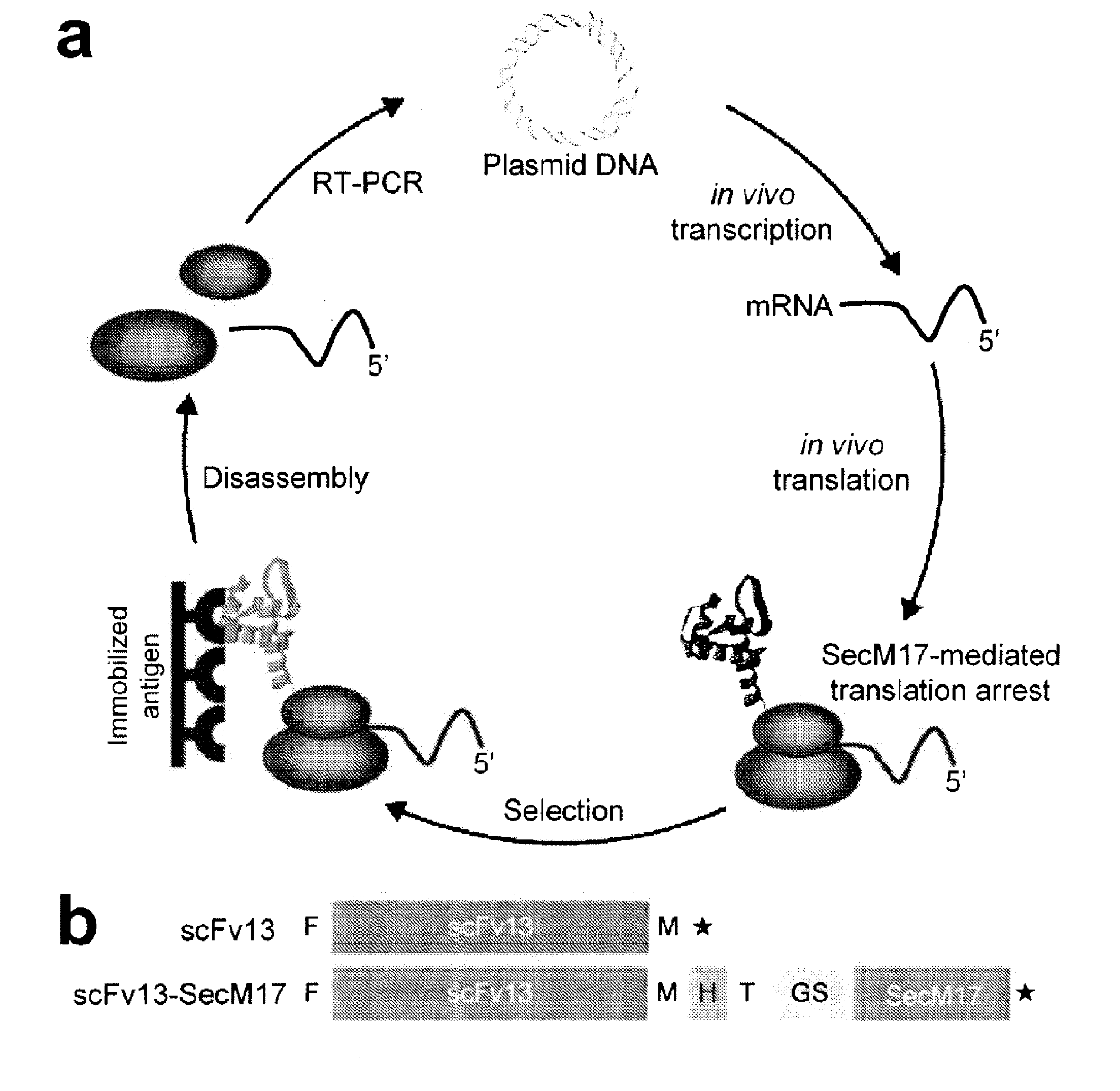
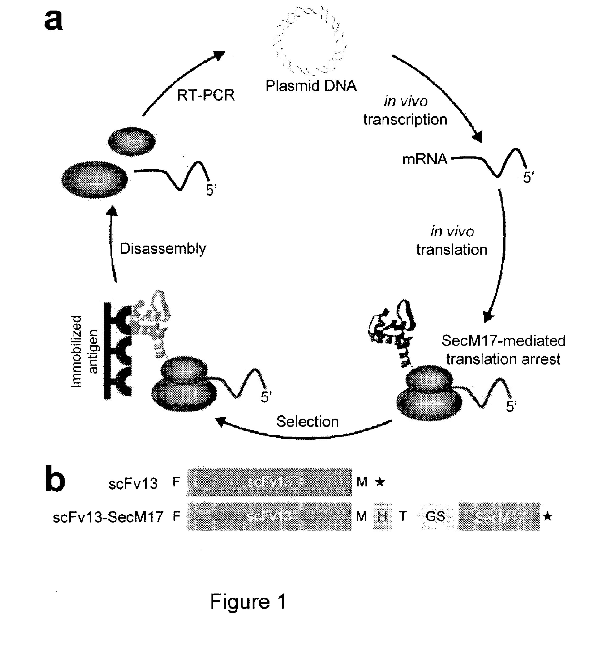
Protein discovery using intracellular ribosome display
a ribosome display and protein technology, applied in the field of protein discovery using intracellular ribosome display, can solve the problems of limiting the usefulness of ribosome display, cytoplasmic expression of scfvs is generally faced with difficulties concerning stability, solubility and aggregation, and requires five rounds of mutagenesis and selection, so as to achieve efficient folding and functional
- Summary
- Abstract
- Description
- Claims
- Application Information
AI Technical Summary
Benefits of technology
Problems solved by technology
Method used
Image

Examples
example 1
Bacterial Strains and Plasmids
[0059]E. coli strain BL21(DE3) was used throughout except for in vivo β-gal activation experiments where the E. coli 959 strain was used which carries the AMEF β-gal gene (Martineau et al., “Expression of an Antibody Fragment at High Levels in the Bacterial Cytoplasm,”J Mol Biol 280:117-27 (1998), which is hereby incorporated by reference in its entirety). Plasmids encoding the SecM stall sequence fusions were constructed as follows. First, a 285-nucleotide segment of the secM gene containing the 17-amino acid stall sequence (FSTPVWISQAQGIRAGP (SEQ ID NO: 1)), plus additional downstream regions, was amplified from plasmid pNH21 by PCR using primers (5′-CTCATGGTCGACTTCAGCACGCCCGTCTGG-3′ (SEQ ID NO: 5)) and (5′-CTCATGCTCGAGTTAAAGCTTCTGCGCAACTGTTGGGAAGC-3′ (SEQ ID NO: 6)) to introduce a SalI restriction site at the 5′ end and an XhoI-HindIII restriction site at the 3′ end. This PCR product was SalI-XhoI digested and ligated into the same sites of pET28a (N...
example 2
Random Mutagenesis of scFv13 Sequence
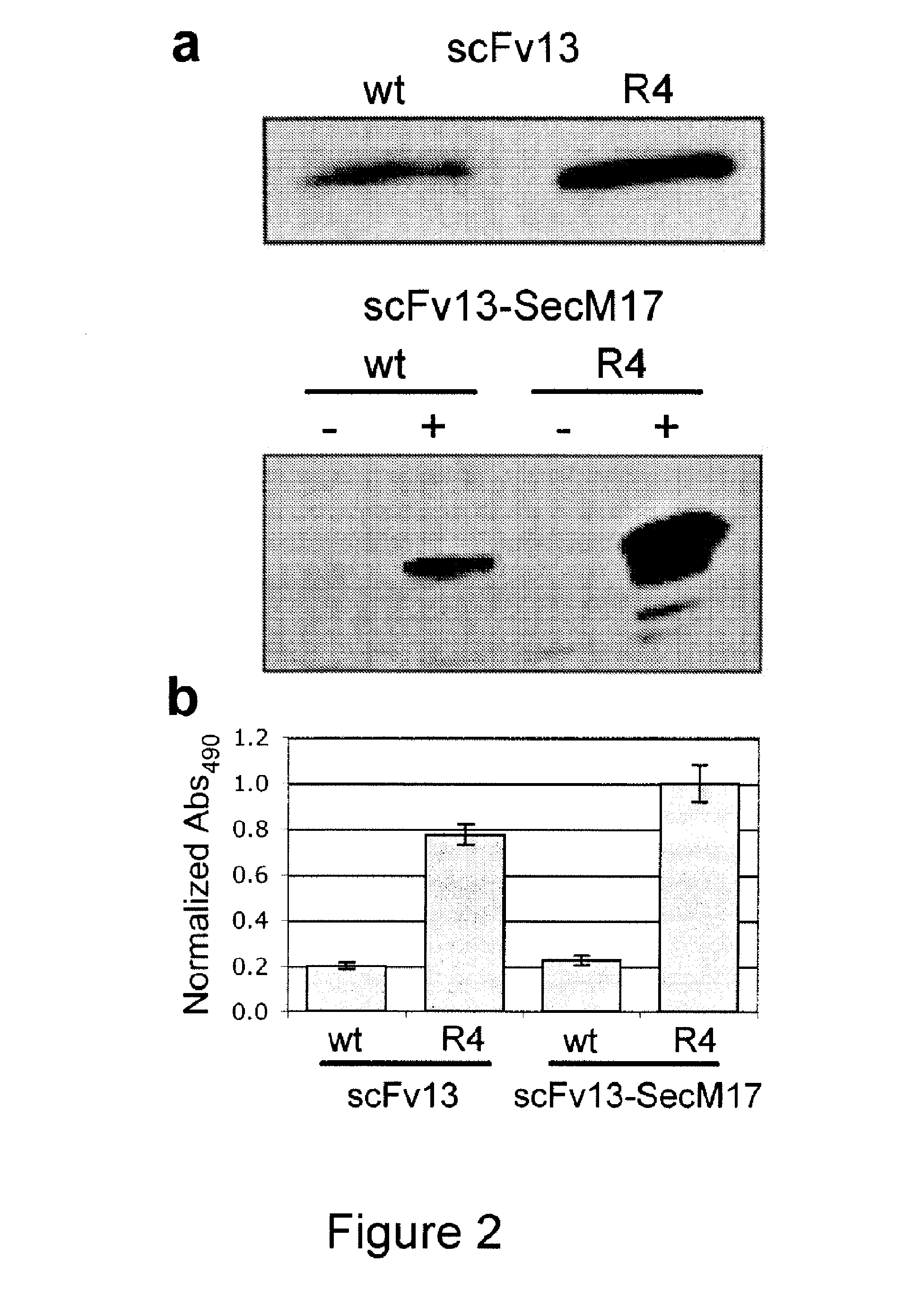
[0060]A library of random mutants was constructed by error-prone PCR of the scFv13 gene sequence using pET-scFv13-SecM17 as template and skewing the nucleotide and magnesium concentrations as described (DeLisa et al., “Genetic Analysis of the Twin Arginine Translocator Secretion Pathway in Bacteria,”J Biol Chem 277: 29825-31 (2002) and Fromant et al., “Direct Random Mutagenesis of Gene-Sized DNA Fragments Using Polymerase Chain Reaction,”Anal Biochem 224:347-53 (1995), which are hereby incorporated by reference in their entirety) to generate a 1.5% error-rate library. Error-prone PCR products were amplified with using primers (5′GCGATGCCATGGCCGACTACAAGGACGATGACGACAAGGGAGCCGAG GTGCAGCTG-3′ (SEQ ID NO: 22) and 5′-GCGATGGTCGACTGCGGCCCCATTCAG-3′ (SEQ ID NO: 23)), restriction digested with NcoI-SacI and ligated into the pET-scFv13-SecM17 that had previously been digested with NcoI-SacI to excise the wt scFv13 gene. Reaction mixtures were electroporate...
example 3
[0061]Cells transformed with the pET28a-derived scFv constructs were grown in 10-ml cultures at 37° C. in Luria-Bertani (LB) supplemented with kanamycin (50 μg / ml). Protein synthesis was induced by adding 1 mM isopropyl-β-D-thiogalactopyranoside (IPTG) when cells reached to mid log phase (OD600˜0.5). Cells were harvested after 1 hour of induction and pelleted by centrifugation for 15 min at 4° C. and 3,500 rpm. The soluble fraction was prepared by resuspending the pellet in 300 ml of phosphate buffered saline (PBS) solution followed by sonication (Branson Sonifier). The sonicant was spun for 15 min at 4° C. and 1,300 rpm and the resulting supernatant was collected as the soluble fraction.
PUM
| Property | Measurement | Unit |
|---|---|---|
| pH | aaaaa | aaaaa |
| volume | aaaaa | aaaaa |
| affinity | aaaaa | aaaaa |
Abstract
Description
Claims
Application Information