
Diagnostic method and kit

a diagnostic method and kit technology, applied in the field of diagnostic methods and kits, can solve the problems of inability to reliably diagnose the presence or absence of tuberculosis, low diagnostic accuracy, and low specificity of the current method used to monitor tuberculosis in animals, so as to achieve the effect of strong immune response, improved tuberculosis testing sensitivity, and reliable diagnosis of the presence or absen

Inactive Publication Date: 2011-01-27
ENFER TECH +1
View PDF1 Cites 3 Cited by
  • Summary
  • Abstract
  • Description
  • Claims
  • Application Information

AI Technical Summary

Benefits of technology

[0021]Each of the specific polypeptides is capable of eliciting a strong immune response in the absence of adjuvant. The characterized proteins / fragments which are immunogenic (or early antigens) and specific to one or other mycobacterial strains can be used reliably for the diagnosis of tuberculosis infection. As described in the examples, such polypeptides elicit a strong immune response in serum from animals immunised with Mycobacterium Bovis. Significantly, the inventors have found that, when these polypeptides are used together in an assay, the sensitivity of tuberculosis testing can be vastly improved over the sensitivity obtained using prior art methods. Thus, the invention, for the first time, enables the use of a single test to reliably diagnose the presence or absence of tuberculosis infection in many different populations of animals across differing geographical areas and thus may form the basis of a universal test.
[0155]DNA vaccines can easily be administered through intradermal application e.g. using a needle-less injector.

Problems solved by technology

Thus, infection of the animal population not only places a strain on economic resources but also presents a threat to human health.
However, the methods currently used to monitor tuberculosis in animals suffer from a number of drawbacks.
However, in 5% of cases, the CFT test may result in false-positive test results (due to exposure to or infection with other closely related bacteria, such as M. avium and M. paratuberculosis) or, in 15% of cases, in false-negative test results—where a very early stage of infection with bovine TB is not detected.
The Gamma-Interferon test is not used as a primary test for mass screening on its own because it does not detect all skin test-positive infected animals; it is relatively expensive and is less specific than the CCT.
Due to their modes of action, the specificity of both the intradermal skin test and the gamma interferon test will always be an issue.
The cross reactions induced by different mycobacteria strains and environmental mycobacteria such as M. microti and M. africanum and the conflicting requirements between specificity and sensitivity of the test antigens, all accrue to the difficulties in establishing a satisfactory serological protocol for bovine TB.
One of the problems associated with the complex antigenic nature of mycobacteria is the definition of those proteins which are important targets of the immune system and are thus likely present in large numbers of field samples.
However, as described herein the mere identification of such markers does not equate with practical utility in a diagnostic method.
Many markers are not expressed sufficiently to reliably be used in the identification of a strain.
In general however, antibody (Ab) tests based upon PPD tuberculins are characterised by a low discriminating power, with the distribution of the antibody titers between infected and non-infected animals being widely overlapping (Amadori et al.
However, although a great many antigens associated with tuberculosis infection have been identified in the prior art, to date, the use of particular combinations has not resulted in a test with an acceptable level of sensitivity and specificity to avoid the many false positive and false negative test results associated with conventional TB testing.
However, although relatively high sensitivity has been reported for some prior art tuberculosis kits, the sensitivity may only be maintained across selected populations of animals, for example within particular herds or in defined geographical regions, where variation in prevalent strains of tuberculosis is limited and thus variation between animals in their response to infection with such strains is limited.
Presently available diagnostic kits do not provide specific combinations of antigens with sufficient coverage over different populations, herds and geographical areas for the reliable diagnosis of tuberculosis.
Although many antigens have been identified, by simply increasing the number of antigens in a test kit, the sensitivity and specificity of the test may not necessarily be improved.
As well as the problems discussed above with respect to identification of antigens for reliable diagnosis of tuberculosis infection, the formats of many diagnostic kits currently used may contribute to the lack of reliability.
Infect. Immun. 66:1445-1452). the necessity of using multiple antigens in an assay, has, however, introduced another challenge.
Evaluation of the standard type of ELISA has shown sensitivity and specificity are reduced when multiple antigens are combined for analysis in a single well thus limiting the way a conventional ELISA can be used (Lyashchenko, et al 2000.
However, a number of problems persist with many of these methods.

Method used


Image

  • Diagnostic method and kit
  • Diagnostic method and kit
  • Diagnostic method and kit

Examples


example 1

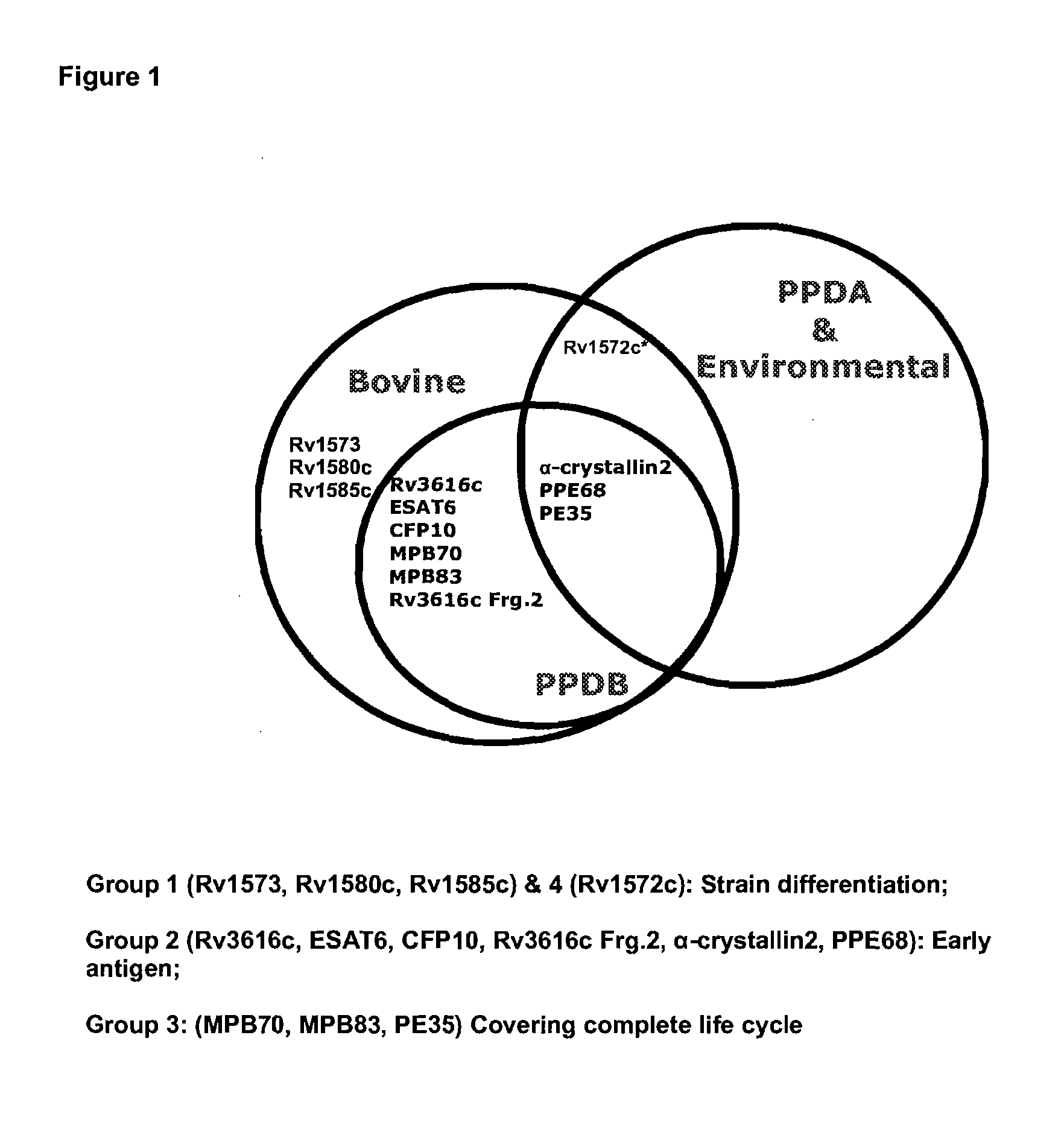
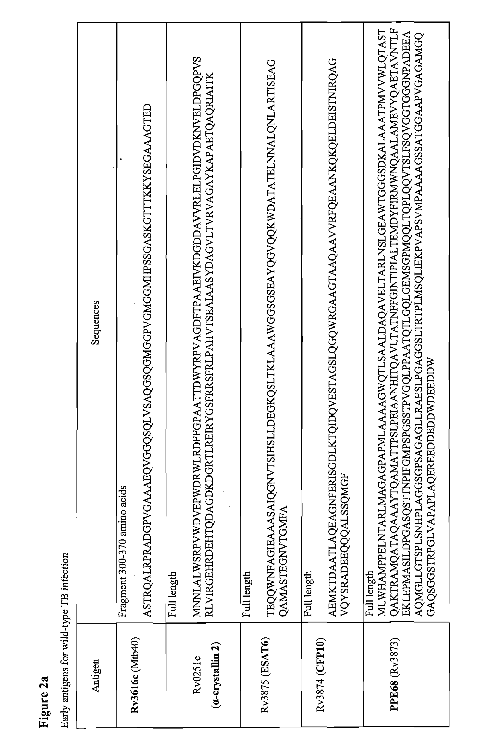
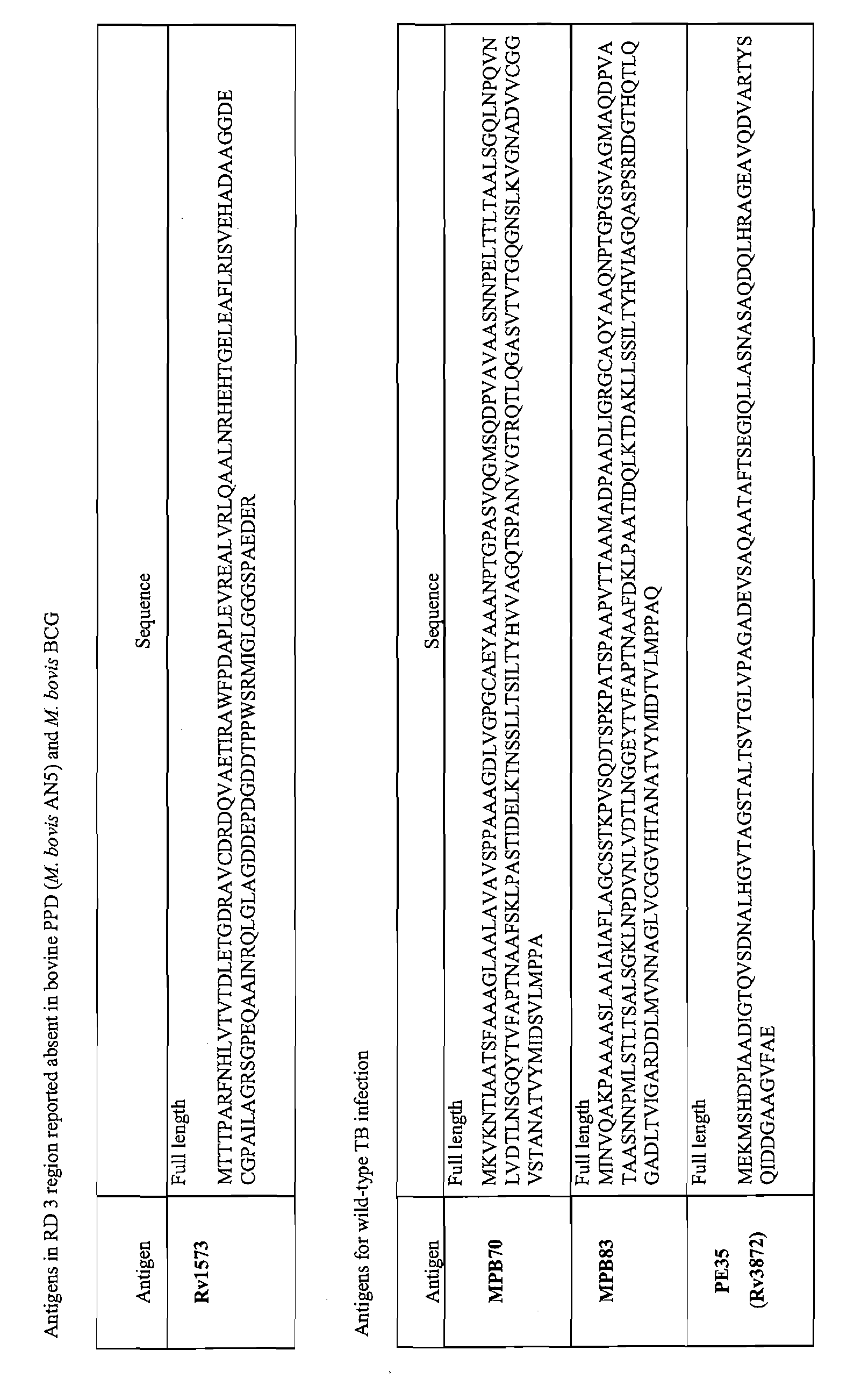
[0163]The inventors have used bioinformatics software to identify and characterise proteins which are specific to one or other mycobacterial strains. Using a genome alignment strategy the group has identified point mutations or deletions which allow differentiation between M bovis, PPD (M. bovis AN5 and M. avium), M. bovis BCG and environmental mycobacteria (FIG. 1). They have identified a panel of proteins capable of strain identification (FIG. 2(a).

[0164]As described below, the inventors have surprisingly found that by using a panel of seven or eight of these antigens, compared to known techniques, the accuracy of detection of tuberculosis may be greatly improved.

[0165]Significantly, a high degree of sensitivity demonstrated using this particular panel of antigens is obtainable across cohorts of tested animals from different geographical regions, giving a level of coverage which is unprecedented in tuberculosis testing.

Methodology

Cloning, Expression and Purification of Mycobacteri...

example 2

Multiplex Assays

Methods

Serum Samples

[0203]Serum samples used in the study were obtained from several sources. Blood samples were taken into serum tubes (Vacuette, serum clot activator tubes, Greiner-bio-one), transported at room temperature and then stored at 2-8° C. until processing. Following centrifugation (3000 g, 30 minutes at 2-8° C.) the serum was removed, aliquoted and stored at −20° C.

[0204]Two sets of sera were obtained, one from a negative sample bank including sera from herds of animals with a known history of being free of Mbv. A third group of sera were collected from animals that were proven to be positive for Mbv infection at the time of slaughter based on subsequent histopathological / bacteriological examination.

[0205]The fourth set of serum samples was part of an infectivity study. The animals were non-vaccinated and challenged via the intra-tracheal route with a low dose of a virulent strain of Mbv (approximately 5,000 CFU). Sera were collected prior to challenge a...

example 3

Identification of Panel of Antigens for Diagnosis of TB in Bovines

[0217]The inventors used the multiplex platform described above to analyse antigen expression from 522 cattle, which had been confirmed TB positive by histopathology and culture methods, using 1489 cattle, which had been confirmed TB negative using tuberculin skin tests, as the controls. The inventors identified a panel of five antigens which when assayed together enable diagnosis of tuberculosis infection with very high sensitivity and selectivity. The assaying for the presence of at least two of the five antigens ESAT-6+Rv3616c+pep1 (MPB70)+MPB83+MPB70 gave a sensitivity of 93.7%, compared to 71.4% obtained with ESAT-6+Rv3616c+pep1 (MPB70)+MPB83, 25.5% obtained using ESAT-6+Rv3616c+pep1 (MPB70). The specificity obtained with ESAT-6+Rv3616c+pep1 (MPB70)+MPB83+MPB70 was 97.6%.


PUM

PropertyMeasurementUnit
volumeaaaaaaaaaa
volumeaaaaaaaaaa
volumeaaaaaaaaaa

Abstract

The invention relates to methods for diagnosis of tuberculosis in an animal, the methods comprising determining the presence or absence of, or an immune response to a group of antigens comprising (i) the group consisting of Rv3616 antigen, MPB70 antigen, MPB83 antigen, and ESAT 6 antigen; or (ii) at least seven of the antigens of the group consisting of Rv3616 antigen, MPB70 antigen, MPB83 antigen, ESAT 6 antigen, CFP10 antigen, α crystalline 2 antigen, PE35 antigen, and PPE68 antigen. Also provided is a method for the diagnosis of the presence of tuberculosis in a reservoir animal e.g. a badger, said method comprising determining the presence or absence of, or an immune response to Rv3616 antigen, MPB70 antigen, and MPB83 antigen. Kits for carrying out the methods are also provided.

Description

FIELD OF THE INVENTION[0001]The invention relates to methods and kits for the diagnosis and / or herd profiling of Mycobacterium infected or contaminated animals.BACKGROUND TO THE INVENTION[0002]Tuberculosis (TB), one of the most widespread infectious diseases, is the leading cause of death due to a single infectious agent among adults in the world. Mycobacterium tuberculosis is the most common cause of human TB.[0003]However, an unknown proportion of cases of zoonotic tuberculosis are due to M. bovis with some due to Mycobacterium avium. Thus, infection of the animal population not only places a strain on economic resources but also presents a threat to human health. The requirement for successful diagnostic assays and potential vaccines has increased due to the recent rise in TB levels in the cattle population of countries such as Great Britain and the wild life reservoirs of the world.[0004]Great Britain performs some 4.6 million tests on bovine TB, costing the tax payer £88 m per ...

Claims


Application Information

Patent Timeline
Patent Type & Authority Applications(United States)
IPC IPC(8): A61K39/04C12Q1/04C12Q1/02G01N33/53C12Q1/68A61P31/06
CPCG01N33/5695A61K39/04A61P31/06Y02A90/10
Inventor OLWILL, SHANE A.JOHNSTON, JAMES A.KWOK, HANG FAIWHELAN, CLARECLARKE, JOHN
Owner ENFER TECH
Learn More