
Stable amyloid beta monomers and oligomers

Active Publication Date: 2011-03-17
ALZINOVA
View PDF9 Cites 1 Cited by
  • Summary
  • Abstract
  • Description
  • Claims
  • Application Information

AI Technical Summary

Benefits of technology

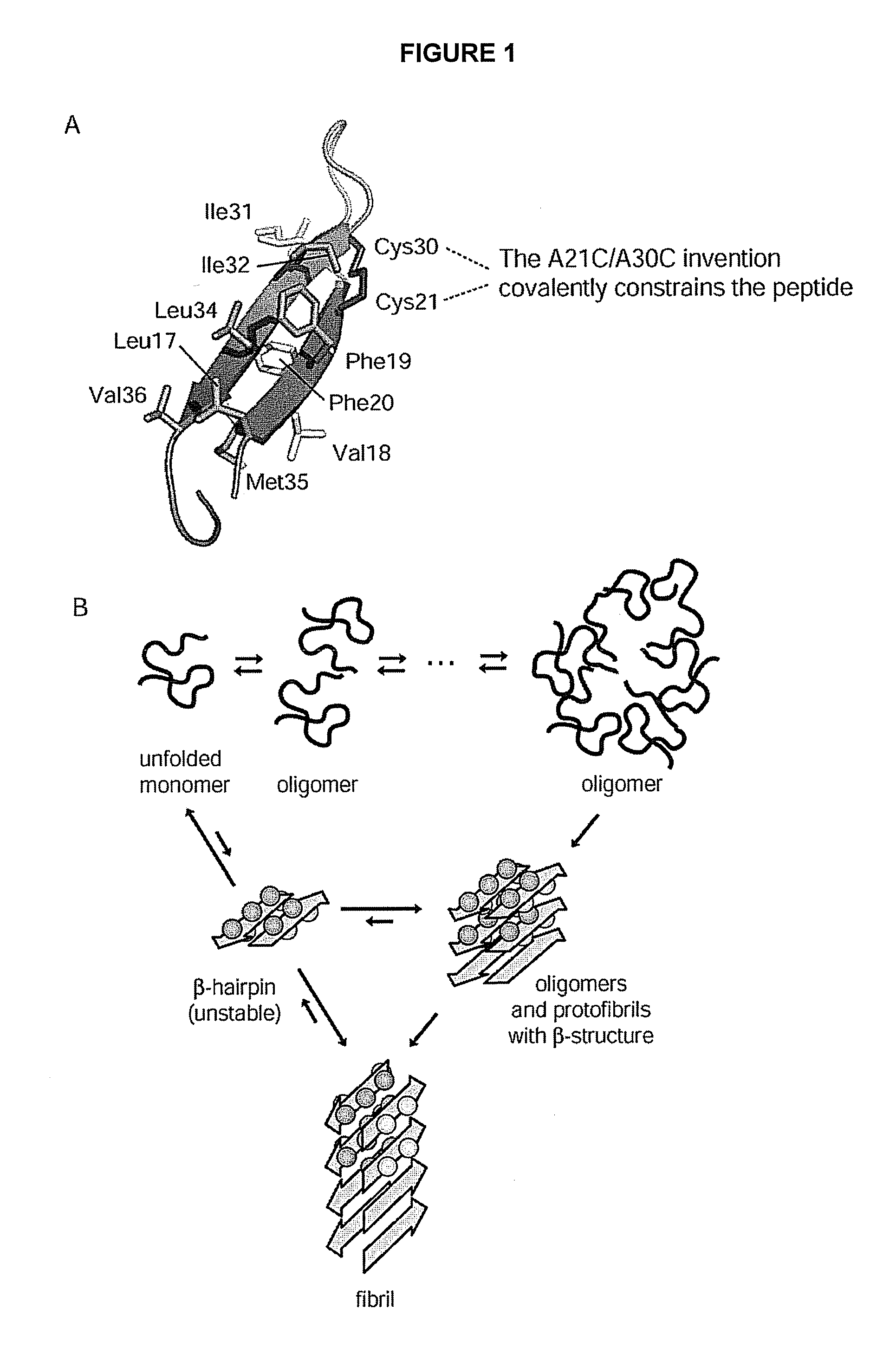
[0011]Because of the constraining disulphide bond present in the peptides according to the invention, other methods to stabilize oligomers are not needed, including (i) non-physiological (elevated) pH to increase repulsion of the peptides, (ii) using additives including 2,2,2-trifluoroethanol (TFE), sodium dodecyl sulphate (SDS), glycerol, or cell membrane glycolipids in the form of GM1 gangliosides, to induce oligomeric structure, (iii) adding anions to screen charges thus increasing solubility, and (iv) using an intermolecular cross-link that covalently stabilizes the oligomers once they are prepared by one or several of alternatives (i)-(iii). The Aβ peptides according to the invention are in fact the first examples of Aβ peptides forming stable oligomers under physiological conditions, this being obtained by arresting the fibrillation step. Furthermore, long incubation times and unpredictable outcomes of oligomeric preparations are avoided, as the Aβ peptides according to the invention can be purified and rapidly concentrated into the oligomeric states.

Problems solved by technology

There are at present no effective treatments against AD and most of the other protein misfolding diseases, partly owing to the fact that it is still unclear if and how these deposits are toxic.
However, both monomeric and oligomeric Aβ proteins have a high tendency to aggregate further into fibrils.
In consequence, fibrillogenesis seriously limits the longevity of the oligomeric preparations, and also the concentrations at which they can be kept.
USA 93: 1125-1129 (1996)), any increase in the concentration of peptide above 100 μM will be detrimental to the stability of the oligomer preparation.
Oligomer preparations therefore have a very limited and unpredictable (aggregation nucleation is a spontaneous event) lifespan once they are formed.
The fibrillation process is thus a serious drawback when, as in screening assays for medicaments, stable proteins are required at relatively high concentration.

Method used


Image

  • Stable amyloid beta monomers and oligomers
  • Stable amyloid beta monomers and oligomers
  • Stable amyloid beta monomers and oligomers

Examples


example 1

Fibrillation Assay of Wild-Type Aβ(40) and Aβ(40) A21C / A30C

[0077]All concentration determinations were carried out spectrophotometrically using an extinction coefficient of 1424 cm−1 M−1 for the difference in absorption at 280 nm and 300 nm. The buffered solution used in all examples was 50 mM K+ phosphate, 50 mM NaCl, pH 7.2, unless otherwise stated. This buffer was also the storage buffer for the Aβ peptides. All peptide samples were prepared fresh, kept at 4° C., and used within 3-4 days.

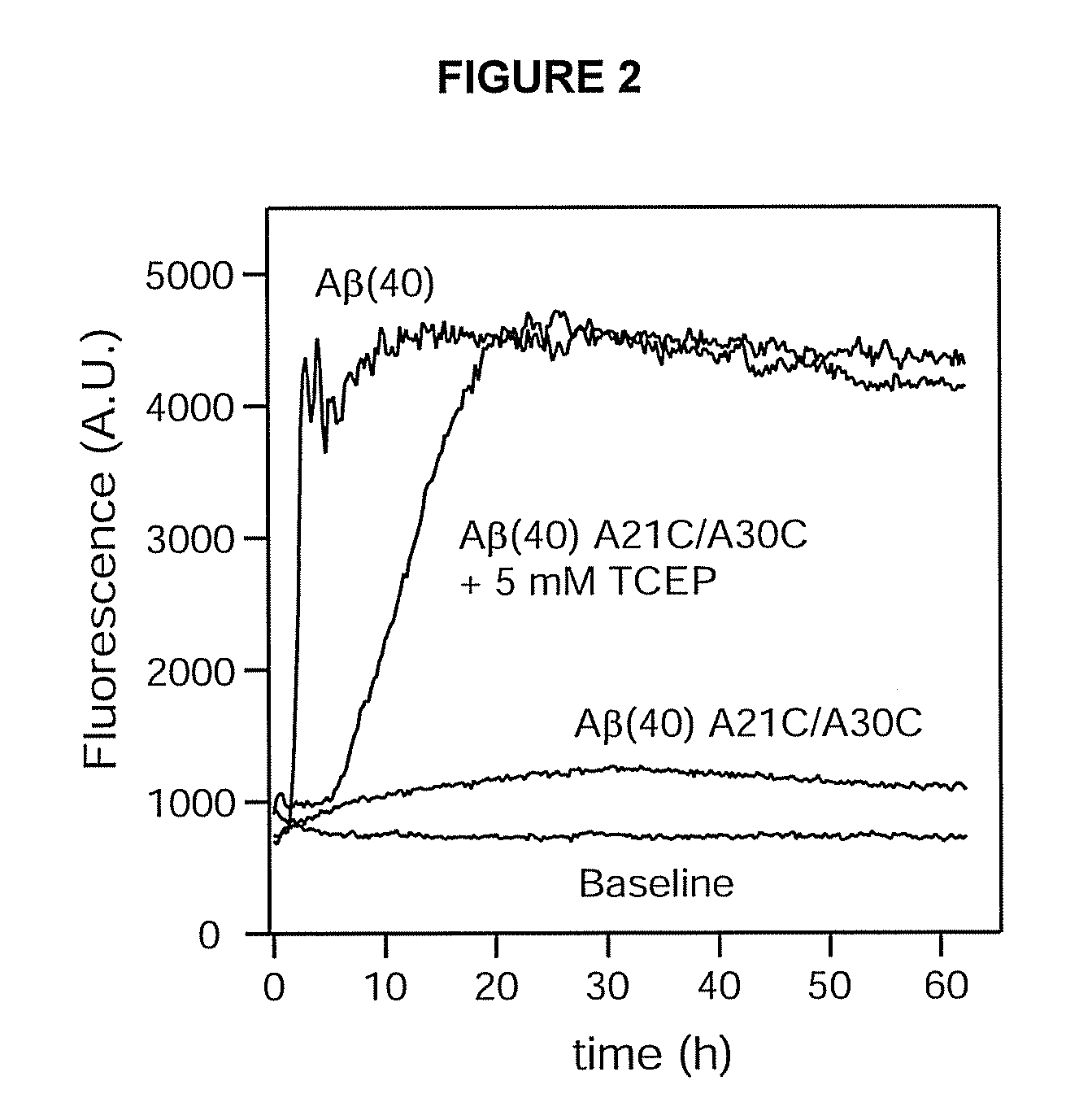
[0078]Fibrillation assays were carried out by monitoring the enhanced fluorescence of the dye TFT upon binding to the fibrils (Levine, H., Methods Enzymol. 309: 274-284 (1999)). Fluorescence was recorded in 96-well plates (Nunc) using a FLUOstar Optima reader (BMG) equipped with 440 nm excitation and 480 nm emission filters. The peptide samples were kept at 30 μM and were supplemented with 10 μM TFT. In addition, 5 mM TCEP was added to one reference sample of the invention to assay the effect of ...

example 2

Fibrillation Assays of Aβ(40) A21C / A30C at Increasing Concentrations

[0080]The fibrillation assay was carried out as in example 1, except that the concentration of peptide was also assayed at 50, 150, and 250 μM, and the TCEP concentration was increased to 10 mM TCEP in the reference samples. Also, data points were recorded every 15 min with 5 min of orbital shaking (width 5 mm) preceding each measurement. Dual samples were analyzed at each concentration, and the result is presented in FIG. 3.

[0081]The result is similar to the result obtained in Example 1. However, there is a rapid increase in fluorescence for the samples with an intact disulphide bond at the 150 and 250 μM concentrations, which then decline to similar levels with much slower rates. As demonstrated in Examples 7 and 8 below, the coil-like oligomers can be made to form protofibrillar-like structures when subjected to elevated temperatures, and these do bind TFT weakly thus giving rise to enhanced fluorescence. As time...

example 3

Wild-Type-Like Structure of Aβ(40) A21C / A30C

[0083]SEC was carried out on a Superdex 75 10 / 300 column (GE Healthcare) equilibrated with 50 mM K+ phosphate, 50 mM NaCl, pH 7.2. The flow rate was 0.8 ml min−1, and the column was operated by an ÄKTA Explorer unit (GE Healthcare). All column runs were carried out at room temperature (21° C.). The column was calibrated with a low molecular-weight gel filtration calibration kit (Amersham Biosciences) under similar conditions. Typically, 500 μl of peptide was injected into this column during each run.

[0084]At low concentrations of peptide, e.g. 25 μM, the Aβ(40) A21C / A30C peptide typically elutes as an approximately 8-10 kDa protein. Even though the Aβ peptide is approximately a 4.5 kDa protein, we do not interpret this as this peptide is eluting as a dimer because of the potentially extended structures of unfolded coil-like proteins compared to the well-folded proteins used in the calibration kit (ribonucelase A, chymotrypsinogen A, ovalbu...


PUM


Abstract

The invention provides monomeric and oligomeric amyloid beta peptide isomers that are resistant towards fibrillogenesis and their use as screening reagents or antigens in methods and pharmaceutical preparations for the treatment of Alzheimer's disease and other conditions related to protein misfolding. The alanines at positions 21 and 30, in wild type amyloid beta peptide amino acid sequence, are according to the invention replaced by cysteins, which results in an intra molecular disulphide bond. The invention further provides transgenic animals expressing modified amyloid precursor proteins or amyloid beta peptides.

Description

FIELD OF THE INVENTION[0001]The invention relates to monomeric and oligomeric amyloid beta peptides that are resistant towards fibrillogenesis for use as antigens or screening reagents for the treatment of Alzheimer's disease and other conditions related to protein misfolding.BACKGROUND OF THE INVENTION[0002]Alzheimer's disease (AD) is an age-related neurodegenerative disease characterized by the accumulation of insoluble amyloid deposits of the amyloid-β (Aβ) protein generated by the cleavage of the amyloid precursor protein (APP) (Blennow, K., et al., Lancet 368: 387-403 (2006)). AD is one of several diseases caused by protein misfolding, which includes over 22 other known ailments such as Parkinson's disease and Type II diabetes (Dobson, C. M., Protein Pept. Lett. 13: 219-227 (2006)). There are at present no effective treatments against AD and most of the other protein misfolding diseases, partly owing to the fact that it is still unclear if and how these deposits are toxic. In f...

Claims


Application Information

Patent Timeline
IPC IPC(8): A61K39/395C07K5/00C07K7/06C07K7/08C07K14/00C07K16/18G01N33/53A61K38/16A01K67/00C07H21/00A61P25/28
CPCC07K14/4711A61K38/00A61P3/00A61P25/00A61P25/16A61P25/28A61P37/04G01N2800/2821G01N2800/52
Inventor HARD, TORLEIFSANDBERG, ANDERS
Owner ALZINOVA
Learn More