Directed Evolution and In Vivo Panning of Virus Vectors
a virus vector and in vivo panning technology, applied in the field of directed evolution and in vivo panning of adenoassociated virus vectors, can solve the problems of inability to select for both of these properties, inability to identify mutants optimized by vitro screening methods, and inability to detect mutants with different forms of md
- Summary
- Abstract
- Description
- Claims
- Application Information
AI Technical Summary
Benefits of technology
Problems solved by technology
Method used
Image
Examples
example 1
Materials & Methods
[0323]Generation of Chimeric AAV Library with Shuffled Capsid Genes
[0324]For construction of random chimeric AAV capsid gene libraries, AAV serotypes 1, 2, 3B, 4, 6, 7, 8, 9 were used as PCR templates. The capsid genes were amplified by primers CAP-5′ (5′-CCC-AAGCTTCGATCAACTACGCAGACAGGTACCAA-3′; SEQ ID NO:139) and CAP-3′ (5′-ATAAGAAT-GCGGCCGC-AGAGACCAAAGTTCAACTGAAACGA-3′; SEQ ID NO:140) and mixed in equal ratio for DNA shuffling (Soong et al., (2000) Nat Genet. 25: 436-9). In brief, 4 μg of the DNA templates were treated by 0.04 U of DNase I at 15° C. briefly. DNA fragments in size of 300-1000 by were purified by agarose gel electrophoresis, denatured, re-annealed and repaired by pfu DNA polymerase to reassemble random capsid genes. Amplification was done by use of pfu DNA polymerase and CAP5′ / CAP3′ primers. The PCR program was 30 cycles of 94° C. 1 min, 60° C. 1 min and 72° C. 4.5 min. The PCR products were then digested with Hind III and Not I and ligated into a...
example 2
Direct In Vivo Panning of DNA-Shuffled AAV Library for Muscle-Targeting Capsids
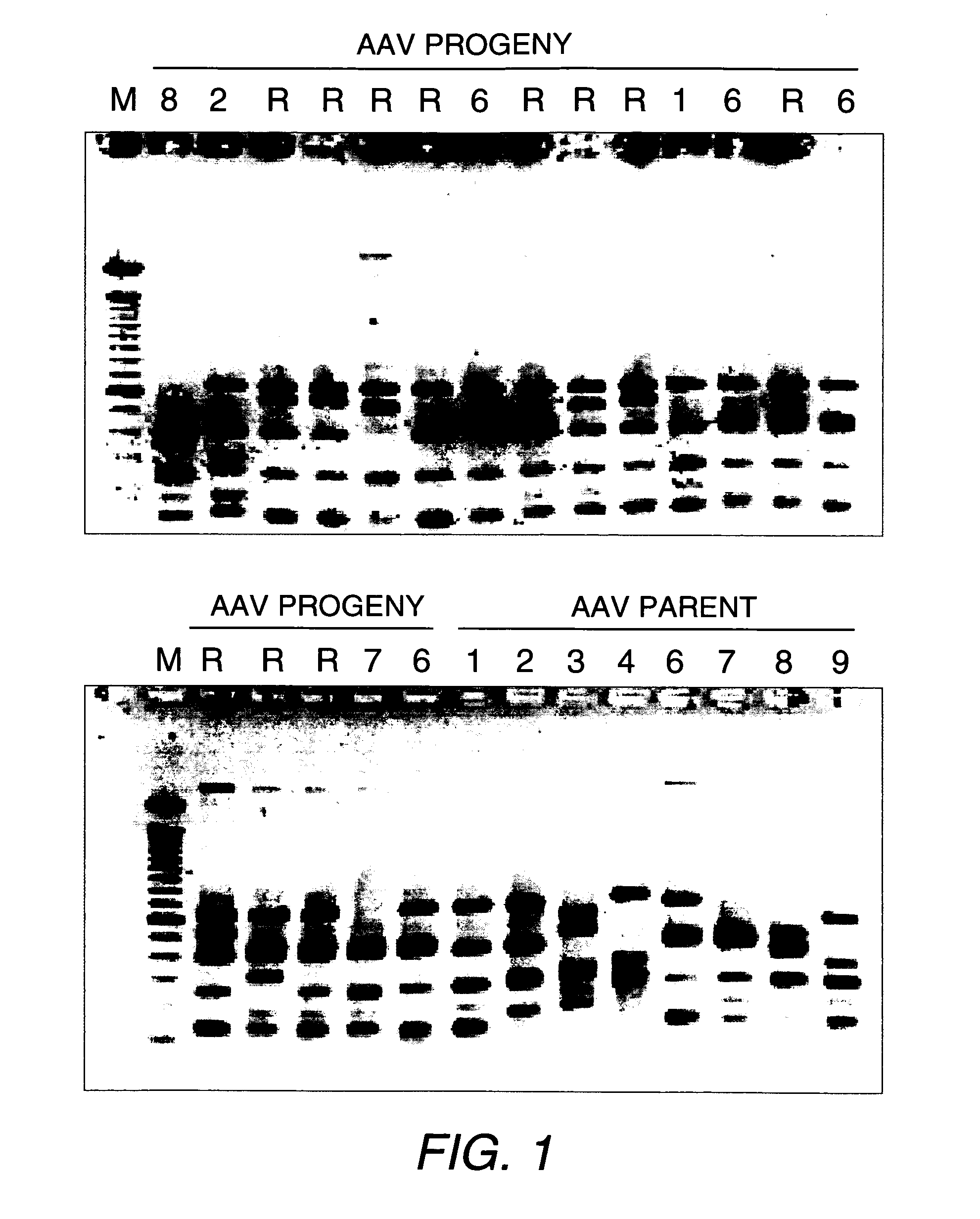
[0330]We constructed a chimeric AAV library by DNA shuffling of the capsid genes of AAV1, 2, 3, 4, 6, 7, 8 and 9, in order to select for combinations of characteristics. The infectious AAV library with shuffled capsid genes was packaged by the method of Muller et al. (Nature Biotechnol. (2003) 21:1040-1046). DNA analysis by restriction digestions on randomly picked mutant AAV clones showed unique patterns and indicated that the vast majority were recombinants and viable in producing AAV particles (FIG. 1).
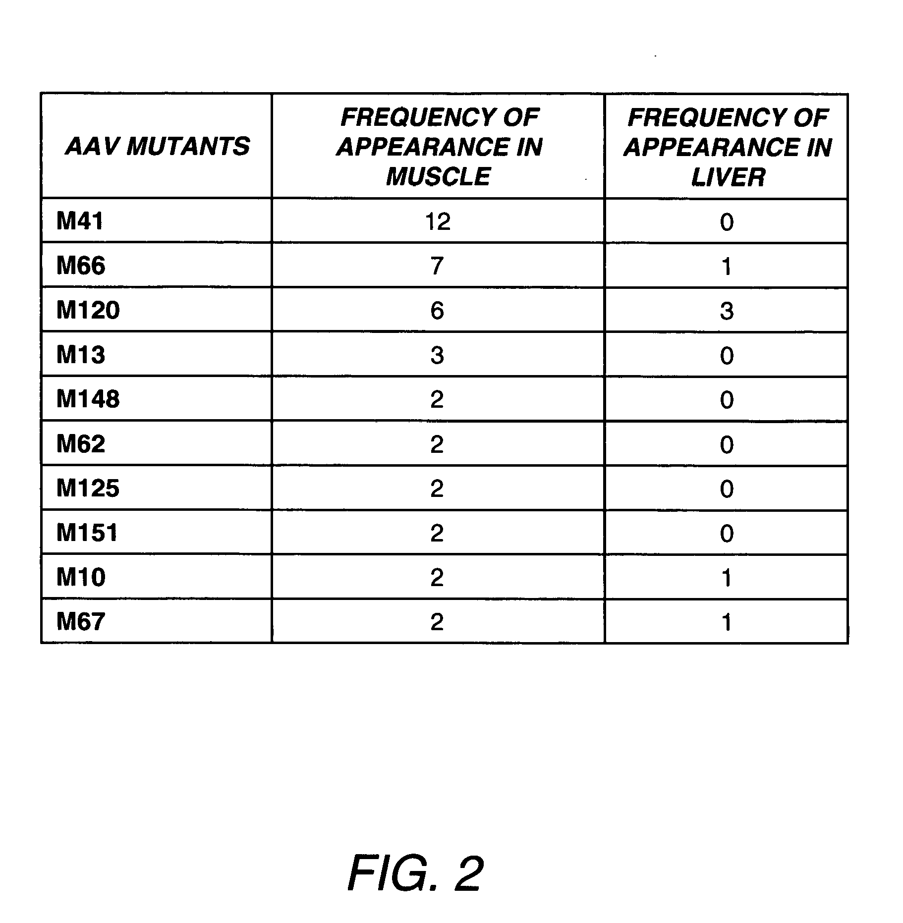
[0331]Although it is known to use in vitro cell culture systems to screen for desirable mutant AAVs, here we solely relied on a direct in vivo screening method, because no cell culture system could simultaneously mimic the in vivo conditions (e.g., the tight endothelial lining, the differentiated muscle cells and the liver, etc.). We used adult mice for in vivo biopanning of the AAV library. Following ta...
example 3
M41 Vector Preferentially Transduces Myocardium after Systemic Administration
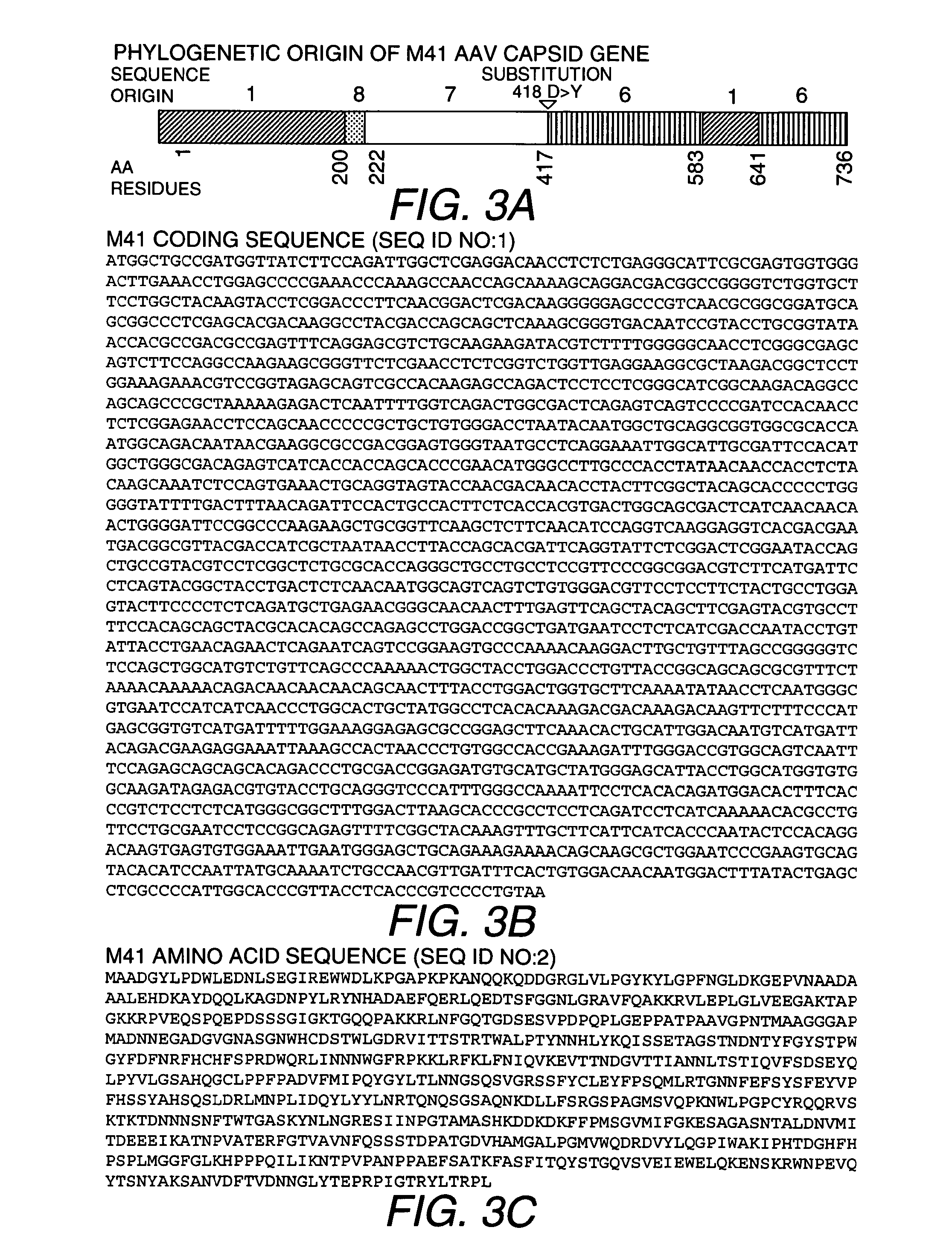
[0333]We next investigated systemic gene delivery efficiency and tissue tropism of AAVM41. The luciferase reporter gene was packaged into viral capsids of M41, AAV9, and AAV6 for a side-by-side comparison in vivo. At 2 weeks post i.v. injection in young adult C57BJ / 6L mice (6-8 wk), luciferase activities and vector DNA copy numbers in various tissues were analyzed. Consistent with previous reports (Inagaki et al. (2006) Mol Ther 14: 45-53), the AAV9 vector efficiently transduced mouse heart, skeletal muscles, and particularly the liver, which had the highest luciferase activity (FIG. 4A) and vector DNA copy numbers (FIG. 4B). Similar to AAV9, the M41 vector also transduced the heart efficiently with slightly lower luciferase activity and vector copy numbers (FIG. 4B). In contrast, M41 showed dramatically reduced gene transfer in the liver, with the luciferase activity 81.1 fold lower and DNA copy number 11....
PUM
| Property | Measurement | Unit |
|---|---|---|
| pH | aaaaa | aaaaa |
| δ-SG | aaaaa | aaaaa |
| nucleic acid | aaaaa | aaaaa |
Abstract
Description
Claims
Application Information
 - R&D
- Intellectual Property
- Life Sciences
- Materials
- Tech Scout
- Unparalleled Data Quality
- Higher Quality Content
- 60% Fewer Hallucinations
Browse by: Latest US Patents, China's latest patents, Technical Efficacy Thesaurus, Application Domain, Technology Topic, Popular Technical Reports.
© 2025 PatSnap. All rights reserved.Legal|Privacy policy|Modern Slavery Act Transparency Statement|Sitemap|About US| Contact US: help@patsnap.com


