Targeting of innate immune response to tumor site

Active Publication Date: 2011-05-26
HADASIT MEDICAL RES SERVICES & DEVMENT +1
View PDF15 Cites 25 Cited by
  • Summary
  • Abstract
  • Description
  • Claims
  • Application Information

AI Technical Summary

Problems solved by technology

Binding of antibodies to tumor, native or as carriers of various toxins may damage the tumor cells.
Nevertheless, the number of cells in solid tumors is huge, and continue to proliferate and adjust to the treatment during the period of the treatment.
Innate immune responses may affect tumor mass, but in most cases

Method used


Image

  • Targeting of innate immune response to tumor site
  • Targeting of innate immune response to tumor site
  • Targeting of innate immune response to tumor site

Examples


example 1

Production of Microbeads Carrying Anti-HER2 mAb and LPS

(i) Binding of Antibodies to Avidin-coated Beads

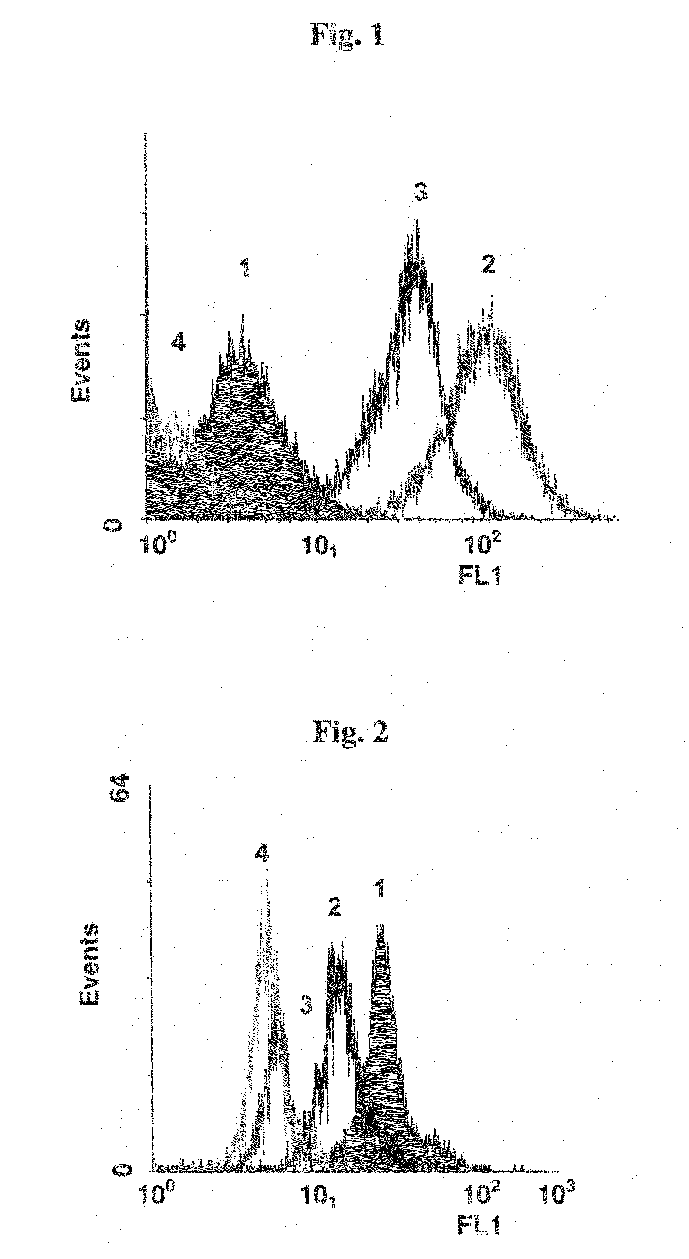
[0099]Monoclonal anti-HER2 antibody (anti-HER2, Herceptin) was biotinylated using the Sulfo-NHS-LC-Biotin kit (Pierce) according to the manufacturer's instructions. Biotinylation rate was determined by HABA test (Acros, USA) according to the manufacturer's instructions. The ability of the biotinylated mAb (Biot-anti-HER2) to detect HER2 was examined by incubation with target cells (SK-BR-3 human breast cancer cells expressing HER2, ATCC) that were stained with STAVF (streptavidin-FITC, Jackson ImmunoResearch). Incubated samples were then examined using Flow Cytometry (FACS). FIG. 1 demonstrates binding of the biotinylated mAb Biot-anti HER2 to SK-BR-3 cells (HER2 positive).

(ii) Biotinylation of Lipopolysaccharide (LPS) [SUPPLIER]

[0100]LPS (Sigma L2654) was biotinylated (Biot-LPS) using biotin hydrazide (Pierce) according to the manufacturer's instructions. Biotinylation efficiency ...

example 2

Binding of Microbeads Carrying Anti-HER2 and LPS to SK-BR-3 Cells

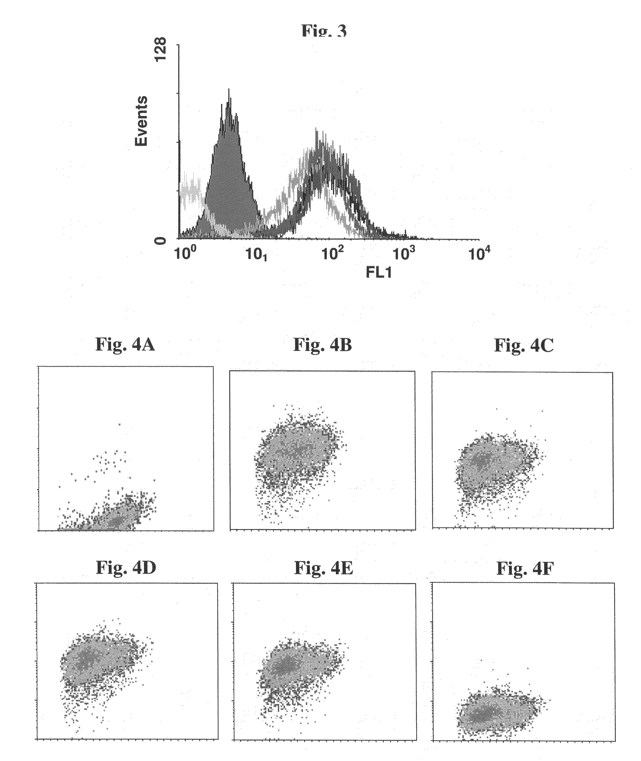
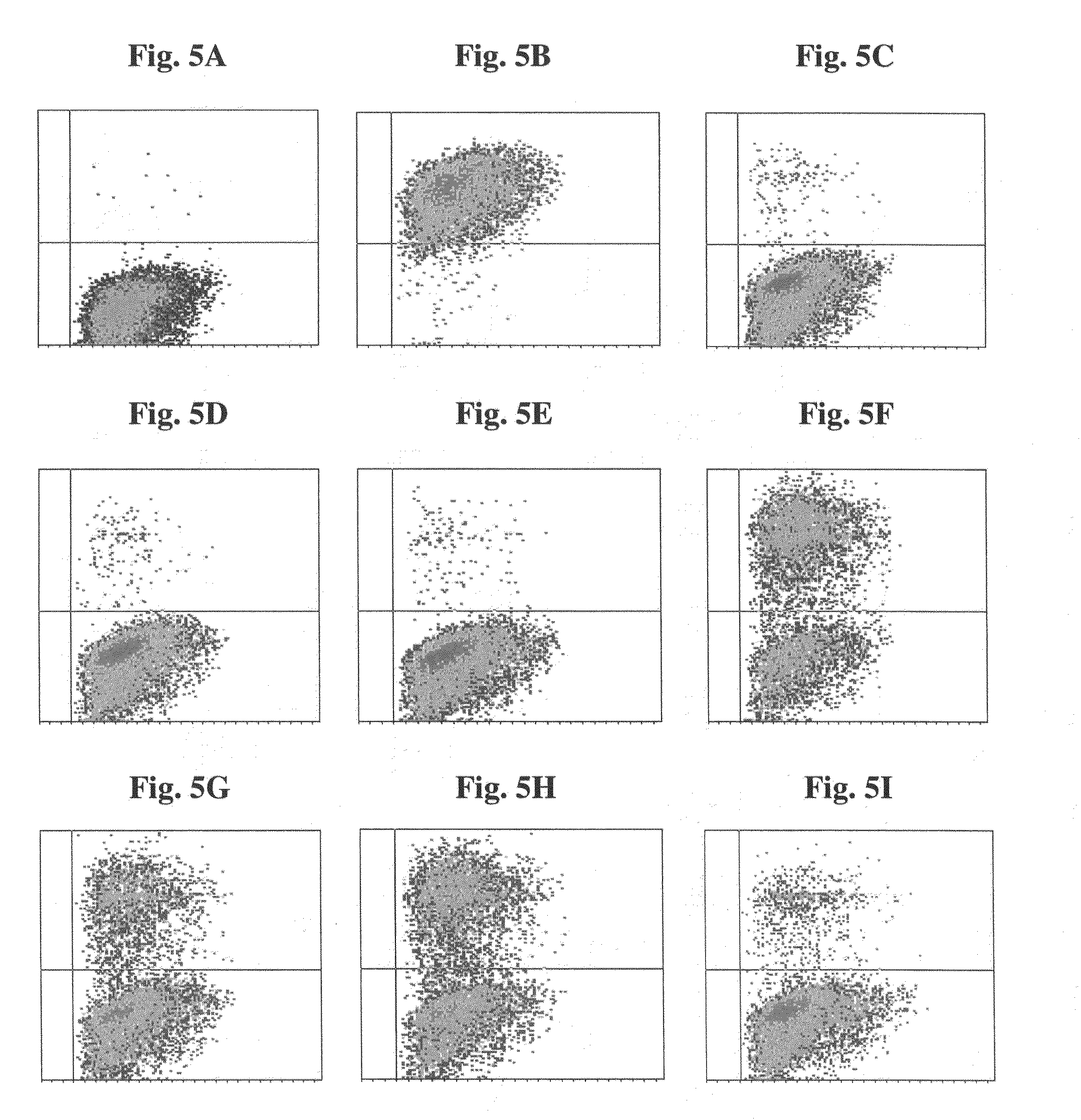
[0102](a) Mixtures of biot-anti-HER2 and biot-LPS, at various ratios, were attached to fluorescent polystyrene beads 2 μm in diameter (F-Beads) (Fluoresbrite YG Polysciences) or Biomag™ particles (approximately 1.5 μm) (Pierce) covalently coated with avidin, and the beads were incubated with SK-BR-3 cells. Samples were fluorescently stained with goat anti human Fc-FITC Abs and examined by FACS. Both Biomag™ particles (FIGS. 3 and 4) and F-Beads complexes (FIG. 5) demonstrated ability to bind SK-BR-3 target cells when anti-HER2 mAbs were attached to them.

[0103](b) SK-BR-3 cells were incubated overnight in 24-well plates. Biomag™ particles of (a) were added and incubated for one hour, followed by two washes. Samples were fluorescently stained with goat anti-human Fc-FITC for one hour, followed by two washes. Microbeads carrying anti-HER2 mAb and LPS attached to cells were detected by fluorescence microscope. FIG. 6 shows...

example 3

THP-1 Monocytes Response to Beads Carrying Anti-HER2 mAb and LPS

(i) Induction of Uptake

[0105]Human monocytic THP-1 cells (ATCC) were incubated overnight in 24-well plates. Ten-μm polystyrene beads carrying anti-HER2 mAb and LPS particles were added and incubated for 3 hours. FIG. 8 shows the uptake of the 10 μm polystyrene beads by THP-1 monocytes.

(ii) Induction of IL-1 Transcription

[0106]THP-1 cells were incubated overnight in 6-well plates. Biomag™ particles carrying anti-HER2 mAb and LPS were added and incubated for 3 hours. Growth medium was discarded. Cells were centrifuged, RNA was extracted using trisol reagent (Sigma). RT-PCR reaction (Promega kit) was performed using Oligo dT primers (Sigma), and the resulted cDNA was used in PCR reaction (Bioline kit) to propagate the IL-1 gene product. The PCR product was run on agarose gel and compared to GAPDH (glyceraldehyde-3-phosphate dehydrogenase, a constitutively expressed gene). DNA was stained with ethidium bromide. Analysis of ...


PUM

PropertyMeasurementUnit
Sizeaaaaaaaaaa
Sizeaaaaaaaaaa

Abstract

The invention provides microparticles or nanoparticles for treatment of tumors comprising: (i) a targeting agent to the tumor or the tumor environment; and (ii) at least one inducer that stimulates a desired immune response in the tumor environment, leading to tumor apoptosis, wherein components (i) and (ii) are non-covalently or covalently attached to the surface of said microparticles or nanoparticles. The targeting agent is an agent that recognizes and binds to an antigen, a receptor or other molecules found on the surface of tumor cells or in the tumor environment and are preferably antibodies.

Description

FIELD OF THE INVENTION[0001]The present invention is in the field of Immunology and relates to microparticles and nanoparticles and compositions comprising an agent for targeting to malignant tumors and an agent that triggers specific immune cells to cause apoptosis of the tumors. Specifically, the current invention relates to particles carrying a tumor-targeting component and an inducer that stimulates a local immune response aimed at tumor elimination.BACKGROUND OF THE INVENTION[0002]Tumor specific or over-expressed antigens were detected on the surface of various cancer cells. e.g. HER2, specific MUC I, GM2 and MHC class II on melanoma cells. These and other markers may be used to target tumors, using specific antibodies, for example. Antibodies against tumor antigens were developed and are used in immunotherapy. Binding of antibodies to tumor, native or as carriers of various toxins may damage the tumor cells. Nevertheless, the number of cells in solid tumors is huge, and contin...

Claims


Application Information

Patent Timeline
IPC IPC(8): A61K39/395A61K9/14A61P35/00
CPCA61K47/484A61K47/48853A61K47/48584A61K47/6807A61K47/6855A61K47/6921A61P35/00C07K16/32C07K2317/24C07K2317/40A61K9/485A61K9/5052A61K9/5073A61K39/00A61K39/38A61K39/385
Inventor PITCOVSKI, JACOBSHAHAR, EHUDGORODETSKY, RAPHAEL
Owner HADASIT MEDICAL RES SERVICES & DEVMENT
Why Eureka
Try Eureka