
System for Blood Separation with Shielded Extraction Port and Optical Control

a technology of shielded extraction and optical control, which is applied in the direction of separation processes, instruments, centrifuges, etc., can solve the problems of loss of certain blood components, no longer available for collection, and significantly decrease the efficiency so as to reduce the possibility of stagnation, reduce the volume of white blood cell collection, and increase the flow velocity

Active Publication Date: 2011-06-16
TERUMO BCT
View PDF27 Cites 26 Cited by
  • Summary
  • Abstract
  • Description
  • Claims
  • Application Information

AI Technical Summary

Benefits of technology

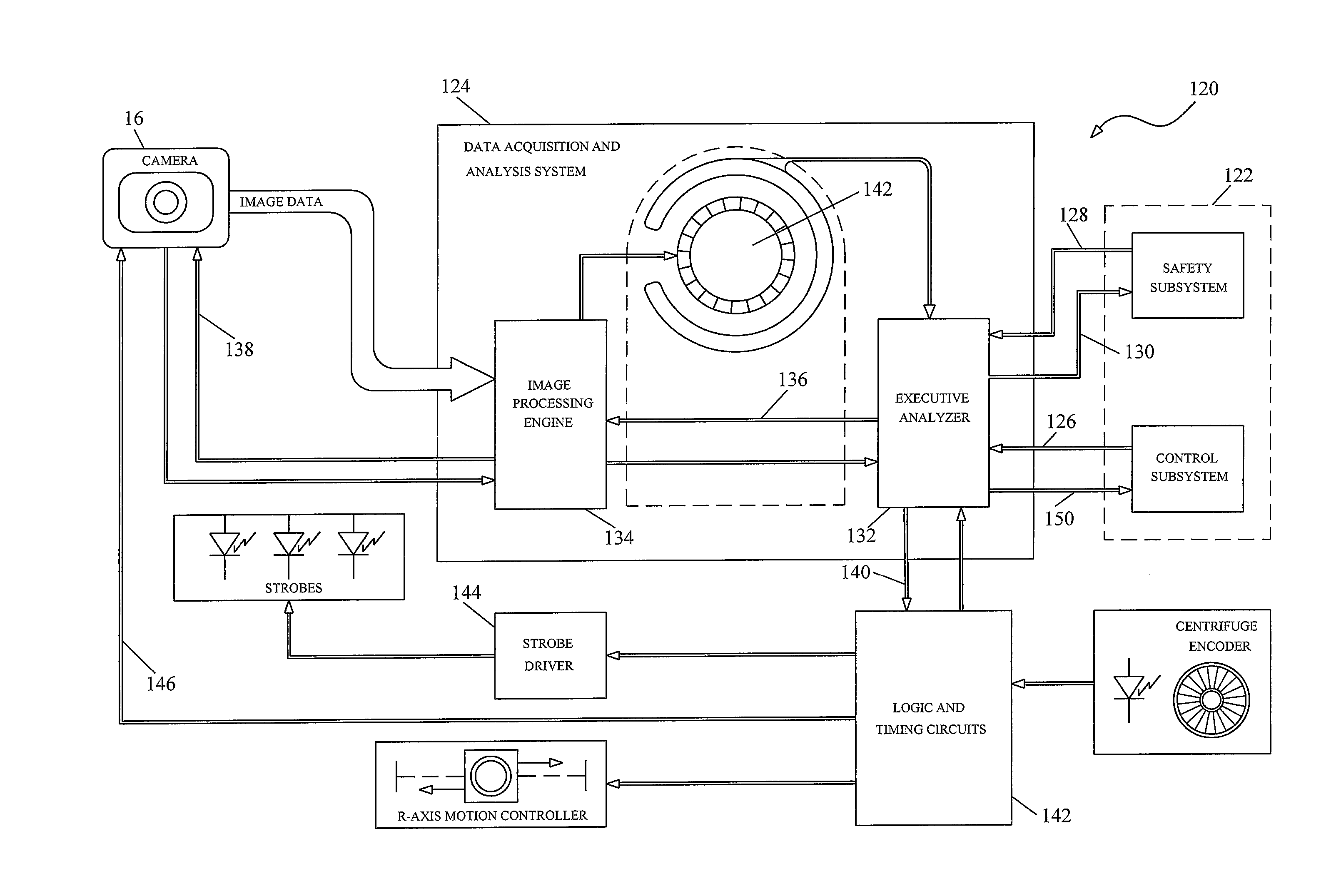
[0010]It has been found that certain transient conditions can cause loss of certain blood components, particularly white blood cells, in an optically controlled blood separation apparatus. Improvements to an optical chamber, wherein blood components are extracted from a separation chamber, have been found to improve the efficiency of white blood cell collection.
[0011]A function of the centrifuge blood processing system described herein may be the collection of white blood cells. It has been found that the collection of white blood cells is highly sensitive to changing flow conditions. If the pumps controlling fluid flow stop, the interface between the red blood cells and the buffy coat generally falls, at least temporarily, to the level of the red blood cell extraction port. In such circumstances, a layer of white cells that had been collected on top of the red blood cell layer is frequently carried into the red blood cell extraction port and returned to the patient or donor. Since such white cells are co-mingled with the total blood volume of the patient / donor, they are no longer available for collection without significant re-processing of the patient's blood. Such losses can significantly decrease the efficiency of white blood cell collection.
[0013]This configuration allows white cell-containing buffy coat to be withdrawn from the optical cell through the buffy coat extraction port for further separation in a fluidized-bed filtration chamber. If the flow conditions are interrupted, for example, by stopping one or more of the pumps, the RBC level falls to the level of the orifice of the RBC extraction port. The dam and ramp, however, prevent the buffy coat layer and white cells from flowing downstream to the third extraction port, thus preserving the white cells for collection when steady state flow conditions are re-established.
[0014]A further feature of the optical cell reduces false detection of red blood cells in the first extraction port, a condition that might cause a loss of collectable white cells. An extraction port comprised a stepped lumen having a larger diameter bore radially outwardly and a smaller diameter bore radially inwardly. The larger diameter bore is needed to allow for a sufficiently large area for optical detection of conditions in flux monitoring regions. The smaller diameter lumen is needed to promote a higher flow velocity through a white cell tube. The volume of white cells collected is comparatively small, compared to the volumes of red blood cells or plasma being processed through the system. A narrow lumen in the white cell tube reduces the possibility of stagnation in the white cell line. Nevertheless, it is believed that vortices in the fluid flowing through the lumen in a prior art design may temporarily capture white blood cells. White cells may continue to accumulate in the vicinity of a lip until a critical volume is dislodged and passes through the small diameter area of the lumen. The accumulation of white cells near the flux monitoring regions impedes the transmission of light through the flux monitoring regions. The darkening of these regions may be erroneously interpreted as red blood cells. To alleviate this condition, a lumen or bore in the first extraction port has a frustroconical taper.

Problems solved by technology

It has been found that certain transient conditions can cause loss of certain blood components, particularly white blood cells, in an optically controlled blood separation apparatus.
Since such white cells are co-mingled with the total blood volume of the patient / donor, they are no longer available for collection without significant re-processing of the patient's blood.
Such losses can significantly decrease the efficiency of white blood cell collection.

Method used


Image

  • System for Blood Separation with Shielded Extraction Port and Optical Control
  • System for Blood Separation with Shielded Extraction Port and Optical Control
  • System for Blood Separation with Shielded Extraction Port and Optical Control

Examples


Embodiment Construction

[0033]Referring to the drawings, like numerals indicate like elements and the same number appearing in more than one drawing refers to the same element. In addition, hereinafter, the following definitions apply:

[0034]The terms “light” and “electromagnetic radiation” are used synonymously. Light useful for the present invention includes gamma rays, X-rays, ultraviolet light, visible light, infrared light, microwaves, radio waves or any combination of these. “Separation axis” refers to the axis along which blood components having different densities are separated in a density centrifuge. As a separation chamber is rotated about a central rotation axis in a density centrifuge, the centrifugal force is directed along separation axes. Accordingly, a plurality of axes rotates about the central rotation axis of a density centrifuge.

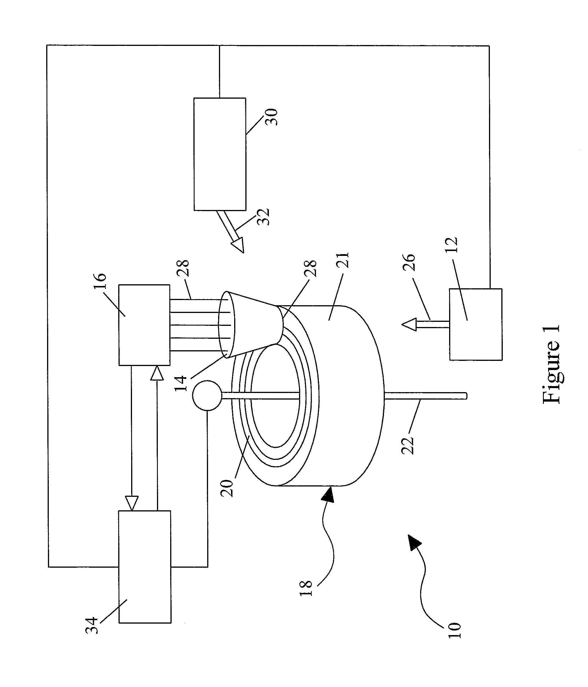
[0035]FIG. 1 schematically illustrates an exemplary embodiment of a blood component separation device 10 with an optical monitoring system capable of measuring ...


PUM

PropertyMeasurementUnit
thicknessaaaaaaaaaa
densityaaaaaaaaaa
diameteraaaaaaaaaa

Abstract

A centrifugal blood separation system comprising a rotor, a light source, an optical sensor, a control system, a separation vessel, and an optical cell on the separation vessel. The optical cell has a first extraction port extending radially outwardly into the optical cell, a red blood cell extraction port downstream from the first extraction port and extending into the optical cell beyond the first extraction port; and a dam between said first extraction port and said red blood cell extraction port, having an upper edge and a lower edge, wherein the first extraction port and the red cell extraction port are radially between the upper edge and the lower edge of the dam. Also, a first extraction port having a bore having a first diameter, a lumen having a second diameter smaller than the first diameter, and a frustro-conical passageway coupling the bore to the lumen.

Description

CROSS-REFERENCE TO RELATED APPLICATIONS[0001]This application is related to U.S. Pat. No. 7,605,388, which is incorporated herein by reference. This application claims the priority of U.S. Provisional Application No. 61 / 285,597, currently pending, filed on Dec. 11, 2009.BACKGROUND OF INVENTION[0002]Blood collection and processing play important roles in the worldwide health care system. In conventional large scale blood collection, blood is removed from a donor or patient, separated into its various blood components via centrifugation, filtration, or elutriation and stored in sterile containers for future infusion into a patient for therapeutic use. The separated blood components typically include fractions comprising red blood cells, white blood cells, platelets, and plasma. Separation of blood into its components can be performed continuously during collection or can be performed subsequent to collection in batches, particularly with respect to the processing of whole blood sample...

Claims


Application Information

Patent Timeline
Patent Type & Authority Applications(United States)
IPC IPC(8): B04B13/00B04B15/00
CPCA61M1/3693A61M2202/0439A61M2205/3306B04B13/00A61M1/3696G01N2035/00495B04B2013/006G01N21/51A61M2202/0071A61M1/3616A61M1/362265A61M1/362266A61M1/362262A61M1/36222A61M1/36225
Inventor KOLENBRANDER, JEREMY P.HOLMES, BRIAN M.FELT, THOMAS J.LADTKOW, JAMES R.
Owner TERUMO BCT
Learn More