
Multi-Layer Slides for Analysis of Urine Sediments

a multi-layer slide and urine technology, applied in the field of urine for particles and sediments analysis, can solve the problems of low surface energy, manual labor, and labor intensive manual microscopy, and achieve the effect of high surface energy

Active Publication Date: 2011-06-23
SIEMENS HEALTHCARE DIAGNOSTICS INC
View PDF33 Cites 8 Cited by
  • Summary
  • Abstract
  • Description
  • Claims
  • Application Information

AI Technical Summary

Benefits of technology

[0034]As will be evident, the urine inspection slide is less expensive to make than the typical microfluidic device. The precision manufacturing needed for microfluidic devices where a defined liquid sample contacts a defined amount of reagents is less important when urine particles and sediments are to be measured. Thus, it is not necessary to make a molded plastic base, polish it, treat it to adjust surface energy to affect liquid flow, and seal the base with a top cover. In contrast, the present three layer inspection slide of the invention is simple to make, provides a clear view of the sample, and yet contains the samples without leaking.

Problems solved by technology

Manual microscopy is accurate, but labor intensive and requires trained technicians.
Flow cytometry and flow cell imaging are less accurate, although they require less manual labor.
However, the slide does not separate the viewing chamber from the sample entry opening, which makes it more difficult to accurately read the images in a microscope.
However, the cost of fabricating such microfluidic devices is rather high and less expensive designs would be desirable.

Method used


Image

  • Multi-Layer Slides for Analysis of Urine Sediments
  • Multi-Layer Slides for Analysis of Urine Sediments

Examples


Embodiment Construction

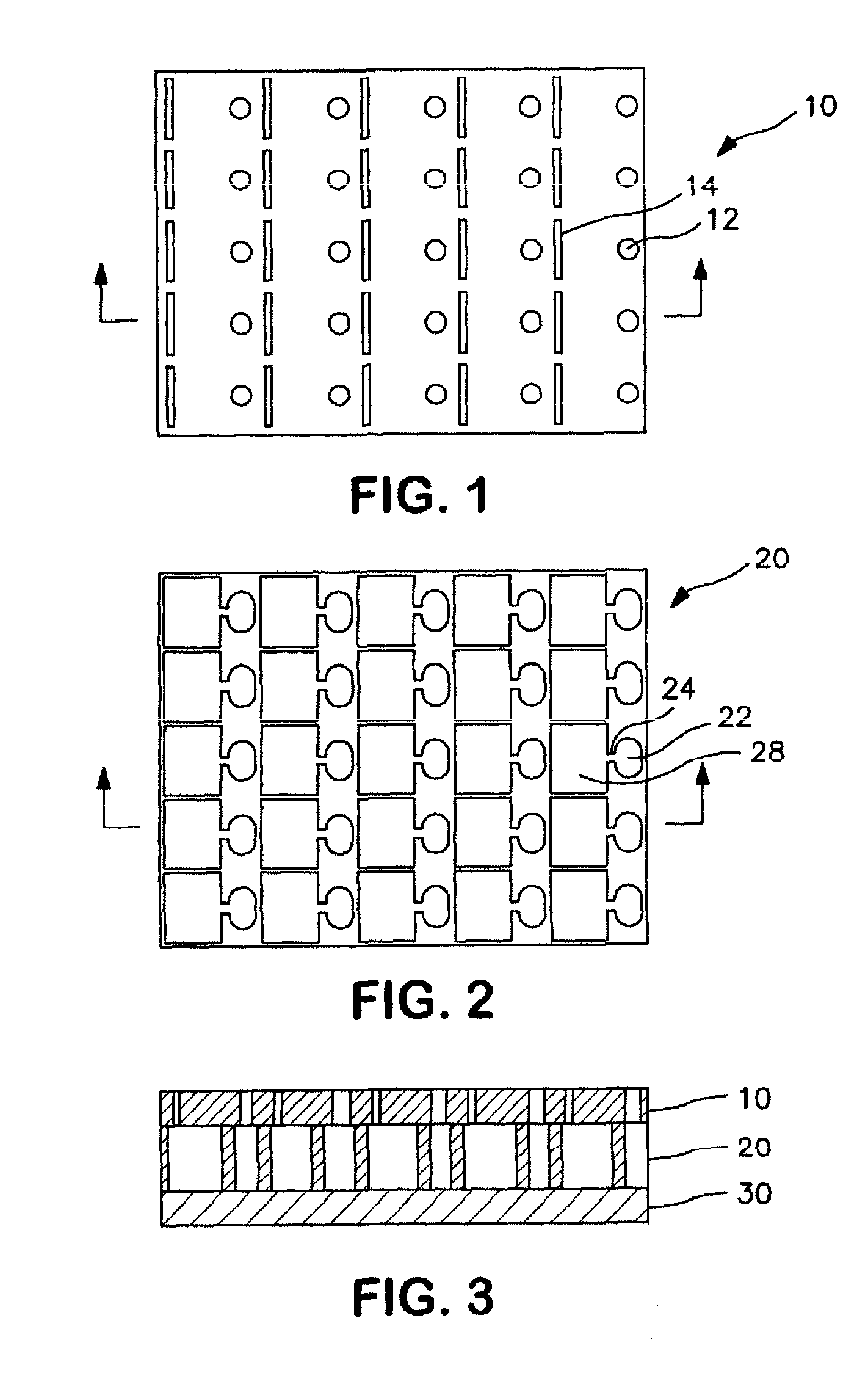
[0012]In general, the invention provides a means for carrying out urine analysis using a slide in which a urine sample is introduced by a pipette and then flows through a capillary passageway into a region in which the sample can be optically examined for the presence of particles and sediments, such as those mentioned above.

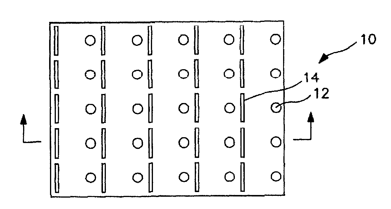
[0013]A preferred embodiment is shown in the FIGS. 1-3. The slide combines three layers (10, 20, and 30 in FIG. 3) and can receive 25 individual samples. The base layer 30 is an optically clear material, with high surface energy relative to the sample, such as cellulose acetate, the top layer 10 is a second sheet of the optically clear material with high surface energy relative to the sample (e.g. cellulose acetate) that has been cut to provide a vent slot 14 for removing air as liquid is introduced and an opening 12 through which the sample is introduced by a pipette. The middle layer 20 is a sheet of polyethylene terephthalate that has been cutout to provide a...


PUM

PropertyMeasurementUnit
surface energyaaaaaaaaaa
thicknessaaaaaaaaaa
thicknessaaaaaaaaaa

Abstract

Visual analysis of urine samples is carried out with the use of a slide consisting of three layers containing an enclosed viewing chamber which receives a urine sample deposited by pipette into an opening on the outer layer of the slide. From the inlet opening the sample enters an inlet chamber in the middle layer and passes through a capillary passageway into the viewing chamber where it is inspected for particles and sediments.

Description

BACKGROUND OF THE INVENTION[0001]This invention generally relates to analysis of urine for particles and sediments of various types. Examples include leukocytes, erythrocytes, epithelial cells, oval fat bodies, hyaline casts, granular casts, leukocytes casts, renal cell casts, waxy casts, fatty casts, uric acid crystals, calcium oxalate crystals, hippuric acid crystals, calcium phosphate crystals, triple phosphate crystals and the like. Such analyses are useful for determining whether a patient requires medical attention.[0002]At present, urine sediment analysis may be done through manual microscopy, flow cytometry, flow cell imaging, and microfluidic slide imaging. Of these, microfluidic slide imaging has advantages over the others. Manual microscopy is accurate, but labor intensive and requires trained technicians. Flow cytometry and flow cell imaging are less accurate, although they require less manual labor. However, follow up with manual microscopy if often required. Examples o...

Claims


Application Information

Patent Timeline
Patent Type & Authority Applications(United States)
IPC IPC(8): G01N21/01
CPCB01L3/5025B01L3/502753B01L2200/0684B01L2300/0819B01L2300/0822G02B21/34G01N21/03G01N21/84G01N33/493G01N2021/0346G01N2021/8405B01L2400/0406
Inventor PUGIA, MICHAEL J.LU, HENRY
Owner SIEMENS HEALTHCARE DIAGNOSTICS INC
Learn More