

Chitosan-based nanoparticles and methods for making and using the same
a technology of chitosan and nanoparticles, applied in the field of biomedical imaging, can solve the problems of limiting the potential use of biomedical imaging applications of these probes, and limiting their application
- Summary
- Abstract
- Description
- Claims
- Application Information
AI Technical Summary
Benefits of technology
Problems solved by technology
Method used
Image

Examples
example 1
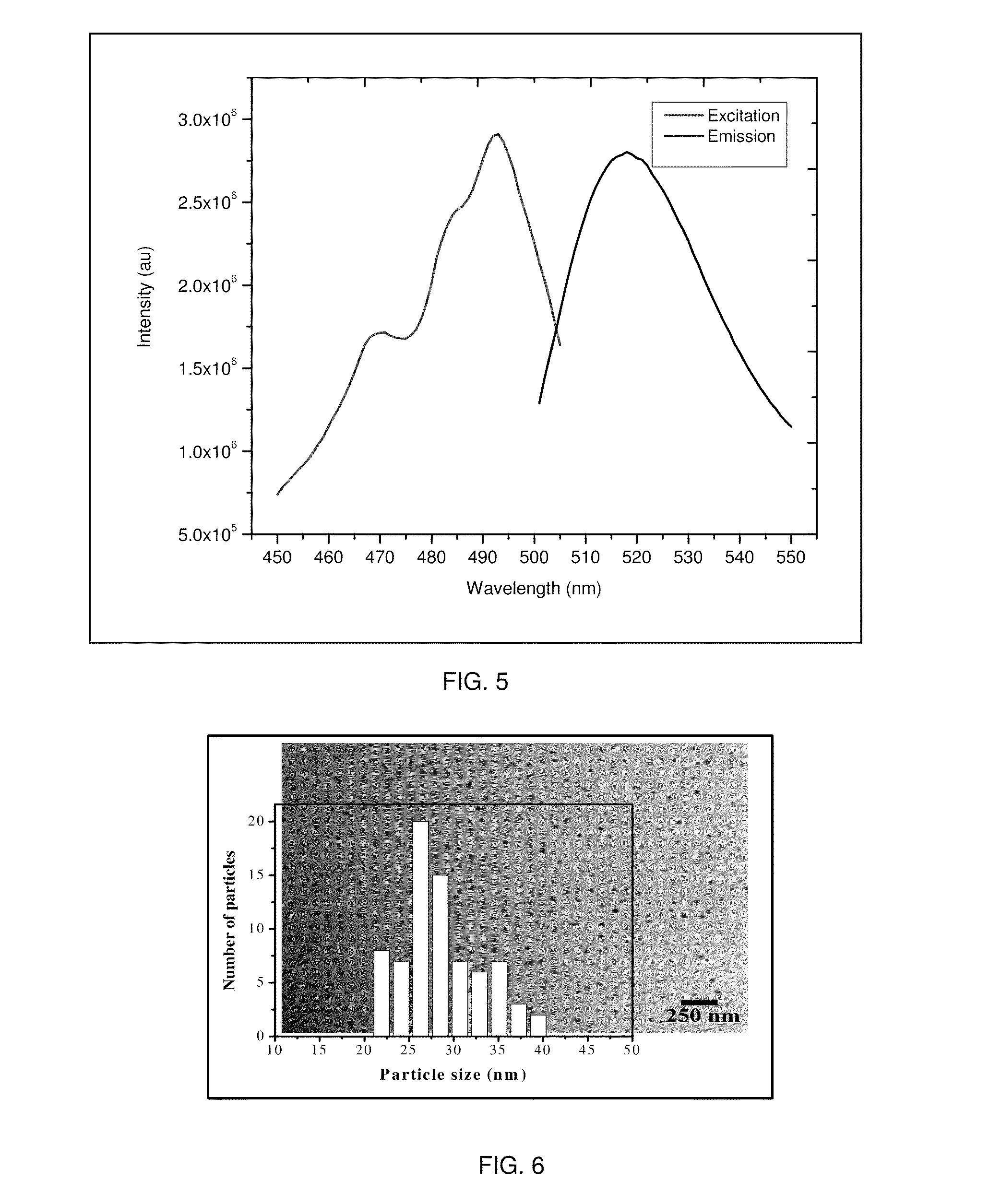
[0053]In this example, fluorescent chitosan nanoparticles (FCNPs) were synthesized using a homogeneous water-in-oil (W / O) microemulsion system consisting of cyclohexane (oil), Triton X-100 (surfactant), n-hexanol (co-surfactant) and water. To retain the particulate integrity, the FCNPs were covalently cross-linked with tartaric acid. Water-insoluble low molecular weight (50-190 kDa) chitosan polymer was first dissolved in 1% acetic acid solution. A part of the chitosan solution was treated with excess amount of amine-reactive fluorescent dye, fluorescein isothiocyanate (FITC), which produced FITC labeled chitosan polymer. Unbound FITC molecules were removed by ethanol / water washing. For the FCNP synthesis, two separate water-in-oil microemulsions, ME and ME U, were prepared. The aqueous phase of ME I contained a mixture of FITC labeled chitosan and pure chitosan polymer solutions. The ME II aqueous phase comprised of a mixture of the crosslinker tartaric acid and water-soluble carbo...
example 2
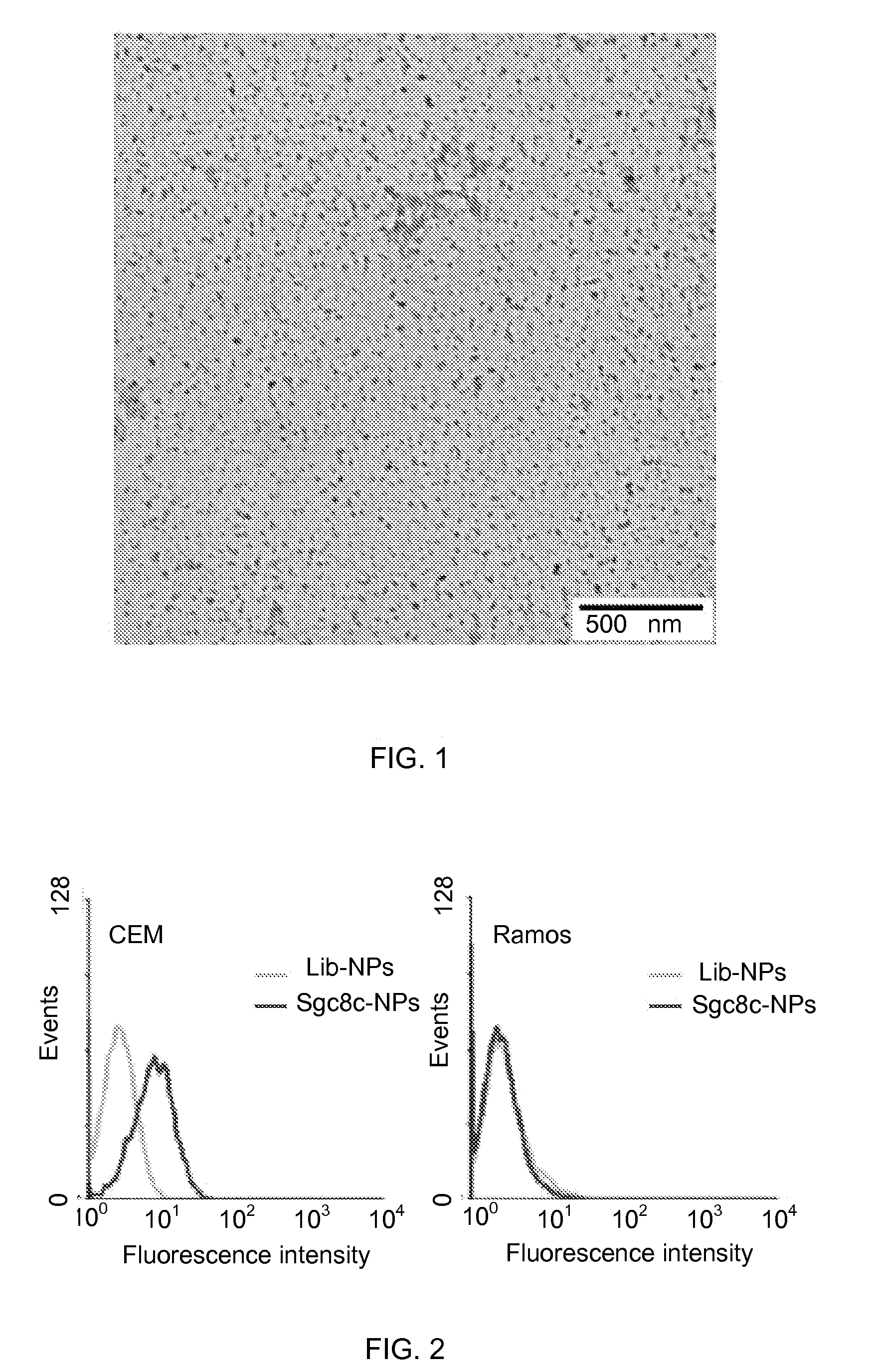
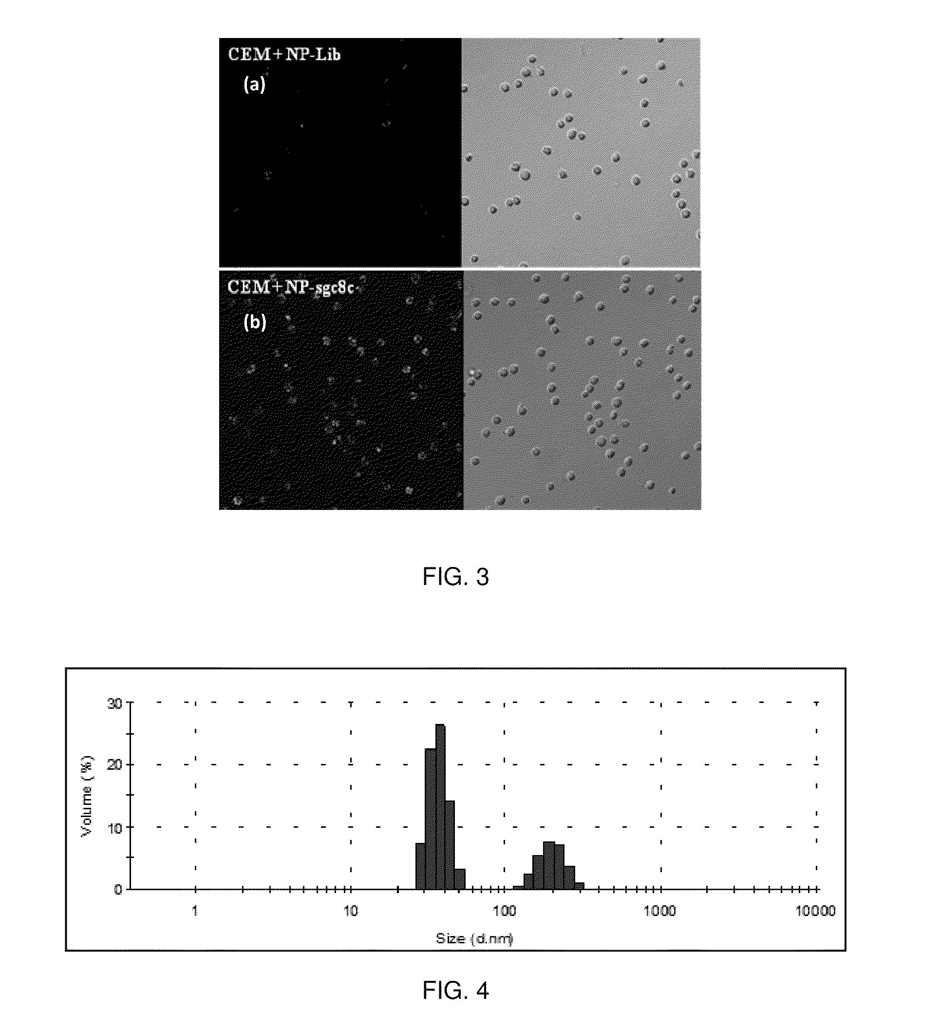
[0055]Identifying cancer is crucial for its early detection and diagnosis. To facilitate this, DNA aptamers specific to cancer biomarkers have become effective specific molecular probes and their conjugation to nanoparticles has given a new dimension to diagnostic and therapeutic applications. To demonstrate specific targeting to the CCRF-CEM cells (CCL-119 T-cell, human acute lymphoblastic leukemia), the FCNPs described above were covalently attached to the DNA aptamer sgc8c (5′-ATC TAA CTG CTG CGC CCC CGG GAA AAT ACT GTA CGG TTA GA-3′). The sgc8c is a strong aptamer with Kd in the nM range. The aptamer was modified at the 5′ position with a carboxyl group. The carboxylated aptamer was then conjugated to the surface amine groups of the FCNPs using water soluble carbodimide chemistry. Similarly, as a negative control, a library of randomized sequence of ssDNA 41 nucleotides was conjugated to the FCNP surface. The aptamer conjugated FCNPs were incubated with CCRF-CEM cells and Ramos ...
example 3
[0057]Example 3 more particularly describes a procedure for making FITC-labeled chitosan nanoparticles set forth in Example 1. Low molecular weight chitosan polymer (75-85% deacetylated), Triton X-100, N-hydroxysuccinimide (NHS), 1-ethyl-3-(3-dimethylaminopropyl carbodiimide hydrochloride) (EDC) were purchased from Sigma-Aldrich Chemical Co., USA; Fluorescein isothiocyanate (FITC), anhydrous ethanol were purchased from Fisher Scientific. Dialysis cellulose membrane (MWCO, 6-8 kD) was purchased from Spectrum Laboratories (Rancho Dominguez, Calif.). Deoxyribonucleotides and 5′-carboxyl modifiers were purchased from Glen Research (Sterling, Va.). All solvents and reagents were obtained from Fisher Scientific and were used without further purification. CCRF-CEM cells (CCL-119 T-cell, human acute lymphoblastic leukemia) and Ramos cells (CRL-1596, B-cell, human Burkitt's lymphoma) were obtained from American Type Culture Association, USA. All of the cells were grown in RPMI-1640 containin...
PUM
| Property | Measurement | Unit |
|---|---|---|
| particle size | aaaaa | aaaaa |
| zeta potential | aaaaa | aaaaa |
| particle size | aaaaa | aaaaa |
Abstract
Description
Claims
Application Information