Medical Image Processing

a technology of medical image and processing, applied in the field of medical imaging and analysis, can solve the problems of large uncertainty inherent in detection problems, limited ability to model the complexity of actual polyps found in human anatomy, etc., and achieve the effects of low cost, low cost, and easy encoded

Active Publication Date: 2011-09-08
SAMSUNG ELECTRONICS CO LTD
View PDF5 Cites 29 Cited by
  • Summary
  • Abstract
  • Description
  • Claims
  • Application Information

AI Technical Summary

Benefits of technology

[0011]The method allows different types of features, particularly appearance features and shape features, to be combined in order to detect objects in a medical image. The advantages of the Bayesian technique disclosed herein are as follows. Firstly, there is a large amount of the uncertainty inherent to the detection problems in medical imaging which can be best accounted for through statistical techniques. Secondly, there often is useful medical knowledge (such as lesion density, size, shape, location, etc.) that can be utilised to constrain the solution of detection problems. This prior medical knowledge can be easily encoded into a Bayesian model. Finally, a Bayesian technique provides a unified framework to incorporate various—often disparate—features into a single statistical model. The probability distribution for each feature tenor is preferably modelled using a parameterised functional form which is based on prior medical knowledge and a training step for the learning of function parameters.
[0012]To the inventors' knowledge, this is the first time that a learning-based Bayesian approach has been used in a CTC CAD system. By deriving the values of the parameters that are used model the various features (such as the shape, appearance and / or anatomical features described below) from training data, the Bayesian framework disclosed herein provides robust and consistent performance. In particular, the Bayesian framework can have a high sensitivity for detecting objects, whilst generating relatively few false positives.

Problems solved by technology

Firstly, there is a large amount of the uncertainty inherent to the detection problems in medical imaging which can be best accounted for through statistical techniques.
Such models have limited capability to model the complexity of actual polyps found in human anatomy, but do not require any training from example data.

Method used


Image

  • Medical Image Processing
  • Medical Image Processing
  • Medical Image Processing

Examples


Embodiment Construction

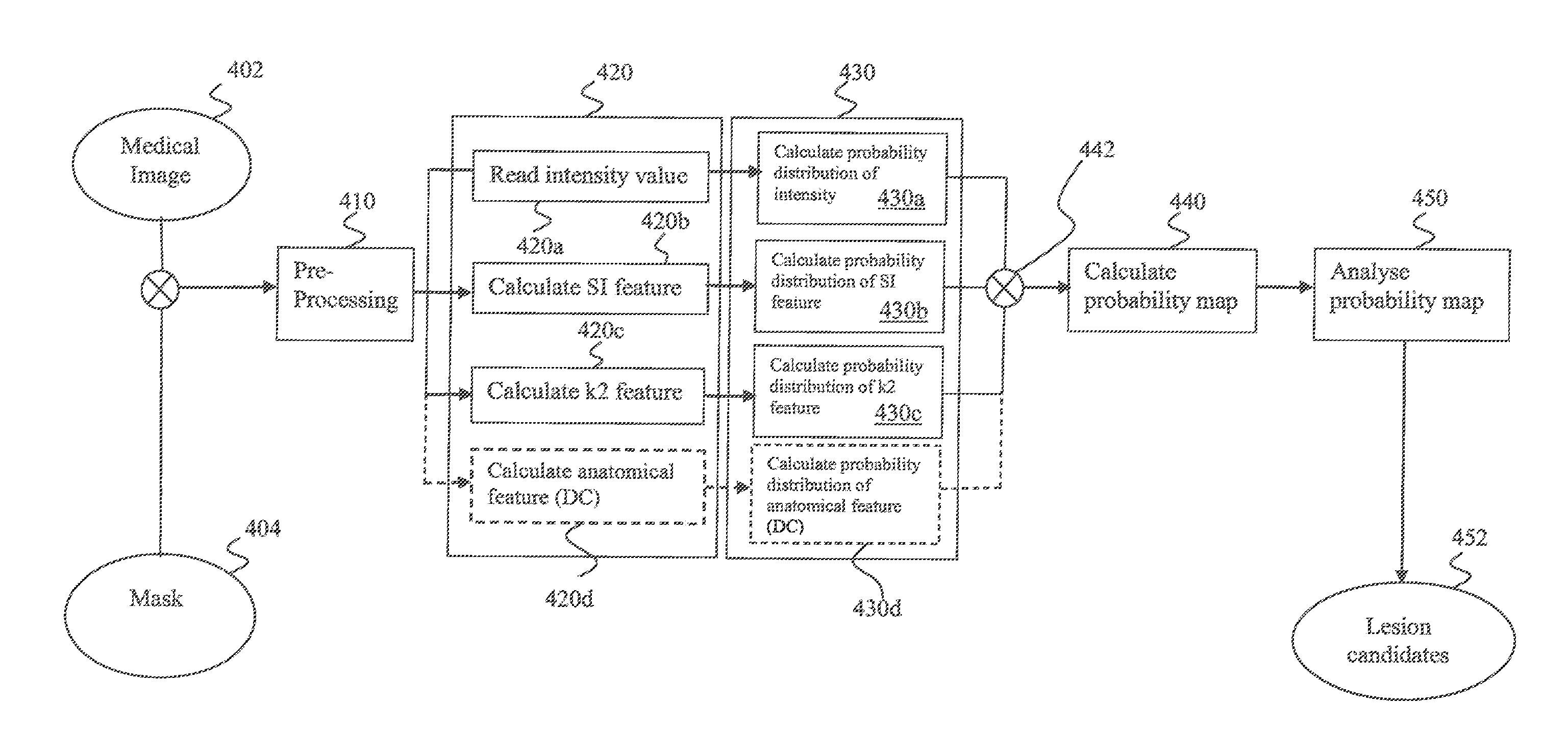
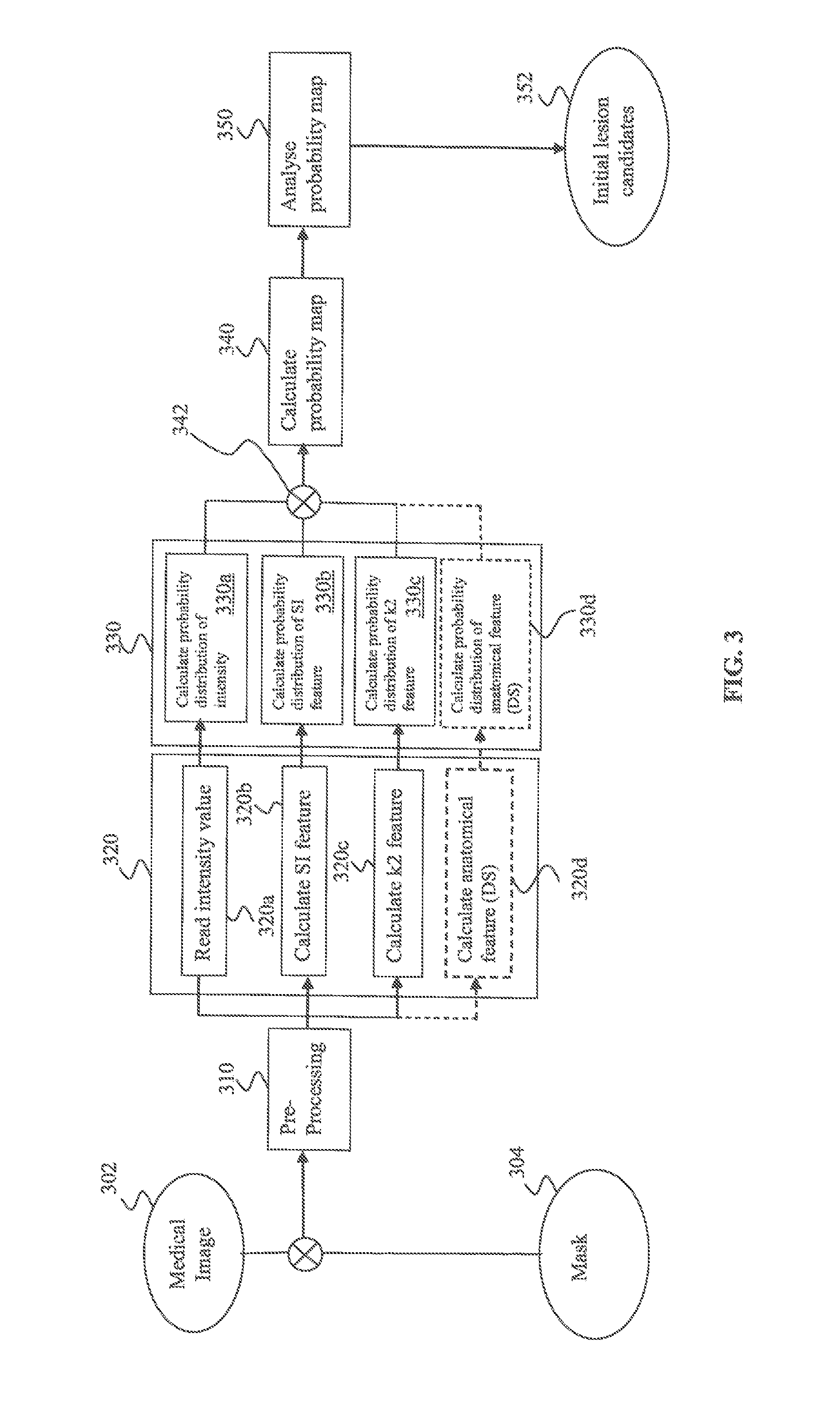
[0041]The invention will now be described, purely by way of example, in the context of a method of identifying polyps in the colon. However, it is to be understood that the invention is not limited solely to the identification of colonic polyps. The invention can also be used to identify other anatomical features such as lung nodules, liver lesions, mammographic masses, brain lesions, any other suitable type of abnormal tissue or suitable types of healthy tissue.

[0042]The invention is directed to processing three-dimensional medical image data using a Bayesian framework. The term “Bayesian framework” as used herein refers to the use of Bayes' law to combine statistical information relating to a plurality of features that characterise properties of a medical image, in order to determine the probability that a particular voxel in the medical image represents a particular object. The three-dimensional medical image data can be generated by a computed tomography (CT) scan, or from a mag...


PUM


Abstract

A computer-implemented method of detecting an object in a three-dimensional medical image comprises determining the values of a plurality of features at each voxel in at least a portion of the medical image. Each feature characterises a respective property of the medical image at a particular voxel. The likelihood probability distribution of each feature is calculated based on the values of the features and prior medical knowledge. A probability map is generated by using Bayes' law to combine the likelihood probability distributions, and the probability map is analysed to detect an object.

Description

FIELD OF THE INVENTION[0001]The present invention relates to medical imaging and analysis. More particularly, the present invention relates to a method and apparatus for detecting abnormalities (lesions) in medical images. The present invention can be used to process medical images in order to detect potential lesions and / or to reject false positives when potential lesions have already been detected.BACKGROUND TO THE INVENTION[0002]Medical imaging is generally recognised as key to better diagnosis and patient care. It has experienced explosive growth over the last few years due to imaging modalities such as x-ray, Computed Tomography (CT), ultrasound, Magnetic Resonance Imaging (MRI) and Positron Emission Tomography (PET). Conventionally, medical images have been inspected visually by highly-trained medical practitioners in order to identify anatomic structures of interest, such as lesions. However, the process can be tedious, time consuming and must be performed with great care.[00...

Claims


Application Information

Patent Timeline
Patent Type & Authority Applications(United States)
IPC IPC(8): G06K9/00
CPCG06T7/0012G06T7/0087G06T2207/30032G06T2207/20076G06T2207/20081G06T2207/10072G06T7/143G06F18/00
Inventor YE, XUJIONGSLABAUGH, GREGORY GIBRAN
Owner SAMSUNG ELECTRONICS CO LTD
Why Eureka
Try Eureka