Method for surface inclusions detection, enhancement of endothelial and osteoblast cells adhesion and proliferation, sterilization of electropolished and magnetoelectropolished nitinol surfaces

a technology of magnetoelectropolishing nitinol and surface inclusions, which is applied in the direction of halogen oxide/oxyacids, blood vessels, dental tools, etc., can solve the problems of enhanced corrosion behavior, unsatisfactory consequences, and reduced bio, so as to enhance endothelial and osteoblast cell adhesion, reduce or diminish bio, and enhance corrosion behavior

Inactive Publication Date: 2012-04-19
ROKICKI MARGARET
View PDF1 Cites 7 Cited by
  • Summary
  • Abstract
  • Description
  • Claims
  • Application Information

AI Technical Summary

Benefits of technology

[0018]The present invention relates to a method for detection of surface inclusions, enhancement of endothelial and osteoblast cell adhesion, and proliferation and sterilization of electropolished and magnetoelectropolished implantable Nitinol medical devices surfaces. The overlooked surface inc

Problems solved by technology

The overlooked surface inclusions on electropolished or magnetoelectropolished Nitinol implantable medical device surfaces could lead to very undesirable consequ

Method used


Image

  • Method for surface inclusions detection, enhancement of endothelial and osteoblast cells adhesion and proliferation, sterilization of electropolished and magnetoelectropolished nitinol surfaces
  • Method for surface inclusions detection, enhancement of endothelial and osteoblast cells adhesion and proliferation, sterilization of electropolished and magnetoelectropolished nitinol surfaces
  • Method for surface inclusions detection, enhancement of endothelial and osteoblast cells adhesion and proliferation, sterilization of electropolished and magnetoelectropolished nitinol surfaces

Examples


example 1

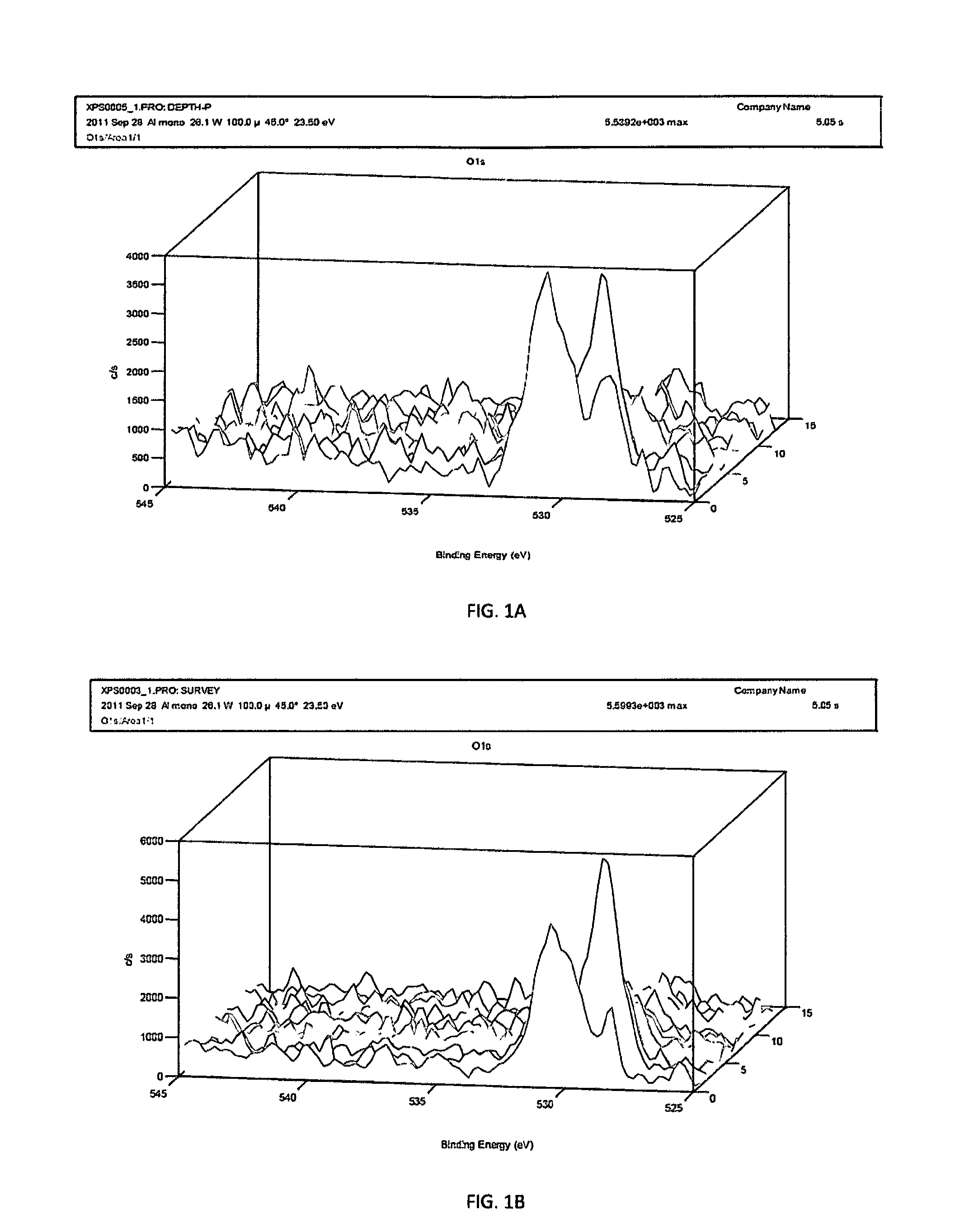
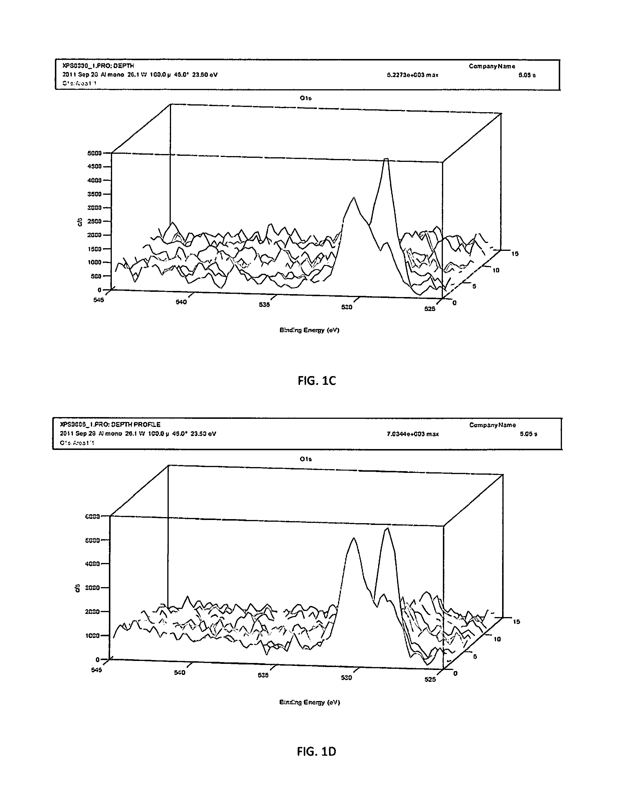
[0057]Ten electropolished Nitinol guidewires were placed separately in glass tubes filled with 6% NaClO of room temperature around 25° C. Almost immediately one wire started to corrode in one place, black flocculent started to develop in corrosion site. Bubbles of oxygen were observed departing upward from corrosion site. After 15 minutes all wires were removed from 6% NaClO and rinsed with water. Visual examination of corroded wire showed a pit in place were black flocculent developed (this place was the place of Nitinol surface inclusion). The nine remaining wires have not shown signs of corrosion.

example 2

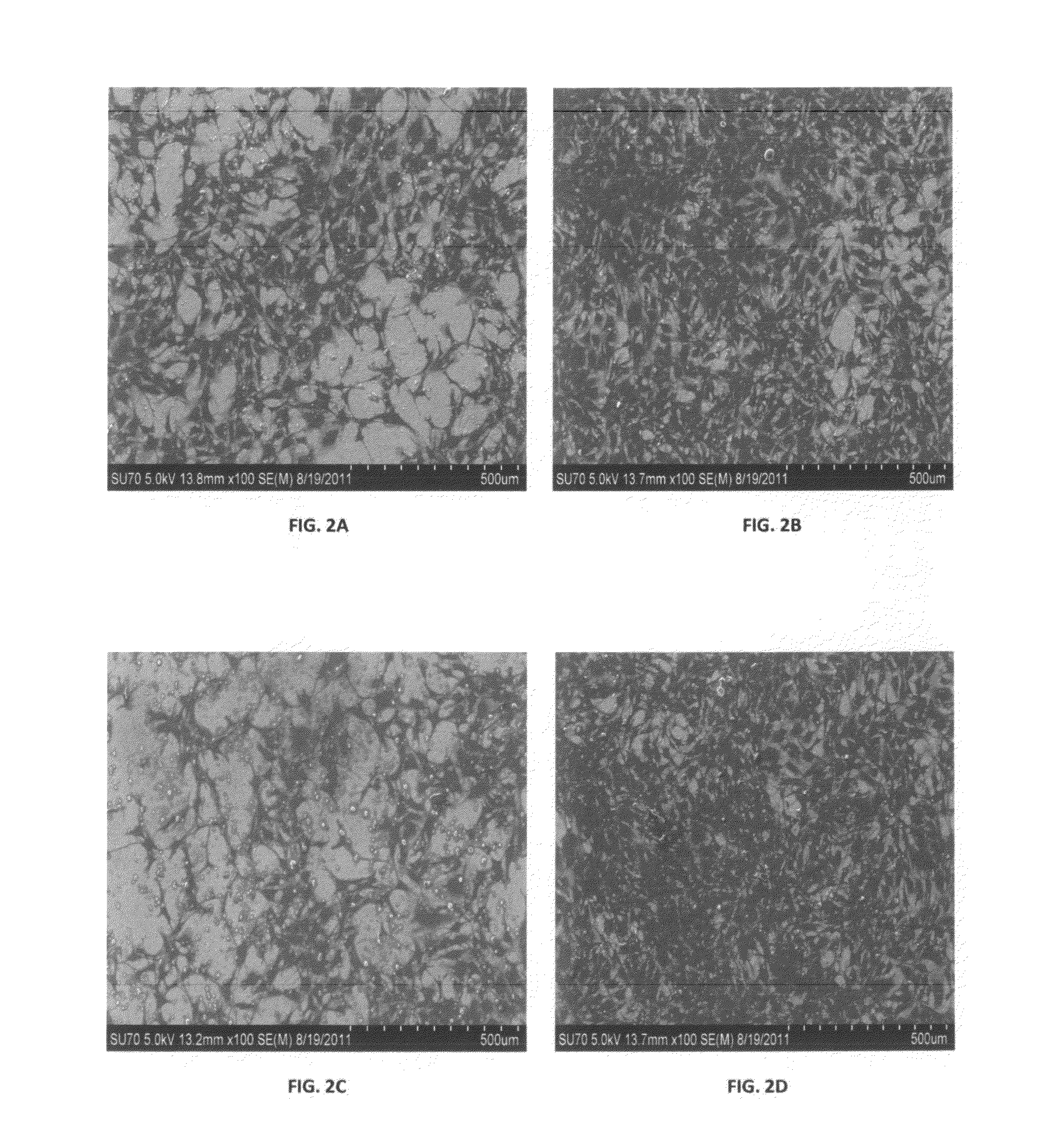
[0058]Four samples (10 mm in diameter 2 mm thick) stamped from the same plate of Nitinol were finished as follow: electropolished (EP)—FIG. 2A, electropolished (EP)+6% NaClO FIG. 2B, magnetoelectropolished (MEP)—FIG. 2C, magnetoelectropolished (MEP)+6% NaClO—FIG. 2D. In order to assess adhesion and proliferation of osteoblast cells on differently finished Nitinol samples MC3T3 osteoblast cells were seeded and incubated for 72 hours at 37° C. After 72 hours of incubation adhesion and proliferation of MC3T3 osteoblast cell were evaluated by scanning electron microscope (SEM). At qualitative level the cells had a healthy response and have a similar morphology on all samples. However at quantitative level anybody can see profound difference between EP (FIG. 2A), MEP (FIG. 2C) and EP (FIG. 2B), MEP (FIG. 2D)+6% NaClO treated samples. EP and MEP+6% NaClO treated samples reached almost 100% confluency compared to about 40% confluency of EP and MEP samples without 6% NaClO treatment. It is ...

example 3

[0059]Four samples (10 mm in diameter 2 mm thick) stamped from the same plate of Nitinol were finished as follow: electropolished (EP)—FIG. 3A, electropolished (EP)+6% NaClO—FIG. 3B, magnetoelectropolished (MEP)—FIG. 3C, magnetoelectropolished (MEP)+6% NaClO—FIG. 3D. In order to assess adhesion and proliferation of HUVEC (human umbilical vein endothelial cell) the ISO 10993 protocols for biological evaluation of medical devices was used. After 72 hours at 37° C., 5% CO2 in cell culture media Nitinol samples were gently washed with DPBS stained with Hoechst dye (to highlight nuclei of the cells) and Mitotracker Red (to highlight mitochondria of the cells). The samples were again incubated for 20 minutes, washed 3 times in DPBS and fixed on the samples surface with 10% formaldehyde and covered by glass slides. The qualitative and quantitative evaluation of adhesion and proliferation of endothelial cells on differently finished Nitinol samples were accomplished by using an Olympus IX81...


PUM

PropertyMeasurementUnit
Temperatureaaaaaaaaaa
Fractionaaaaaaaaaa
Fractionaaaaaaaaaa

Abstract

The method for surface inclusions detection, enhancement of endothelial and osteoblast cells adhesion and proliferation and sterilization of electropolished and magnetoelectropolished Nitinol implantable medical device surfaces uses an aqueous solution of chemical compounds containing halogenous oxyanions as hypochlorite (ClO) and hypobromite (BrO) preferentially 6% sodium hypochlorite (NaClO).

Description

CROSS REFERENCE TO RELATED APPLICATIONS[0001]This application is continuation-in-part and claims the benefit of priority to U.S. patent application Ser. No. 12 / 660,904 with filling date of Mar. 5, 2010, which claims priority to U.S. provisional application No. 61 / 214,380, filed on Apr. 23, 2009.BACKGROUND OF THE INVENTION[0002]The present invention relates to the field of chemical testing, modifying the surface of a material to promote enhancement of endothelial and osteoblast cells monolayer coverage and sterilization of intermetallic materials. More specifically, it is for testing, surface modifying and sterilization of the electropolished and magnetoelectropolished nickel titanium intermetallic compound known as Nitinol.[0003]The purpose of this test is the detection of Nitinol surface inclusions (except of titanium dioxides TiO2 native inclusions originated during melting process, which do not dissolve in sodium hypochlorite (NaClO) and are harmless to endothelial and osteoblast...

Claims


Application Information

Patent Timeline
IPC IPC(8): A61F2/82B32B15/01B32B5/18A61L2/18C01B11/06C01B11/20A01N59/08C22C19/03A61C3/00
CPCA61B17/0401Y10T428/12479A61B17/12022A61B17/12113A61B17/1214A61B17/32A61B19/34A61B2017/00526A61B2017/00867A61F2/24A61F2/82A61L27/06A61L29/02A61L31/022C22C19/007C22C19/03G01N33/20Y10T428/12806A61B17/064A61B90/70G01N33/208G01N33/2028
Inventor ROKICKI, RYSZARD
Owner ROKICKI MARGARET
Why Eureka
Try Eureka